The scimitar syndrome is a constellation of cardiopulmonary abnormalities consisting of anomalous right pulmonary venous drainage of the right lung to the inferior caval vein, an anomalous systemic arterial blood supply from the abdominal aorta to the inferior segments of the right lung, variable degrees of hypoplasia of the right lung and pulmonary artery, dextroposition of the heart and anomalies of the right bronchial tree including sequestration.Reference Dusenbery, Geva and Seale 1 , Reference Midyat, Demir and Aşkin 2 In ~2/3 of the patients, the anomalous pulmonary “scimitar” vein provides drainage for the entire right lung, but in one-third only the lower segments, usually the middle and lower lobes, are involved and the right upper lobe venous drainage is then to the left atrium.Reference Midyat, Demir and Aşkin 2
Many terms have been used by a number of authors to describe those cases that, although a close cousin, lack all the elements of the complete scimitar syndrome.Reference Dusenbery, Geva and Seale 1 , Reference Valdez-Davila, Avila-Varguez, Castaneda-Zuniga, Probst and Amplatz 3 – Reference Yoo, Al-Otay and Babyn 12 There is, however, no general agreement about an exact classification of these variants. In this study, we report our experience of scimitar syndrome variants since 1992.
Our aim was to describe those patients with variants of the classical scimitar syndrome presenting to a single institution over a period of 22 years.
Materials and methods
We undertook a retrospective review of every case of scimitar syndrome and its variants diagnosed at our institution between January, 1992 and December, 2013.
The medical records, operation notes, and the investigative findings including cardiac catheterisation and angiography, thoracic CT, cardiovascular MRI, and bronchoscopy or bronchogram were reviewed.
Scimitar syndrome was definedReference Dusenbery, Geva and Seale 1 , Reference Midyat, Demir and Aşkin 2 as partial or total anomalous right pulmonary venous drainage to the inferior caval vein with varying degrees of right pulmonary artery and lung hypoplasia; one or more anomalous abdominal aortopulmonary collateral to the right lung, usually right lower lobe; and variable degrees of dextroposition of the heart.
The anomalous pulmonary vein may also connect to the inferior part of the right atrium below or just above the diaphragm. Associated features are as follows: horseshoe lung, secundum atrial septal defect, pulmonary vein stenosis, abnormalities of the bronchial branching pattern, and lung segmentation. Also included in this group was a patient with partial right anomalous venous drainage to the inferior caval vein and to the right atrium.
This was because the main features of the scimitar syndrome were present, and these types of cases have been included in previously published large series as well.Reference Dusenbery, Geva and Seale 1
We arbitrarily proposed the following criteria for the variants of the scimitar syndrome:
-
∙ “Left variant”: aortopulmonary collateral to the left lung, degrees of left lung and pulmonary artery hypoplasia, at least one anomalous vein draining to various sites.
-
∙ Presence of an anomalous right pulmonary vein that appears identical to the usual scimitar vein, draining to the inferior caval vein, but without other features of the scimitar syndrome.
-
∙ Presence of these three features of scimitar syndrome: abdominal aortopulmonary collateral, hypoplastic right lung, and hypoplastic or absent right pulmonary artery with normal or anomalous pulmonary venous drainage to a site different from the inferior caval vein.
Results
A total of 55 patients were identified according to our inclusion criteria for scimitar syndrome and its variants in the period of time considered. A variation in the usual pattern for scimitar syndrome was identified in 10 patients (18%). There were 10 patients (18% of total) with mixed pulmonary venous drainage to the left atrium and inferior caval vein who were included into the group of patients with scimitar syndrome. This decision was made in view of the presence of at least one of the right pulmonary veins draining to the inferior caval vein, which is the hallmark of the syndrome, associated with other features of the syndrome.
There were four additional patients not included in this report despite some similarities with the scimitar syndrome. The features common to these four patients were a single or multiple large aortopulmonary collateral(s) connected to the lower part of the left or right lower lobe with hypoplasia of the ipsilateral lung. There were no other features of the scimitar syndrome, and in particular the course and connection of the pulmonary veins were entirely normal; one of these patients also had a congenital cystic adenomatous malformation of the left lower lobe with a systemic supply from the abdominal aorta, whereas three others had sequestration of the left or right lower lobe perfused by the abdominal aortopulmonary collateral. In one of these cases, the heart was located in the right hemithorax.
Presentation
There were four (40%) females; three patients (30%) had an antenatal diagnosis of suspect scimitar syndrome, and the median age at postnatal diagnosis in the remainder was 45 months – with a range from 11 to 319 months. With the exception of one asymptomatic patient with an antenatal diagnosis, the remainder (90%) presented with tachypnoea, with respiratory distress in 3, recurrent chest infections or wheezing in 5, and exercise intolerance with haemoptysis in 1; one patient was lost to follow-up, whereas the remainder were alive at the time of data analysis.
Investigations
All the patients underwent cardiac catheterisation at a median age of 42 months (3–312 months of age); five of them had a CT scan, five had a bronchoscopy,Reference Dusenbery, Geva and Seale 1 – Reference Herer, Jaubert, Delaisements, Huchon and Chretien 4 , Reference Morgan and Forker 6 and two of them a cardiac MRI (patients n. 5 and 8).
Morphology
-
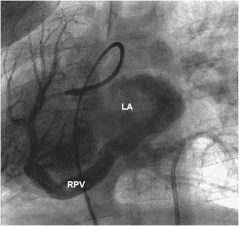
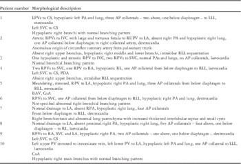
∙ The morphological diagnosis is summarised in Table 1. Among all, three patients had a normal pulmonary venous drainage to the left atrium. In one, the right pulmonary venous drainage to left atrium was formed by a single, “meandering”, tortuous, right pulmonary vein (Fig 1).
-
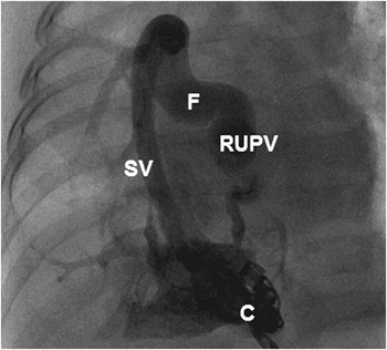
∙ In one patient, there was a virtually atretic right pulmonary vein, severely stenosed at its insertion into the inferior caval vein; this vein was connected to the right upper pulmonary vein via a large and tortuous “fistula” (Fig 2).
-
∙ In three patients with total anomalous right pulmonary venous drainage, the pulmonary veins drained to the superior caval vein, the superior caval vein and the right atrium , and the superior and inferior caval veins, respectively.
-
∙ Only one patient had a mixed drainage of the right pulmonary veins to the left atrium and the superior caval vein.
-
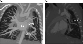
∙ Only one patient had total anomalous left pulmonary venous drainage to the coronary sinus (Fig 3a and b).
-
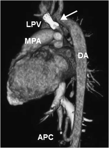
∙ Only one patient had a stenosed anomalous left upper pulmonary vein draining to the innominate vein (Fig 4).
Patient 5, laevophase of a right pulmonary arteriogram showing a single, meandering right pulmonary vein (RPV), draining the entire right lung to the left atrium (LA) with dextroposition of the heart.

Patient 2, selective angiogram into the aortopulmonary collateral during coil embolisation (C). There is an anomalous right scimitar vein (SV) draining to the inferior caval vein (IVC), which was stenosed at the junction with IVC and connected to the right upper pulmonary vein (RUPV) draining to left atrium by a large and tortuous fistula (F). There is dextroposition of the heart.

( a and b ) Patient 1, coronal and sagittal CT views showing tortuous and irregular left pulmonary veins (LPVs) draining to the coronary sinus (CS), left lung hypoplasia, an aortopulmonary collateral (APC) arising above the diaphragm to the inferior part of the left lung. LA=left atrium.

Patient 10, CT 3D reconstruction shows a stent (arrow) positioned at the level of a left upper pulmonary vein stenosis. APC=aortopulmonary collateral; DA=descending aorta; LPV=left pulmonary vein; MPA=pulmonary trunk; 3D=three dimensional.

There were four patients with pulmonary vein stenosis (patients n. 2, 3, 5, and 10); the obstruction was at the junction of the right pulmonary vein with the left atrium, via a single, tortuous, abnormal vessel in one, in the left upper pulmonary vein above the left pulmonary artery in the second (Fig 4), and at the junction of the right pulmonary vein with the inferior caval vein in the third patient. Another patient had a virtual atresia of the anomalous right pulmonary vein to the inferior caval vein that was connected to the right upper pulmonary vein via a “fistula”.
We termed this “fistula” rather than anomalous vein as this looked like a connection between the anomalous pulmonary vein to the inferior caval and the right upper pulmonary vein. The mechanism that we hypothesised was that the fistula was secondary to the severe stenosis of the scimitar vein.
In the latter, there was general hypoplasia of the true pulmonary vein.
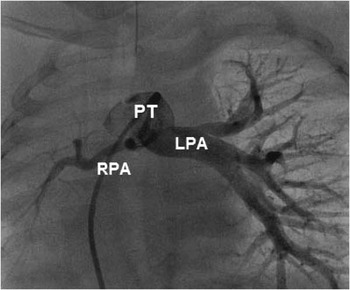
Among all, one patient had normal pulmonary arteries, the right pulmonary artery was absent in three patients, and was hypoplastic in four patients (Fig 5); two patients with a left-sided variant had a hypoplastic left pulmonary artery.
Patient 9, selective pulmonary arteriogram showing a severely hypoplastic right pulmonary artery (RPA) with an abnormal branching pattern. LPA=left pulmonary artery; PT = pulmonary trunk.

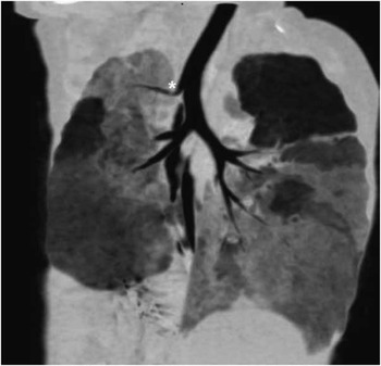
Moreover, four patients had a solitary abdominal aortopulmonary collateral, five patients had multiple collaterals, (Figs 6 and 7) and one patient had no collateral.
Patient 6, descending aortogram showing a large aortopulmonary collateral (APC) arising from the descending aorta (DA) above coeliac axis (CA) and supplying the lower part of the right lung. The DA distal to the origin of the APC is considerably smaller than that above. SMA=superior mesenteric artery.

Patient 10, thoracic CT reformat showing hypoplastic right lung with abnormal right bronchial branching pattern, hypoplastic right bronchi, and a pig bronchus (*).

When the collaterals were in multiples, the smaller additional collaterals were observed in three patients arising from the thoracic aorta. The connection of the collateral(s) was always ipsilateral to the other features.
There was a total of 21 collaterals in nine patients.
In the two patients with a left variant, the collaterals were directed to the lower part of the left lung. Although the origin of the collateral was below the diaphragm in the majority, in six patients it was above the diaphragm – five from the descending thoracic aorta and one from the aortic arch.
There was laevocardia in four patients, mesocardia in two cases, and dextrocardia in the remainder of patients.
Overall, five patients had associated cardiac abnormalities, including coarctation of the aorta (in 2), bicuspid aortic valve (in 1), left superior caval vein to coronary sinus (in 2), patent ductus arteriosus, and anomalous origin of the circumflex coronary artery from the bifurcation of the pulmonary trunk.
In addition, five patients underwent lung CT scan and five had a bronchoscopy, and the bronchopulmonary anatomy was clarified in a total of seven patients.
In this latter group, only two patients had a normal bronchopulmonary anatomy, two had intralobar sequestration of the right lower lobe, hypoplastic right bronchi was found in three patients (Fig 7), absent right upper bronchus in two patients, and a diffuse abnormal parenchyma with bronchiectasies and parenchymal cysts in one patient. In addition, one patient had a pig bronchus in which the origin of the right upper lobe bronchus arises directly from the trachea rather than the right main bronchus .Reference Dolittle and Mair 13
Summary of patients’ morphological description.

AP=aortopulmonary; BAV=bicuspid aortic valve; CoA=coarctation of the aorta; CS=coronary sinus; IVC=inferior caval vein; LA=left atrium; LLL=left lower lobe; LPV=left pulmonary vein; PA=pulmonary artery; PDA=patent ductus arteriosus; PV=pulmonary vein; RL=right lung; RLL=right lower lobe; RPV=right pulmonary vein; RUPV=right upper pulmonary vein; SVC=superior caval vein
Treatment
At the time of cardiac catheterisation, five patients underwent percutaneous embolisation of one or more haemodynamically significant aortopulmonary collaterals; the remainder had an absent or small aortopulmonary collaterals, and one of them underwent right pneumonectomy.
Repeated transcatheter balloon dilatation and stenting of the left upper pulmonary vein stenosis were carried out in one patient (number 10).
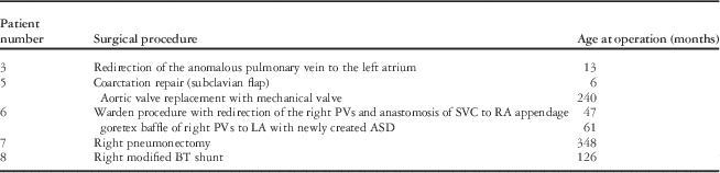
Moreover, four patients had one or more surgical procedures aimed to repair or palliate the venous malformation part of the Scimitar variant.
In addition, one patient (number 5) was treated with an aortopulmonary collateral occlusion and also had a coarctation repair and an aortic valve replacement.
Additional procedures performed to treat associated malformations are listed in Table 2.
Surgical procedures.

ASD=atrial septal defect; BT=Blalock–Taussig; LA=left atrium; PV=pulmonary vein; RA=right atrium; SVC=superior caval vein
Outcome
In this study, one patent was lost to follow-up, whereas this was from 1 to 252 months (median 42) in the remainder. Every patient with available follow-up information was alive at the end of the study. Among the five patients treated only with transcatheter embolisation of the aortopulmonary collateral, two of them were symptom-free, two had mild wheezing, and one had exercise intolerance.
Among the four patients who underwent surgical procedures, one had redirection of the anomalous pulmonary vein to the left atrium and was symptom-free. Patients n. 6 and 7 had subjective exercise intolerance, whereas patient n. 8 had wheezing.
Discussion
The term scimitar variant has been used to describe the spectrum of malformations that lack all the typical features of the scimitar syndrome and sometimes manifest additional features.
Some authors have included cases with the right pulmonary veins draining to both the inferior caval vein and the left atrium,Reference Valdez-Davila, Avila-Varguez, Castaneda-Zuniga, Probst and Amplatz 3 but we preferred to consider the latter as part of the classical syndrome.
A scimitar vein draining to the left atrium is a rare form of variant with only a few cases described in the literature.Reference Yoo, Al-Otay and Babyn 12 , Reference Gao, Burrows, Benson, Rabinovitch and Freedom 14 , Reference Cantinotti, Giordano and Spadoni 15 Left-sided variants of the scimitar syndrome have also been described with hypoplasia of the left lung and pulmonary artery, an anomalous systemic blood supply from the abdominal aorta to the lower segments of the left lung, and an anomalous left pulmonary venous drainage to the inferior caval vein or azygos vein.Reference Bratincsák, Rao and El-Said 9 , Reference Ben-Menachem, Kuroda, Kyger, Brest, Copeland and Coan 16 Bilateral partial anomalous pulmonary venous drainage is also encountered, the most common type being an anomalous drainage of the left superior pulmonary vein to the left innominate vein and right superior pulmonary vein to the superior caval vein.Reference Papamichael, Ikkos, Alkalais and Yannacopoulos 17 Furthermore, there have been a few reports of bilateral anomalous single pulmonary veins; in one case, both vessels formed a common venous trunk connecting to the left atrium.Reference Bratincsák, Rao and El-Said 9
We have described 10 cases of scimitar syndrome variants, which to our knowledge is the largest series of patients from a single centre. The small number and the variation in the morphology make it difficult to come to any generalised conclusion.
Nonetheless, the type of pulmonary venous drainage was the most variable feature. This had a normal course to the left atrium in two cases, but one additional patient had a meandering right pulmonary vein to the left atrium. In addition to the left-sided variants, there was partial or total anomalous pulmonary venous drainage to the superior caval vein in the remaining three cases.
When a common right pulmonary vein drains normally to the left atrium with an abnormal and tortuous course, it can still produce the classical “scimitar sign” on X-ray, and this was observed in our patients. This anomaly was first described by Morgan and Forker,Reference Morgan and Forker 6 and was later called “meandering right pulmonary vein” by Goodman et al.Reference Goodman, Jamshidi and Hipona 7 Although the right-sided variants have been described previously, to our knowledge, at the time of this study, there have been no reports of the left-sided variants.
There were nine patients in whom the major aortopulmonary collateral arose in a typical manner from the abdominal aorta in close proximity to the origin of the coeliac axis, although smaller additional collaterals were observed in three patients arising from the thoracic aorta. The only patient without collaterals had partial anomalous right pulmonary venous drainage to the inferior caval vein at the classical site for scimitar syndrome and the right upper lobe drained anomalously to the right superior caval vein.
With the exception of this patient, every other case had some degree of hypoplasia of the right or left pulmonary artery. The bronchopulmonary abnormalities were present in the majority of patients.
There were four additional patients not included in this report despite some similarities to the scimitar syndrome, as previously mentioned in the results. It is arguable whether or not these four patients represent a variation of the scimitar syndrome, but they are probably best described merely as pulmonary sequestration despite the similarities.
The clinical outcome was variable and depended, primarily, on the presence and extent of anomalous pulmonary venous drainage and the degree of lung hypoplasia, unless there were other associate congenital anomalies such as aortic coarctation, aortic valve disease, and anomalous origin of the left coronary artery.
Although some patients were still symptomatic after transcatheter aortopulmonary collateral occlusion or surgical repair, clinical performance was markedly improved; the main residual symptoms were wheezing and recurrent chest infections. Any exercise intolerance was usually mild and subjective.
Acknowledgement
None.
Financial Support
This research received no specific grant from any funding agency, commercial, or not-for-profit sectors.
Conflicts of Interest
None.