INTRODUCTION
Free-living amoebae of the genus Acanthamoeba are distributed worldwide and are frequently isolated from dust, air, soil, and a variety of water samples, including tap water and recreational water systems (for reviews see: Marciano-Cabral and Cabral, Reference Marciano-Cabral and Cabral2003; Khan, Reference Khan2006; Ibrahim et al. Reference Ibrahim, Boase and Cree2007; Thomas et al. Reference Thomas, McDonnell, Denyer and Maillard2010). Acanthamoeba can cause both opportunistic infections (e.g. Acanthamoeba granulomatous encephalitis, sinusitis, and disseminated cutaneous lesions) and non-opportunistic infections such as keratitis, a disease principally associated with wearing contact lenses that may lead to severe corneal damage (reviews: Marciano-Cabral and Cabral, Reference Marciano-Cabral and Cabral2003; Clarke and Niederkorn, Reference Clarke and Niederkorn2006; Khan, Reference Khan2006, Reference Khan2008).
Acanthamoeba grow and multiply as phagotrophic trophozoites that avidly predate on a variety of bacterial species in the environment (Bottone et al. Reference Bottone, Perez, Gordon and Qureshi1994; Weekers et al. Reference Weekers, Bodelier, Wijen and Vogels1993; De Moraes and Alfieri, Reference De Moraes and Alfieri2008). Adverse conditions cause trophozoites to encyst; cysts are double-walled, resistant stages that remain viable (and infective) for several years (Mazur et al. Reference Mazur, Hadas and Iwanicka1995; Aksozek et al. Reference Aksozek, McClellan, Howard, Niederkorn and Alizadeh2002; Sriram et al. Reference Sriram, Shoff, Booton, Fuerst and Visvesvara2008).
Acanthamoeba spp can be easily identified at the genus level, and are classified according to cyst size and morphology into 3 morphological groups (I, II, and III) (Pussard and Pons, Reference Pussard and Pons1977). However, identification at the species level is more problematic, and is currently based on phylogenetic analyses of SSU rRNA gene sequences. Of the 17 genotypes (T1 to T17) identified in the genus (Stothard et al. Reference Stothard, Schroeder-Diedrich, Awwad, Gast, Ledee, Rodriguez-Zaragoza, Dean, Fuerst and Byers1998; Horn et al. Reference Horn, Fritsche, Gautom, Schleifer and Wagner1999; Gast, Reference Gast2001; Hewett et al. Reference Hewett, Robinson, Monis and Saint2003; Corsaro and Venditti, Reference Corsaro and Venditti2010; Nuprasert et al. Reference Nuprasert, Putaporntip, Pariyakanok and Jongwutiwes2010), four (T7, T8, T9, and T17) are linked to the more rarely investigated species classified in the morphological group I, a group thus far represented by Acanthamoeba astronyxis, A. comandoni, A. echinulata, A. tubiashi (Ray and Hayes, Reference Ray and Hayes1954; Lewis and Sawyer, Reference Lewis and Sawyer1979; Page, Reference Page1988), and a small number of environmental isolates (Lorenzo-Morales et al. Reference Lorenzo-Morales, Lindo, Martinez, Calder, Figueruelo, Valladares and Ortega-Rivas2005a–Reference Lorenzo-Morales, Ortega-Rivas, Foronda, Martínez and Valladaresc; Ertabaklar et al. Reference Ertabaklar, Türk, Dayanir, Ertug and Walochnick2007; Nuprasert et al. Reference Nuprasert, Putaporntip, Pariyakanok and Jongwutiwes2010). The distinctive feature of group I species is that they form large (diameter >18 μm), characteristically star-shaped cysts; and while Acanthamoeba genotypes are usually distinguished by ∼5% sequence variation, the SSU rRNA sequences of isolates currently assigned to genotypes T7, T8, T9, and T17 are highly divergent from other sequence types (Stothard et al. Reference Stothard, Schroeder-Diedrich, Awwad, Gast, Ledee, Rodriguez-Zaragoza, Dean, Fuerst and Byers1998; Nuprasert et al. Reference Nuprasert, Putaporntip, Pariyakanok and Jongwutiwes2010). The species of morphological group I remain poorly investigated and are less frequently found in the environment. We have isolated Acanthamoeba from Brazilian environmental samples and have established axenic cultures of 7 new isolates that form large (diameter >20 μm) star-shaped cysts. The SSU rRNA-based genotyping, growth parameters, and physiological features of these new isolates are reported here.
MATERIALS AND METHODS
Acanthamoeba isolation and axenic culture
The following Brazilian Acanthamoeba isolates are characterized in this study: ARO01 and ARO03 (both from river water, Monte Negro City, Rondonia State), ASP03 (water, Jurumirim's reservoir, Avaré City, São Paulo State), TMS02 (soil, Campo Grande City, Mato Grosso do Sul), TSC01 (soil, Joinville City, Santa Catarina State), TSP01 (humus garden, São Paulo City, São Paulo State), and TSP07 (soil, polluted part of Billings reservoir, São Paulo Metropolitan region, São Paulo State).
Soil and water samples were collected in sterilized polypropylene centrifuge tubes. Soil was directly seeded on 0 2% soy extract-agar plates, and water samples were filtered through 0 45-μm Millipore membranes and processed as described by Magliano et al. (Reference Magliano, Maia da Silva, Teixeira and Alfieri2009). Cultures that were positive for Acanthamoeba were axenized and cloned twice by limiting dilution (Magliano et al. Reference Magliano, Maia da Silva, Teixeira and Alfieri2009), and are now kept at 28°C or 37°C without antibiotics in ATCC 712 growth medium (PYG: proteose peptone, yeast extract, glucose; Alfieri et al. Reference Alfieri, Correia, Motegi and Pral2000). Aliquots were frozen and stored under liquid nitrogen.
Growth kinetics and osmotolerance assays
Trophozoites of log-phase 3- to 4-day-old cultures were resuspended at a density of ∼1×10⁵/ml in ATCC 712 medium, and incubated (20 ml of each cell suspension in 250-ml Erlenmeyer flasks) at 28, 37 or 40°C with shaking (100 rpm). The trophozoites and cysts were enumerated in a Fuchs-Rosenthal chamber under a phase-contrast light microscope.
Osmotolerance assays were performed at 28°C in 24-well tissue-culture plates. Cultures were started with 5×10⁴ trophozoites in 0 5 ml of ATCC 712 medium with or without mannitol (concentration range: 0 25–1 0 m). Growth was monitored under an inverted microscope and, on day 4, the trophozoites and cysts were enumerated.
Acanthamoeba conditioned medium and cytopathic assays
Trophozoite suspensions (1×10⁶ cells/ml) prepared in serum-free DMEM (Dulbecco's Modified Eagle Medium) were incubated at 37°C for 48 h. Following centrifugation (400 g, 10 min), the Acanthamoeba-conditioned medium was filtered and processed as described by Alfieri et al. (Reference Alfieri, Correia, Motegi and Pral2000). SIRC (Statens Seruminstitut Rabbit Cornea) and HeLa cells distributed in the wells of 96-well microplates were grown at 37°C (5% CO2 atmosphere) in DMEM supplemented with 10% fetal bovine serum. Confluent monolayers were washed and incubated overnight (∼18 h) with dilutions of Acanthamoeba-conditioned medium or non-conditioned DMEM (control cell monolayers), all containing fetal bovine serum. Cultures were washed with PBS, and then fixed and stained with crystal violet. The proportion of cells remaining was visually estimated.
DNA extraction, PCR amplification and sequencing of SSU ribosomal genes
Log-phase trophozoites were washed twice by centrifugation in PBS (pH 6 5), and pellets (∼1×10⁷ trophozoites) were frozen at −20°C or immediately processed for DNA extraction by phenol-chloroform (Kilvington et al. Reference Kilvington, Beeching and White1991). The oligonucleotides JAP1U (5′-CGGGGATGTATTTATTAG-3′) and JAP5D (5′-GAAAACATCCTTGGC-3′) were used to amplify ∼1200-bp fragments under previously established conditions (Alves et al. Reference Alves, Gusmão, Teixeira, Freitas, Foronda and Affonso2000; Magliano et al. Reference Magliano, Maia da Silva, Teixeira and Alfieri2009). PCR products were purified in Spin-X columns (Costar), cloned into E. coli DH5α (TA cloning kit, Invitrogen), and at least 3 clones were automatically sequenced with the Big Dye Terminator kit. The nucleotide sequences determined in this study are available at GenBank under the Accession numbers JF 325883-JF 325889.
Alignment of SSU ribosomal sequences and phylogenetic inferences
Partial SSU rRNA gene sequences (∼1200 bp) of the Brazilian Acanthamoeba isolates were aligned with 31 Acanthamoeba sequences retrieved from GenBank using GeneDoc Software and visual adjustment. Phylogenetic trees were inferred with maximum likelihood (ML), neighbour-joining (NJ), maximum parsimony (MP), and Bayesian (B) methods. ML trees were generated with RAxML v.7.0 (Stamatakis, Reference Stamatakis2006), using the GTRGAMMA model, gamma shape parameter and proportion of invariable sites. Model parameters were estimated in RAxML over the duration of the tree search. Nodal supports were estimated with 600 replicates, also in RAxML, using the rapid bootstrapping algorithm. MP trees were inferred using PAUP* v. 4.0b10 (Swofford, Reference Swofford1998) with 1000 replicates of random addition taxa and TBR branch swapping; all positions were of equal weight. NJ (Saitou and Nei, Reference Saitou and Nei1987) was also carried out using 1000 replicates of bootstrap analyses in PAUP* v.4.0b10 with the GTRGAMMA model parameters, gamma shape parameter and proportion of invariable sites. Bayesian inferences were carried out with MrBayes v.3.1.2 (Huelsenbeck and Ronquist, Reference Huelsenbeck and Ronquist2001), using the GTR model. The first 100 trees from 1 000 000 generations were discarded as burn-in, and posterior probability values were calculated on the consensus of last tree samples. A divergence matrix was generated with Point Replacer v.2.0. The trees were rooted with the sequences of Balamuthia mandrillaris V039 (AF019071).
RESULTS
Genotyping of the Brazilian Acanthamoeba isolates and phylogenetic inferences
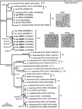
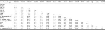
Genotyping of the new Brazilian isolates was based on analyses of partial (∼1200-bp fragments) SSU rRNA gene sequences. When the sequences were aligned with 31 other Acanthamoeba sequences retrieved from GenBank, phylogenetic analyses positioned the Brazilian isolates in the robustly supported clade composed of the species of morphological group I (Fig. 1; Table 1). The overall sequence divergence among the species in this clade is ∼12 7%; the divergence of group I species from the remaining taxa is ∼35%. Analyses placed Acanthamoeba TSP07 in genotype T9 and indicated that it is phylogenetically closely related to A. astronyxis 30901, Acanthamoeba PJ and PSH (1 0% divergence); it is 8 2% divergent from A. comandoni ATCC 30135, the reference species for this genotype. On the other hand, Acanthamoeba TSP07 is very divergent (∼16 9%; range: 16 6–17 1%) compared to the other 6 Brazilian Acanthamoeba isolates. The isolates ASP03, ARO01, TMS02, TSC01, TSP01, and ARO03 were homogeneous to each other (∼1 9% divergence within their sequences; range: 0 4–2 9%) and clustered with 2 isolates recently assigned to the novel genotype T17 (Nuprasert et al. Reference Nuprasert, Putaporntip, Pariyakanok and Jongwutiwes2010).
Maximum likelihood tree based on partial SSU rRNA Acanthamoeba sequences. Bootstrap values for ML and NJ analyses and posterior probabilities for a Bayesian inference are indicated at the tree nodes; values lower than 50% (ML and NL) or 0 7 (Bayesian inference) are not shown. The 7 Brazilian Acanthamoeba isolates characterized in this study are in bold. The Accession numbers of the sequences analysed and the Acanthamoeba genotypes are indicated. Balamuthia mandrillaris V039 was used as the outgroup. The characteristically star-shaped cysts of the Acanthamoeba isolates TSP07, TSP01, and ARO01 are shown in the figure (Scale bar=10 μm).

Divergence within the SSU rRNA gene sequences of Acanthamoeba species of morphological group I

Overall, trees generated by different methods displayed similar topologies, and nodal values consistently supported the relative positions of the group I species analysed. ML analyses separated the morphological group I Acanthamoeba isolates into 3 main groups: the first, with an internal sequence divergence of ∼14 4%, is composed of the species of genotypes T7 (A. astronyxis 30137) and T8 (A. tubiashi 30867); the second (internal sequence divergence of ∼3 2%; range: 0 4–8 2%) is composed of A. comandoni 30135 (genotype T9) and A. comandoni-related isolates, including Acanthamoeba TSP 07 (present study), A. astronyxis 30901, and isolates AE2a, PJ, and PSH; and the third group (internal sequence divergence: ∼2 0%; range: 0 1–2 9%) is composed of 2 recently characterized isolates of genotype T17 (Nuprasert et al. Reference Nuprasert, Putaporntip, Pariyakanok and Jongwutiwes2010) and 6 of the 7 new Brazilian environmental isolates.
Our analyses are in general agreement with previously reported Acanthamoeba phylogenies (Gast, Reference Gast2001; Gast et al. Reference Gast, Ledee, Fuerst and Byers1996; Stothard et al. Reference Stothard, Schroeder-Diedrich, Awwad, Gast, Ledee, Rodriguez-Zaragoza, Dean, Fuerst and Byers1998). The results indicate a close relationship among genotypes T3, T4 and T11 (5–7% divergence) and an interspecific polymorphism in genotype T4 (∼4 1% internal divergence). In addition and corroborating an earlier study (Corsaro and Venditti, Reference Corsaro and Venditti2010), analyses separately resolved 2 A. astronyxis strains, yielding ∼20% divergence: A. astronyxis 30137, the reference species of genotype T7, and A. astronyxis 30901; the latter was positioned in the clade of genotype T9.
Growth kinetics, thermotolerance and osmotolerance of the Brazilian Acanthamoeba isolates
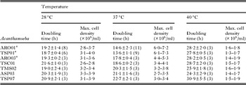
Growth kinetics of the Brazilian Acanthamoeba isolates was studied at 28°C, 37°C, and 40°C, with the results indicating similar growth rates at 28°C (Table 2). All 7 isolates grew at 37°C and 40°C, although at the latter temperature, doubling times significantly increased. Interestingly, the results indicated that Acanthamoeba ARO01, TSP01 and ARO03 grow better at 37°C than at 28°C (Table 2).
Growth kinetics of the Brazilian Acanthamoeba isolates at different temperatures

a Non-paired t-test: doubling times at 37°C significantly lower than at 28°C (P value <0 05).
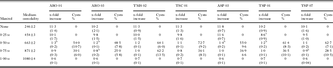
The ability of trophozoites to multiply under and/or resist hyperosmotic conditions was examined in experiments carried out at 28°C in ATCC 712 medium supplemented with 0 25, 0 5, 0 75 and 1 m mannitol (Table 3). At the lowest concentration assayed, mannitol did not appreciably interfere with the growth of trophozoites, although the growth of Acanthamoeba TSP01 was moderately reduced (P<0 05) compared to the growth of the control without mannitol. Trophozoites became rounded and vacuolated in medium containing 0 5 m mannitol (663 mOsm/kg.H2O), a condition that markedly reduced (isolates ARO01, ARO03, ASP03 and TSP01) and even eliminated (isolates TMS02, TSC01, and TSP07) the growth response. From those cultures, high numbers of cysts were recovered on day 4 (Table 3). Higher mannitol concentrations impaired growth and encystment. Cyst recovery was lower in 0 75 m than in 0 5 m mannitol-treated cultures, and no cysts were visualized until day 4 in cultures treated with 1 m mannitol, a concentration that also significantly reduced viability (Table 3). To confirm the viability of cells remaining on day 4, cultures of all Acanthamoeba isolates were successfully re-started after removal of 1 m mannitol from the medium (results not shown); trophozoites exhibiting typical morphology were evident within 2–6 days.
Growth and encystment of the Brazilian Acanthamoeba isolates under osmotic stress conditions
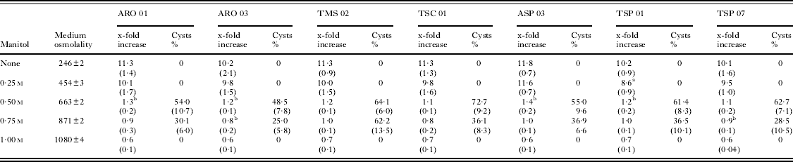
a Significantly different from the respective control (P<0 05).
b Significantly different from 1 0 (P<0 05).
Cytophatic effects produced by Acanthamoeba conditioned medium
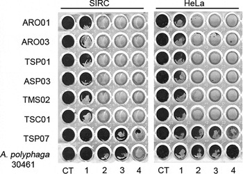
SIRC and HeLa cell monolayers were markedly affected when incubated with medium conditioned by trophozoites (Fig. 2). Cell monolayers were almost completely destroyed by medium conditioned by the isolates of genotype T17 (ARO01, ARO03, TSP01, ASP03, TMS02, and TSC01), while medium conditioned by Acanthamoeba TSP07 (genotype T9) produced an effect comparable to that observed with A. polyphaga ATCC 30461, a low virulent keratitis isolate (Visvesvara et al. Reference Visvesvara, Jones and Robinson1975).
In vitro cytophatic effects produced by different Acanthamoeba-conditioned media. SIRC and HeLa cell monolayers were incubated for 18 h at 37°C with medium conditioned by each of the Brazilian Acanthamoeba isolates and by A. polyphaga ATCC 30461. Conditioned media were previously diluted in complete DMEM (1: 10%; 2: 25%; 3: 50%) or directly added to cell cultures (4). CT: control cells incubated with non-conditioned DMEM. Cells were fixed and stained with crystal violet.

DISCUSSION
Acanthamoeba phylogenies are traditionally based on SSU rRNA gene sequences. Although more sequence data have become available in recent years, we are far from understanding the diversity and evolutionary relationships among the organisms within this genus. Of the 17 genotypes (T1–T17) identified thus far, 4 (T7, T8, T9, and T17) accommodate the more rarely examined species of morphological group I, a group formed by relatively few species: 2 isolates recently assigned to the novel genotype T17; A. astronyxis ATCC 30137, A. tubiashi ATCC 30867, and A. comandoni ATCC 30135 (the reference species of genotypes T7, T8, and T9, respectively); and some T7 and T9 environmental isolates (Ray and Hayes, Reference Ray and Hayes1954; Lewis and Sawyer, Reference Lewis and Sawyer1979; Pussard and Pons, Reference Pussard and Pons1977; Stothard et al. Reference Stothard, Schroeder-Diedrich, Awwad, Gast, Ledee, Rodriguez-Zaragoza, Dean, Fuerst and Byers1998; Lorenzo-Morales et al. Reference Lorenzo-Morales, Lindo, Martinez, Calder, Figueruelo, Valladares and Ortega-Rivas2005a–c; Ertabaklar et al. Reference Ertabaklar, Türk, Dayanir, Ertug and Walochnick2007; Nuprasert et al. Reference Nuprasert, Putaporntip, Pariyakanok and Jongwutiwes2010).
Here, we report the molecular and physiological characterization of 7 new environmental Acanthamoeba isolates, all morphologically classified in the group I. We have sequenced ∼1200-bp fragments of the SSU rRNA genes from the 7 new Acanthamoeba isolates to infer their genetic relatedness to known Acanthamoeba species, particularly those currently assigned to genotypes T7, T8, T9 and T17. In this regard, phylogenetic inferences with the species of group I have been limited by the small number of isolates and sequences available. The genotypes T7, T8 and T9 were defined on the basis of highly divergent sequences of single species (respectively A. astronyxis 30137, A. tubiashi 30867, and A. comandoni 30135), and genotype T17 was identified during the characterization of 2 environmental isolates from Thailand (Nuprasert et al. Reference Nuprasert, Putaporntip, Pariyakanok and Jongwutiwes2010). Phylogenetic analysis by several different methods placed the 7 Brazilian isolates in the robustly supported clade composed of the species of morphological group I and congruently resolved the group I isolates analysed here. Acanthamoeba TSP07 clustered with A. comandoni 30135, the reference species of genotype T9, but the other 6 Brazilian isolates (ASP03, ARO01, TMS02, TSC01, TSP01 and ARO03) clustered with 2 non-thermotolerant isolates that were recently assigned to the novel genotype T17 (Nuprasert et al. Reference Nuprasert, Putaporntip, Pariyakanok and Jongwutiwes2010). With the inclusion of these 6 new isolates, genotype T17 now forms a homogeneous (∼2 0% sequence divergence), well-supported group of isolates that likely represents a new Acanthamoeba species.
Acanthamoeba species of genotypes T7, T8, T9 and T17 have not yet been clearly implicated in human diseases (Khan, Reference Khan2006), but A. astronyxis was tentatively identified as the possible agent of a non-lethal case of human meningitis (Callicott et al. Reference Callicott, Nelson, Jones, dos Santos, Utz, Duma and Morrison1968). A study by Hadas and Mazur (Reference Hadas and Mazur1993) also indicated the pathogenicity and virulence for mice of a soil-derived isolate morphologically characterised as A. astronyxis. Studies with A. astronyxis, A. comandoni and A. tubiashi are scarce. Based on indirect markers of virulence (temperature tolerance, osmotolerance, and cytopathic effects), A. astronyxis reference strains (ATCC 30137, ATCC 30901, and CCAP 1534/1) and A. comandoni CCAP 1501/5 have been characterized as non-pathogenic (Hurt et al. Reference Hurt, Neelam, Niederkorn and Alizadeh2003; Khan, Reference Khan2001; Khan et al. Reference Khan, Jarroll, Panjwani, Cao and Paget2000, Reference Khan, Jarroll and Paget2002), but some studies indicate the pathogenic potential of environmental A. astronyxis-related strains (Lorenzo-Morales et al. Reference Lorenzo-Morales, Monteverde-Miranda, Jiménez, Tejedor, Valladares and Ortega-Rivas2005b,Reference Lorenzo-Morales, Ortega-Rivas, Foronda, Martínez and Valladaresc). Growth of all 7 Brazilian Acanthamoeba isolates was observed at temperatures up to 40°C, with the results indicating that 3 isolates of genotype T17 grow better at 37°C than at 28°C. Additionally, experiments with mannitol indicated that the Brazilian isolates resist/survive, at least for a few days, under hyperosmotic conditions; although high concentrations of mannitol impaired growth and encystment and 1 m mannitol significantly affected viability, cultures of all isolates could be successfully re-started from cells incubated for 4 days in 1 m mannitol. Finally, and indicating the secretion of cytotoxic molecules, incubation of SIRC and HeLa cells with medium conditioned by trophozoites resulted in pronounced cytophatic effects. Although the results suggest the pathogenicity of the new Acanthamoeba isolates, their potential to cause human and/or animal disease is yet to be demonstrated.
The Acanthamoeba phylogeny is still incomplete. The genus presently includes the highly divergent species of morphological group I, thus far represented by a small number of ATCC reference species and environmental isolates, 7 of which were characterized in this study. Based on the high degree of sequence divergence, the creation of one or more novel genera to accommodate A. astronyxis, A. tubiashi and A. comandoni has been suggested (Stothard et al. Reference Stothard, Schroeder-Diedrich, Awwad, Gast, Ledee, Rodriguez-Zaragoza, Dean, Fuerst and Byers1998). We are, however, just beginning to unravel the genetic variability of the Acanthamoeba species that form star-shaped cysts, and there is hidden diversity yet to be disclosed. The addition of new isolates and new SSU rRNA sequences, preferably combined with studies of other gene markers, will contribute to a better understanding of this group of Acanthamoeba isolates. Their phylogenetic relationships with the other species within the genus and their potential pathogenicity remain to be determined.
ACKNOWLEDGEMENTS
We thank Dr S. Jongwutiwes for sending us his SSU sequences before they were available in GenBank and Carmen S. Takata and Marcio M. Yamamoto for their assistance at the sequencing facility. We are indebted to students (Elen G. dos Santos and Katia P. de Azevedo) and many colleagues for their help in sample collection and culture from the very beginning of this project.
FINANCIAL SUPPORT
This work was supported with funds from FAPESP (Fundação de Amparo à Pesquisa do Estado de São Paulo, Brazil, Grant No 2009/53574-9) and CNPq (Brazil). A.C.M.M. received a Ph.D. Fellowship grant from CNPq (Brazil).