Case report
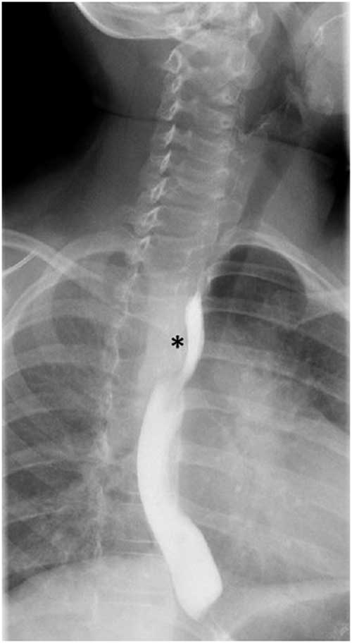
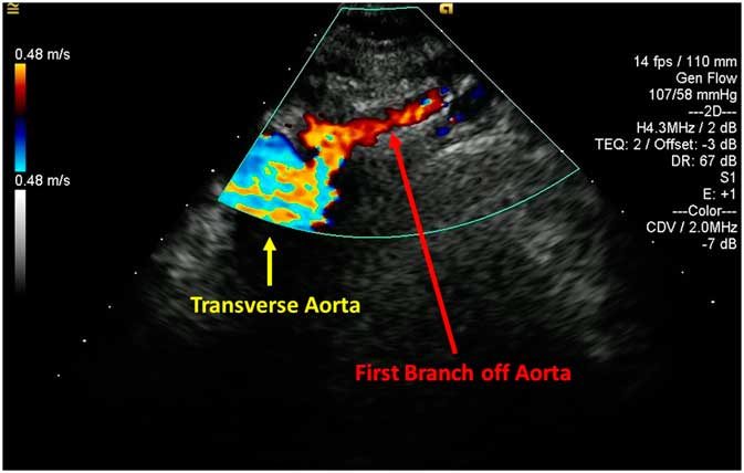
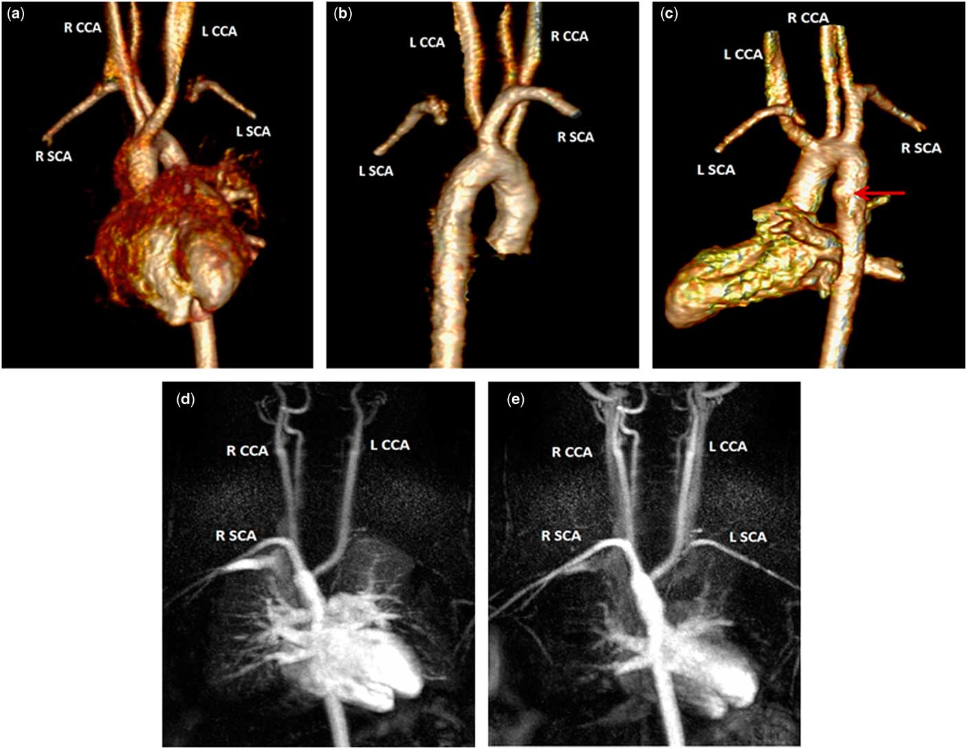
An 8-year-old girl was referred to paediatric cardiology for evaluation and management of a possible vascular ring. She had a history of asthma, gastroesophageal reflux, dysphagia, and emesis. An upper gastrointestinal series demonstrated possible narrowing in her mid-oesophagus (Fig 1). An echocardiogram showed normal intracardiac anatomy and function; however, owing to image quality, the aortic arch images were non-diagnostic (Fig 2). A cardiac MRI was therefore obtained that clearly demonstrated a right aortic arch giving rise to the left carotid, right carotid, and right subclavian arteries (Fig 3a–d). The left subclavian artery had no clear origin from the aorta and instead appeared to be supplied retrograde by vessels from the head and neck (Fig 3b–e). These findings were consistent with an isolated left subclavian origin from a previous left ductus arteriosus, now supplied by the left vertebral artery, and therefore not a substrate for a vascular ring.
Image obtained from upper gastrointestinal series. Contrast is seen in the oesophagus and it demonstrates possible narrowing in the mid-oesophagus region on the right (*). This narrowing was initially thought to be attributed to a right aortic arch.

Images obtained from echocardiogram. Echocardiographic image with colour demonstrates a leftward first branch off the aortic arch, supportive of a right aortic arch.

( a – e ) Images obtained from cardiac MRI. Anterior ( a ), posterior ( b ), and lateral ( c ) views via 3D reconstruction. A right aortic arch is seen giving rise to the left carotid, right carotid, and right subclavian arteries. The left subclavian artery has no clear origin from the aortic arch. A subtle aortic diverticulum is seen in image C as denoted by the arrow. Magnetic resonance angiography maximum image projections ( d and e ). Initial contrast is seen filling the left carotid, right carotid, and right subclavian arteries; however, no contrast is seen in the area where the left subclavian should be ( d ). In a subsequent 4.2 second delayed image, the left subclavian artery can now be seen and appears to be supplied by collaterals arising from the head and neck ( b ). (LCCA=left common carotid artery; LSCA=left subclavian artery; RCCA=right common carotid artery; and RSCA=right subclavian artery).

The patient’s clinical picture remained highly suggestive of a vascular ring. Review of her echocardiogram and MRI, however, could neither confirm nor refute this diagnosis. Despite the lack of a definitive imaging diagnosis, she was referred to surgery because of her persistent symptoms and strong clinical concern.
She subsequently underwent a left thoracotomy and her anatomy was determined to be a right aortic arch with aberrant left subclavian artery and left ligamentum arteriosus. The left subclavian artery was found to have a 6 cm long atretic portion proximally. Significant compression of her oesophagus was noted and the diagnosis of a vascular ring was confirmed. The ligamentum arteriosus was ligated and her oesophagus was dissected away from the compressive tissues. A subsequent review of her MRI images, with the surgical diagnosis established, demonstrated a very subtle diverticulum in the proximal descending aorta (Fig 1c) which likely represented the atretic origin of the aberrant left subclavian artery. Her post-operative course was uneventful, and her symptoms had completely resolved on follow-up evaluation.
Discussion
Vascular rings occur embryologically when the aortic arch and its branches fail to regress normally, and create a ring of vascular tissue that surrounds the oesophagus and trachea.Reference Tola, Ozturk and Yildiz 3 These formations can lead to compression of the oesophagus and trachea,Reference Naimo, Fricke and Donald 2 , Reference Tola, Ozturk and Yildiz 3 with concomitant respiratory and gastrointestinal symptoms. They are rare and constitute only 1–3% of congenital cardiac disease, with a right aortic arch and aberrant left subclavian artery being the second most common type.Reference Hanneman, Newman and Chan 1 , Reference Tola, Ozturk and Yildiz 3 There is no non-invasive gold standard for the diagnosis of vascular rings.Reference Tola, Ozturk and Yildiz 3 Current options for non-invasive imaging to identify abnormalities include upper gastrointestinal series, echocardiography, cardiac MRI, and CT angiography.Reference Hanneman, Newman and Chan 1 Pre-operative identification of the anatomy is important for surgical planning,Reference Hanneman, Newman and Chan 1 which is the standard treatment for symptomatic patients.Reference Tola, Ozturk and Yildiz 3
This is an unusual case of a vascular ring because the proximal portion of the left subclavian artery was atretic. Although echocardiography and MRI are commonly sufficient investigative tools to diagnose vascular rings, they were not diagnostic in this patient since neither modality can optimally demonstrate an atretic segment of artery. In such patients, secondary signs become essential for making the diagnosis.Reference Gould, Rigsby and Donnelly 4 In this patient’s case, slight angulation of the subclavian artery segment towards a subtle diverticulum in the proximal descending aorta (Fig 1c) were the only imaging clues. This specific type of rare vascular ring has previously been described in a few other case reports.Reference Shuford, Sybers and Gordon 5 – Reference Borushok, White and Oh 7 These, however, have primarily described symptoms associated with subclavian steal syndrome. To our knowledge, this is the first such case reported to present primarily with respiratory and gastrointestinal symptoms.
The decision to refer the patient for surgery with the definitive diagnosis unconfirmed was ultimately made with heavy consideration to the patient’s history. In this era of technology and test-heavy medical practice, this case illustrates the need for physicians to maintain a high index of suspicion when diagnosing vascular rings based on the patient’s clinical symptoms, even when the standard imaging modalities are deemed inconclusive.
Financial Support
None.
Conflict of Interest
The authors declare that he/she have no conflict of interest.
Ethical Standards
This article does not contain any studies with human or animal participants performed by any of the authors.
Acknowledgements
None.