Introduction
The development of mammalian preimplantation embryos generally arrests at specific developmental stages when they are cultured from the 1-cell stage in vitro. The developmental arrest occurs at the 2-cell stage in the mouse, rat and hamster (Goddard & Pratt, Reference Goddard and Pratt1983; Mayer & Fritz, Reference Mayer and Fritz1974; Schini & Bavister, Reference Schini and Bavister1988), at the 4-cell stage in the pig (Petters et al., Reference Petters, Johnson, Reed and Archibong1990) and at the 8- to 16-cell stages in the cow (Pinyopummintr & Bavister, Reference Pinyopummintr and Bavister1991). In the mouse, rat and hamster, the phenomenon of developmental arrest at the 2-cell stage is termed ‘2-cell block’ (Goddard & Pratt, Reference Goddard and Pratt1983; Mayer & Fritz, Reference Mayer and Fritz1974; Schini & Bavister, Reference Schini and Bavister1988).
Addition of ethylenediamine tetraacetic acid (EDTA) (Abramczuk et al., Reference Abramczuk, Solter and Koprowski1977; Fissore et al., Reference Fissore, Jackson and Kiessling1989) or superoxide dismutase (SOD) (Noda et al., Reference Noda, Matsumoto, Umaoka, Tatsumi, Kishi and Mori1991; Johnson & Nasr-Esfahani, Reference Johnson and Nasr-Esfahani1994) to the in vitro culture medium and culture at a low oxygen concentration (Kwon et al., Reference Kwon, Yang, Hwang, Yoo, Kim, Lee, Ryu and Oh1999) have been utilized for overcoming the 2-cell block. Removal of phosphate from the culture medium has also been shown to enable recovery from the 2-cell block in some mouse strains (Haraguchi et al., Reference Haraguchi, Naito, Azuma, Sato, Nagahama, Yamashita and Toyoda1996). In vitro developmental capacity of mouse preimplantation embryos has been found to be different among many strains (Suzuki et al., Reference Suzuki, Asano, Yamamoto, Takano and Koura1996). The development of embryos of AKR/N (AKR) mouse strain cultured in a medium with phosphate is arrested at the 2-cell stage, whereas the 2-cell block is rescued when embryos are cultured in a phosphate-free medium (Haraguchi et al., Reference Haraguchi, Naito, Azuma, Sato, Nagahama, Yamashita and Toyoda1996). The removal of phosphate from the culture medium can also rescue the 2-cell block of rat (Miyoshi et al., Reference Miyoshi, Funahashi, Okuda and Niwa1994) and hamster (Schini & Bavister, Reference Schini and Bavister1988) embryos. Furthermore, co-culture of mouse 1-cell embryos with oviductal epithelial cells has been demonstrated to enable in vitro development to proceed to the blastocyst stage (Minami et al., Reference Minami, Utsumi and Iritani1992; Liu et al., Reference Liu, Chan, Ho and Yeung1995). However, the molecular mechanisms by which the 2-cell block is prevented by removal of phosphate from the culture medium or by using a co-culture system have not been elucidated.
It has been shown that oocytes from the DDK mouse strain fertilized with non-DDK sperm cause 95% lethality, although they develop to full term when fertilized with the same DDK sperm (Wakasugi et al., Reference Wakasugi, Tomita and Kondo1967; Wakasugi, Reference Wakasugi1974). Since embryonic lethality occurred when RNA extracts derived from DDK oocytes were injected into non-DDK oocytes or blastomeres, the maternal lethal factor of DDK is thought to be present as RNA molecules (Renard et al., Reference Renard, Baldacci, Richoux-Duranthon, Pourmin and Babinet1994). The locus responsible for embryonic lethality of DDK oocytes in combination with sperm from the other strains has been mapped to chromosome 11 (Baldacci et al., Reference Baldacci, Richoux, Renard, Guenet and Babinet1992; Sapienza et al., Reference Sapienza, Paquette, Pannunzio, Albrechtson and Morgan1992).
The factor of 2-cell block in mouse preimplantation embryos has been suggested to be transmitted maternally because cross experiments between strains with different developmental capacities have shown that the 2-cell block is controlled by the cytoplasmic factors of oocytes (Goddard & Pratt, Reference Goddard and Pratt1983; Toyoda et al., Reference Toyoda, Azuma, Itagaki and Takada1992). However, a gene possibly contributing to the 2-cell block has not been revealed. Mouse 2-cell blocked embryos have been found to arrest in the transition from the G2 phase to the M phase (Aoki et al., Reference Aoki, Choi, Mori, Yamashita, Nagahama and Kohmoto1992; Haraguchi et al., Reference Haraguchi, Naito, Azuma, Sato, Nagahama, Yamashita and Toyoda1996). Therefore, the phenomenon of 2-cell block would provide a good model for understanding a mechanism of the cell cycle in early embryonic development. In the present study, we investigated the specific factors that rescue AKR embryos from the 2-cell block in a phosphate-free medium and in a system of co-culture with somatic cells. Cytoplasmic factors involved in the 2-cell block of embryos have also been searched for by means of a cross experiment and the production of reconstructed embryos by pronuclear exchange.
Materials and methods
Chemicals and media
All chemicals were purchased from Sigma Chemicals (St Louis, MO) unless otherwise stated. The medium used for handling embryos was M2 medium (Quinn et al., Reference Quinn, Barros and Whittingham1982) without phosphate. The culture medium for embryos was modified Whitten's medium (Whitten, Reference Whitten1971) containing 0.5 mM glucose and 0.1 mM EDTA with or without 1.17 mM KH2PO4.
Embryo collection
AKR, C57BL/6 (C57BL), ddY and ICR female mice (7–8 weeks old; SLC Japan, Hamamatsu, Japan) were mated with males from the same or other strains. The F1 female mice were obtained from reciprocal crosses between AKR and C57BL strains. To produce the backcrossed female progeny, AKR females were crossed to F1 males from the above combination. The backcrossed females obtained were mated with AKR males. Copulating was confirmed by observing vaginal plugs at 08:00–09:00 hours on the day after male and female mice were mated. Females that had copulated were killed at 11:00–12:00 hours and the oviducts were obtained. The ampulla of the oviduct was dissected in phosphate-free M2 medium, and oocytes with a second polar body or with pronuclei, which were defined as 1-cell stage embryos, were collected. One-cell embryos were washed four times in phosphate-free M2 medium before the start of in vitro culture.
All experiments were performed with the approval of the Hokkaido University Institutional Animal Care and Use Committee.
Embryo culture
After washing with phosphate-free M2 medium, 1-cell embryos were washed four times with modified Whitten's medium and transferred into 100μ l drops of the same culture medium covered with mineral oil in a Petri dish (Falcon 1008, Becton Dickinson, Franklin Lakes, NJ). The embryos were cultured for 120 h at 37 °C under 5% CO2 in air. Development to 2-cell, 4-cell, 8-cell, morula and blastocyst stages was observed at 24, 48, 60, 72 and 120 h after the start of in vitro culture, respectively.
Co-culture with buffalo rat liver (BRL) cells and the conditioned medium
BRL cells were cultured in Dulbecco's Modified Eagle's Medium (D-MEM) supplemented with 10% fetal bovine serum (FBS; Biosource International, Camarillo, CA) in 4-well culture dishes (Nunc, Roskilde, Denmark) for 24 h at 37 °C in 5% CO2 in air. After becoming confluent, the cells were washed three times with modified Whitten's medium, incubated in the same medium covered with mineral oil at 37 °C in 5% CO2 in air, and used for co-culturing with embryos. For preparing the conditioned medium, confluent BRL cells in 4-well culture dishes were washed twice with modified Whitten's medium and incubated in the same medium for 48 h at 37 °C in 5% CO2 in air. The supernatant fluid was filtrated through a 0.45μm filter (Millipore, Bedford, MA) to remove cell debris and was used as conditioned medium for culture of embryos. Fractionated media with molecular weights of less than 10, 30 and 100 kDa were prepared by ultrafiltration of the conditioned medium through a Vivaspin 500 (Vivascience, Hannover, Germany).
Enucleation, nuclear transfer and electrofusion
One-cell embryos, collected from oviducts at 11:00–12:00 hours, were placed in modified Whitten's medium containing 5μg/ml cytochalasin B for 40 min at 37 °C in 5% CO2 in air. After the zona pellucida had been slit with a microneedle, both female and male pronuclei were drawn into a micropipette with an approximate inner diameter of 20μm. Karyoplasts with both pronuclei were transferred into another enucleated embryo. Electrofusion was performed in 0.3 M mannitol solution with a electrical pulse (DC pulse of 1.5 kV/cm for 60μs) (Lee et al., Reference Lee, Lee, Yoon, Roh and Kim2001). The reconstructed 1-cell embryos were incubated in modified Whitten's medium for 120 h at 37 °C in 5% CO2 in air.
Assessment of the genetic factor involved in the 2-cell block
To determine the candidate gene involved in the 2-cell block of AKR embryos, the in vitro developmental ability of each embryo from F1 females between AKR and C57BL strains and the backcrossed females was examined. Each F1 and backcrossed female was mated with an AKR male, and the 1-cell embryos obtained were cultured in modified Whitten's medium. The stages at which embryos completed development during in vitro culture were scored as shown in Table 1. The score for each backcrossed female was estimated as the mean of the scores for all the embryos collected per female.
Estimated score corresponding to the stages at which each embryo completed development during in vitro culture

Statistical analysis
All data obtained from in vitro culture of 1-cell embryos were analysed by ANOVA and Duncan's multiple range test using Generalized Linear Models in Statistical Analysis System (SAS). All percentage data were subjected to arcsine transformation before the test. A probability of p < 0.05 was considered to indicate statistical significance.
Results
In vitro development of embryos derived from different strains
As shown in Table 2, all 1-cell embryos derived from four mouse strains (AKR, C57BL, ddY and ICR) developed to the 2-cell stage during in vitro culture. All C57BL embryos developed to the 4-cell stage, and 94.6% progressed to the blastocyst stage. The developmental profile of C57BL embryos was the highest among the four strains. In the ICR and ddY strains, 94.6% and 83.5% of the embryos developed to the 4-cell stage and 73.9% and 31.9% of the embryos progressed to the blastocyst stage, respectively, although embryos of both strains have been reported to show 2-cell block (Suzuki et al., Reference Suzuki, Asano, Yamamoto, Takano and Koura1996; Toyoda et al., Reference Toyoda, Azuma, Itagaki and Takada1992). On the other hand, the development of almost all AKR embryos was arrested at the 2-cell stage, and only 4.3% of the embryos developed to the 4-cell stage. No development to further stages was observed in AKR embryos. Embryos from AKR and C57BL mice were therefore used as 2-cell blocking and non-blocking models, respectively, in the following experiments.
In vitro development of embryos derived from different mouse strains*

* The medium contained 1.17 mM KH2PO4.
† Eight experiments were carried out in each group, and 10–15 embryos were used in each experiment.
‡ Numbers in parentheses indicate hours of in vitro culture.
a–dDifferent superscripts within a column indicate a statistically significant difference (p < 0.05).
Effect of phosphate on in vitro embryonic development
Table 3 shows the effect of phosphate on in vitro development of 1-cell embryos of AKR and C57BL strains. About 95% of C57BL embryos developed to the blastocyst stage regardless of the presence or absence of phosphate. In contrast, the development of almost all AKR embryos cultured in the medium containing phosphate was arrested at the 2-cell stage. However, 83.2% of AKR embryos cultured in the medium without phosphate developed to the 4-cell stage and 42.1% progressed to the blastocyst stage.
Effects of phosphate on in vitro development of AKR and C57BL 1-cell embryos

* The medium contained 1.17 mM KH2PO4 (+) or not (−).
† Eight experiments were carried out in each group, and 10–15 embryos were used in each experiment.
‡ Numbers in parentheses indicate hours of in vitro culture.
a–c Different superscripts within a column indicate a statistically significant difference (p < 0.05).
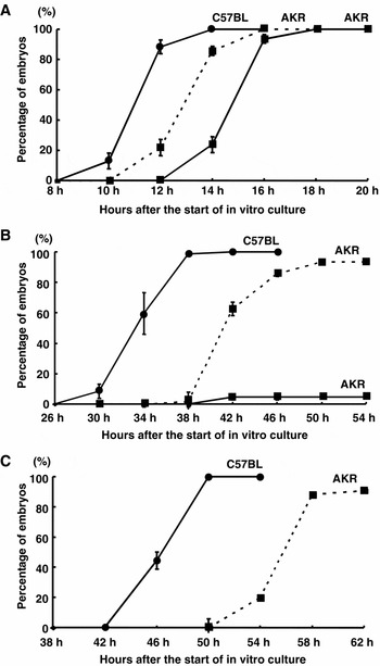
Fig. 1 shows the timetable of development of AKR and C57BL 1-cell embryos to the 8-cell stage. In the medium containing phosphate, C57BL embryos developed to the 2-, 4- and 8-cell stages at 14, 38 and 50 h after the start of in vitro culture, respectively, but the development of AKR embryos stopped after the step to the 2-cell stage at 18 h. On the other hand, in the absence of phosphate, AKR embryos developed to the 2-, 4- and 8-cell stages at 16, 50 and 58 h after the start of in vitro culture, respectively. The first cleavage to the 2-cell stage of AKR embryos cultured in phosphate-free medium was faster than that in the medium containing phosphate, but the first, second and third cleavages of AKR embryos were later than those of C57BL embryos.
Time courses of in vitro development of AKR and C57BL 1-cell embryos to the 2-cell (A), 4-cell (B) and 8-cell (C) stages. Squares indicate the developmental data of AKR embryos, and circles indicate those of C57BL embryos. Ten to 15 1-cell embryos were cultured in a medium with (unbroken lines) or without (broken lines) 1.17 mM KH2PO4. Four experiments were carried out, and values are the means ± SEM.

Effect of co-culture with BRL cells on in vitro embryonic development
To determine whether co-culture with somatic cells influences the 2-cell block of AKR embryos, we examined in vitro development of AKR 1-cell embryos co-cultured with somatic cells. BRL cells were used as co-cultured feeder cells because they have been shown to secrete many products during in vitro culture (Carr et al., Reference Carr, Huang, Itakura, Noel and Marceau1989; Massague et al., Reference Massague, Kelly and Mottola1985; Smith & Hooper, Reference Smith and Hooper1987). As shown in Table 4, 86.8% of the AKR embryos co-cultured with BRL cells developed to the 4-cell stage and 40.7% progressed to the blastocyst stage. The effect of BRL-conditioned medium on in vitro development of AKR embryos was also investigated. As shown in Table 5, 74.4% of the embryos cultured in BRL-conditioned medium developed to the 4-cell stage and 32.2% progressed to the blastocyst stage. The effect of a combination of phosphate-free medium and co-culture with BRL cells or conditioned medium was examined to determine whether they have a different action on in vitro development of AKR 1-cell embryos (Table 6). In the absence of phosphate, 94.4% of the embryos in co-culture with BRL cells and 90.2% of the embryos in the conditioned medium developed to the 4-cell stage, and 90.0% and 79.3%, respectively, progressed to the blastocyst stage.
Effect of co-culture with BRL cells on in vitro development of AKR 1-cell embryos*

* The medium contained 1.17 mM KH2PO4.
† Eight experiments were carried out in each group, and 10–15 embryos were used in each experiment.
‡ Numbers in parentheses indicate hours of in vitro culture.
a,b Different superscripts within a column indicate a statistically significant difference (p < 0.05).
Effect of BRL-conditioned medium on in vitro development of AKR 1-cell embryos*

* The medium contained 1.17 mM KH2PO4.
† Nine experiments were carried out in each group and 10–15 embryos were used in each experiment.
‡ Numbers in parentheses indicate hours of in vitro culture.
a–c Different superscripts within a column indicate a statistically significant difference (p < 0.05).
Effect of combination of phosphate-free medium and co-culture with BRL cells on in vitro development of AKR 1-cell embryos

* The medium contained 1.17 mM KH2PO4 (+) or not (−).
† Nine experiments were carried out in each group, and 10–15 embryos were used in each experiment.
‡ Numbers in parentheses indicate hours after the start of in vitro culture.
a–dDifferent superscripts within a column indicate a statistically significant difference (p < 0.05).
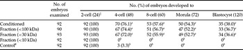
Since the conditioned medium using BRL cells was confirmed to be effective for rescue of the 2-cell block of AKR 1-cell embryos in in vitro development, it was investigated whether the effective factor is protein and whether it can be fractionated molecularly. As shown in Table 7, 72% of the embryos cultured in the medium fractionated below 30 kDa developed to the 4-cell stage and 36% of the embryos progressed to the blastocyst stage. On the other hand, the development of all embryos cultured in the medium fractionated below 10 kDa was arrested at the 2-cell stage. In vitro development of AKR embryos cultured in heat-treated or trypsin-treated medium was also investigated (Table 8). In a preliminary experiment, the developmental rate (36.0%) to the blastocyst stage of AKR embryos cultured in phosphate-free Whitten's medium treated with trypsin-agarose beads was not significantly different from that (37.4%) of the embryos cultured in the non-treated phosphate-free medium, indicating that the trypsin treatment does not affect the embryos and inhibit development. The medium fractionated below 30 kDa was heated at 56 °C for 30 min or treated with trypsin-agarose beads at 37 °C for 5 min to destroy the proteins contained in the medium. The development of almost all embryos cultured in the heat-treated or trypsin-treated medium was arrested at the 2-cell stage.
Effects of different fractionated media on in vitro development of AKR 1-cell embryos*

* The medium contained 1.17 mM KH2PO4.
† Eight experiments were carried out in each group, and 10–15 embryos were used in each experiment.
‡ Numbers in parentheses indicate hours of in vitro culture.
# As a control, AKR embryos were cultured in modified Whitten's medium for 120 h.
a,b Different superscripts within a column indicate a statistically significant difference (p < 0.05).
In vitro development of AKR 1-cell embryos cultured in heat- and trypsin-treated conditioned media*

* The medium contained 1.17 mM KH2PO4.
† Eight experiments were carried out in each group, and 10–15 embryos were used in each experiment.
‡ Fractionated medium was heated in water at 56 °C for 30 min.
# Fractionated medium was treated with trypsin-agarose beads at 37 °C for 5 min.
¶ As a control, AKR embryos were cultured in modified Whitten's medium for 120 h.
∥ Numbers in parentheses indicate hours of in vitro culture.
a,b Different superscripts within a column indicate statistically significant difference (p < 0.05).
In vitro development of reconstructed embryos between AKR and C57BL strains and embryos from F1 females
Table 9 shows the rates of development of 1-cell embryos derived from F1 females crossed between AKR and C57BL strains to the blastocyst stage. Regardless of the strains of males used, almost all embryos derived from C57BL females developed to the 4-cell stage. The rate of development to the blastocyst stage of embryos from C57BL females crossed with AKR males (39.8%) was significantly lower than that of embryos from C57BL females crossed with C57BL males (94.7%). On the other hand, only 4.3% and 14.9% of the embryos derived from AKR females crossed with AKR and C57BL males, respectively, developed to the 4-cell stage, and no morulae and blastocysts were observed in these embryos. Therefore, it was thought that the 2-cell block phenomenon is caused by a maternal factor from AKR oocytes. We also investigated the maternal factor causing the 2-cell block of AKR embryos in in vitro development of reconstructed embryos produced by pronuclear exchange reciprocally between AKR and C57BL strains (Table 10). Regardless of whether the karyoplasts were derived from C57BL or AKR 1-cell embryos, about 90% of the reconstructed embryos produced by fusion with C57BL cytoplasts developed to the 4-cell stage. Reconstructed embryos produced by fusion of C57BL cytoplasts with AKR karyoplasts showed a significantly lower rate of development to the blastocyst stage (39.3%) than did reconstructed embryos produced by fusion of C57BL cytoplasts with C57BL karyoplasts (88.3%). On the other hand, the development of almost all reconstructed embryos produced by fusion with AKR cytoplasts was arrested at the 2-cell stage regardless of whether the karyoplasts were derived from AKR or C57BL strains.
In vitro development of 1-cell embryos from females crossed between AKR and C57BL*

* The medium contained 1.17 mM KH2PO4.
† Eight experiments were carried out in each group, and 10–15 embryos were used in each experiment.
‡ Numbers in parentheses indicate hours of in vitro culture.
a–c Different superscripts within a column indicate a statistically significant difference (p < 0.05).
In vitro development of reconstructed embryos following nuclear exchange among AKR and C57BL pronuclear-stage embryos*

* The medium contained 1.17 mM KH2PO4.
† Eight experiments were carried out in each group, and 15–20 embryos were used in each experiment.
‡ Numbers in parentheses indicate hours of in vitro culture.
a–c Different superscripts within a column indicate a statistically significant difference (p < 0.05).
To determine which phenotypes of 2-cell blocking or non-blocking embryos are dominant, F1 mice were produced by crossing AKR and C57BL strains. As shown in Table 11, more than 97% of the embryos derived from F1 females of the reciprocal cross between AKR and C57BL strains developed to the 4-cell stage and more than 92% progressed to the blastocyst stage. These results suggest that the phenotype of the 2-cell block in AKR embryos is a recessive character and is generated by a nuclear genetic factor of oocytes but not mitochondrial DNA.
In vitro development of embryos from the (AKR × C57BL) F1 females crossed with AKR males*

* The medium contained 1.17 mM KH2PO4.
† Eight experiments were carried out in each group, and 10–15 embryos were used in each experiment.
‡ Numbers in parentheses indicate hours of in vitro culture.
In vitro development of embryos derived from backcrossed females
To determine whether the phenotype of the 2-cell block appears in the backcrossed progeny, F1 females produced reciprocally between AKR and C57BL strains were further crossed with AKR males to obtain backcrossed females. Then the in vitro development of 1-cell embryos derived from the backcrossed females after copulation with AKR males was examined. As a basic experiment, the number of ovulated oocytes from the parental AKR and C57BL females copulated with males of the same strain were checked; the mean numbers of oocytes collected were 15.8 ± 2.3 and 11.8 ± 1.9, respectively. The fertilization rates were 63.3% in AKR oocytes and 86.4% in C57BL oocytes. The mean number of ovulated oocytes in the backcrossed females copulated with AKR males was 13.1 ± 2.7 and the fertilization rate of oocytes was 81.9%.
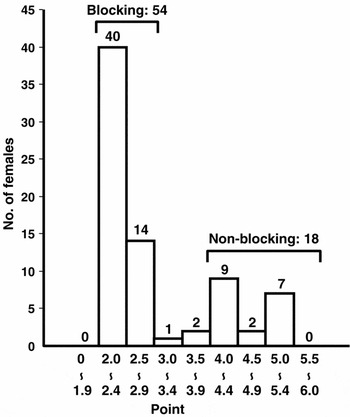
The stage at which each embryo completed development during in vitro culture was scored as shown in Table 1 and the mean value of the scores of all collected embryos per female was calculated. The scores of embryos from parental AKR mice ranged from 2.0 to 2.9 and the mean was 2.2 ± 0.1. The scores of embryos from the other parental C57BL mice ranged from 4.0 to 6.0 and the mean was 5.7 ± 0.1. Seventy-five backcrossed females were copulated and examined: as shown in Fig. 2, the score for embryos from 54 females ranged from 2.0 to 2.9 with a mean of 2.2 ± 0.1, and that from 18 females ranged from 4.0 to 6.0 with a mean of 4.7 ± 0.1. The 54 backcrossed females were considered to be 2-cell blocking type, and the 18 females were considered to be non-blocking type. Since about three-quarters of the backcrossed females were blocking type and about one-quarter were non-blocking type, it was thought that two genes are responsible for the 2-cell block in in vitro development of AKR 1-cell embryos.
In vitro developmental capacity of 1-cell embryos from individual backcrossed females produced between (AKR × C57BL) F1 males and AKR females. The score of each backcrossed female was estimated as the mean of the total points of embryos collected per a female divided by the number of embryos, as described in Materials and Methods.

Discussion
It is well known that the in vitro developmental capacity of mouse preimplantation embryos differs among strains. Embryos from AKR, ddY and ICR mouse strains have been reported to show 2-cell block (Haraguchi et al., Reference Haraguchi, Naito, Azuma, Sato, Nagahama, Yamashita and Toyoda1996; Suzuki et al., Reference Suzuki, Asano, Yamamoto, Takano and Koura1996; Toyoda et al., Reference Toyoda, Azuma, Itagaki and Takada1992). However, it has been demonstrated that ICR embryos cultured in a medium containing EDTA, a cell-membrane-impermeable metal chelator, overcome the 2-cell block but that ddY embryos do not despite the presence of EDTA (Suzuki et al., Reference Suzuki, Asano, Yamamoto, Takano and Koura1996; Toyoda et al., Reference Toyoda, Azuma, Itagaki and Takada1992). In the present study, ICR embryos cultured in media containing EDTA overcame the 2-cell block and developed to the blastocyst stage. Generally, in vitro developmental capacity of rodent embryos is influenced by the glucose concentration in the culture medium (Schini & Bavister, Reference Schini and Bavister1988; Suzuki et al., Reference Suzuki, Asano, Yamamoto, Takano and Koura1996; Miyoshi et al., Reference Miyoshi, Funahashi, Okuda and Niwa1994). Substrates utilized for energy metabolism in the mouse embryo are different between the 1- to 8-cell stages and the morula to blastocyst stages (Cross & Brinster, Reference Cross and Brinster1973). During early development before the 8-cell stage, a high glucose concentration in the medium has a detrimental effect on the embryos, a phenomenon termed the Crabtree effect (Seshagiri & Bavister, Reference Seshagiri and Bavister1991). Large number of ddY and ICR embryos in culture media containing low concentrations of glucose developed to the 4-cell stage in this study. Therefore, both ddY and ICR embryos are thought not to be a strict 2-cell blocking type.
On the other hand, the development of AKR 1-cell embryos was arrested at the 2-cell stage regardless of the presence of EDTA and even with a low glucose concentration. It has been reported that embryos from the mouse, rat and hamster overcome the 2-cell block in phosphate-free culture medium (Schini & Bavister, Reference Schini and Bavister1988; Suzuki et al., Reference Suzuki, Asano, Yamamoto, Takano and Koura1996; Miyoshi et al., Reference Miyoshi, Funahashi, Okuda and Niwa1994). In this study, the 2-cell block of AKR embryos was rescued in the medium without phosphate. Furthermore, the first cell cycle to the 2-cell stage of AKR embryos cultured in the medium with phosphate was later than that in the medium without phosphate. Phosphorylation and dephosphorylation states of M-phase promoting factor, a complex of cyclin-dependent kinase cdc2 and cyclin B, in mouse embryos are known to be altered by the presence of phosphate in the culture medium (Haraguchi et al., Reference Haraguchi, Naito, Azuma, Sato, Nagahama, Yamashita and Toyoda1996; Ohashi et al., Reference Ohashi, Minami and Imai2001). In eukaryotic cell cycles, cdc2 kinase is dephosphorylated and activated by cdc25 phosphatase (King et al., Reference King, Jackson and Kirchner1994; Lew & Kornbluth, Reference Lew and Kornbluth1996), and cyclin B is phosphorylated by Polo-like kinase 1 and translocated into the nucleus (Pines & Hunter, Reference Pines and Hunter1991; Ookata et al., Reference Ookata, Hisanaga, Okano, Tachibana and Kishimoto1992). The development of AKR embryos cultured in a medium with phosphate has been found to be arrested at the G2/M transition of the second cell cycle with the cdc2 phosphorylation being kept (Haraguchi et al., Reference Haraguchi, Naito, Azuma, Sato, Nagahama, Yamashita and Toyoda1996). In that condition, cyclin B is still in a dephosphorylated state and remains in the cytoplasm of 2-cell blocked embryos (Ohashi et al., Reference Ohashi, Minami and Imai2001). Therefore, the concentration of phosphate in the culture medium is considered to change the phosphorylation state of some molecules involved in the cell cycle of embryos and to cause 2-cell block as a result.
In vitro development of ICR 1-cell embryos to the blastocyst stage has been reported to be promoted by co-culture with oviduct organs or oviductal cells (Minami et al., Reference Minami, Utsumi and Iritani1992; Liu et al., Reference Liu, Chan, Ho and Yeung1995). The co-culture might support embryonic development by improving the suboptimal culture conditions; for example, co-cultured cells have been reported to eliminate the influences of hazardous substances such as reactive oxygen species (Bavister, Reference Bavister1992). Furthermore, the production of high-molecular-weight (>10 kDa) or low-molecular-weight (<10 kDa) embryotrophic factors from human and mouse oviductal cells has been suggested to promote in vitro development of mouse embryos, although these factors have not been identified as a specific molecules (Minami et al., Reference Minami, Utsumi and Iritani1992; Liu et al., Reference Liu, Chan, Ho and Yeung1995). In the present study, the 2-cell block of AKR embryos was rescued by both co-culture with BRL cells and culture in BRL-conditioned medium. Culture in a medium fractionated below 30 kDa also promoted in vitro embryonic development, although AKR embryos cultured in a heat- or trypsin-treated medium or in a medium below 10 kDa did not overcome the 2-cell block. Therefore, proteins with molecular weights of 10 to 30 kDa might be specific factors to support the development of AKR embryos from the 2-cell stage to subsequent stages. One possible function of the factors might be control of the uptake of phosphate into embryos.
Results of cross experiments between mouse strains that show different developmental capacities of preimplantation embryos have suggested that the 2-cell block is controlled by maternal factors that are present in the cytoplasm of oocytes (Goddard & Pratt, Reference Goddard and Pratt1983; Toyoda et al., Reference Toyoda, Azuma, Itagaki and Takada1992). Regardless of the strain from which males were derived, the development of 1-cell embryos from females of the blocking type of strain was arrested at the 2-cell stage, whereas embryos from females of a non-blocking strain developed to the 4-cell stage and later stages. In the present study, it was also confirmed that embryos from AKR females crossed with C57BL males showed 2-cell block but those from C57BL females crossed with AKR males developed to the 4-cell stage. Therefore, the 2-cell block of AKR embryos must be generated by maternal factors in the oocyte cytoplasm. Furthermore, the results of a pronuclear exchange experiment clearly indicated that the reconstructed embryos with AKR cytoplasts caused 2-cell block but that the reconstructed embryos with C57BL cytoplasts progressed to the 4-cell stage. In addition, embryos from F1 females obtained by a reciprocal cross between AKR and C57BL strains developed to the 4-cell stage, indicating that the phenotype of 2-cell block is a recessive character and is controlled by some nuclear factor(s) but not mitochondrial DNA.
When backcrossed females were produced by combination between (AKR × C57BL) F1 males and AKR females, three-quarters of the backcrossed females showed the 2-cell blocking phenotype and one-quarter of them showed the non-blocking phenotype. These results suggest that two genes are responsible for the 2-cell block of AKR embryos. Our collaborator has determined by linkage analysis using microsatellite markers that two allelic genes concerning with the 2-cell block of the AKR strain are located in chromosomes 3 and 4 (unpublished data), and over 250 backcrossed females are going to be produced for the detailed analysis of quantitative trait loci. Since the 2-cell block of AKR embryos was demonstrated to occur only in the culture medium with phosphate and since proteins of 10–30 kDa molecular weight fractionated from the conditioned medium of BRL cells had the ability to rescue the 2-cell block, the specific effective factor coded by a candidate gene must be related to an oocyte cytoplasmic molecule whose phosphorylated state is influenced by 10–30 kDa proteins secreted probably from the oviduct. The present study is the first report to have indicated that a candidate gene which controls the development of 2-cell embryos to further stages will be identifiable as a specific molecular factor in future work.