I. TUNNELING-BASED SEQUENCING
The first successful demonstration of translocation and sensing of DNA and RNA strands through an alpha-hemolysin pore was reported by Kasianowicz et al. just over two decades ago in 1996. Reference Kasianowicz, Brandin, Branton and Deamer1 Since then, many advances have been made toward reading the sequence of bases that encode the genetic instructions for life. These approaches, collectively termed nanopore sequencing, began first with biological nanopores followed by synthetically made solid-state nanopores. Now a mature technology, biological nanopore sequencing is capable of sequencing long unlabeled DNA strands with high accuracy and with miniaturized devices that offer both portability and affordable sequencing. Reference Clarke, Wu, Jayasinghe, Patel, Reid and Bayley2 However, limitations resulting from the finite dimensions of both biological and solid-state nanopores remain. Reference Branton, Deamer, Marziali, Bayley, Benner, Butler, Di Ventra, Garaj, Hibbs, Huang, Jovanovich, Krstic, Lindsay, Ling, Mastrangelo, Meller, Oliver, Pershin, Ramsey, Riehn, Soni, Tabard-Cossa, Wanunu, Wiggin and Schloss3 Even nanopores in atomically thin membranes formed from 2D materials, such as graphene and MoS2, suffer from these limitations as the effective channel length is increased by stray electric fields. Reference Garaj, Liu, Golovchenko and Branton4,Reference Lindsay5 Sensitivity of the nanopore is also decreased by the formation of an electrical double layer and noise caused by the mechanical vibrations of the membrane. Reference Arjmandi-Tash, Belyaeva and Schneider6,Reference Li, Zhang, Yang, Bi, Ni, Li and Chen7
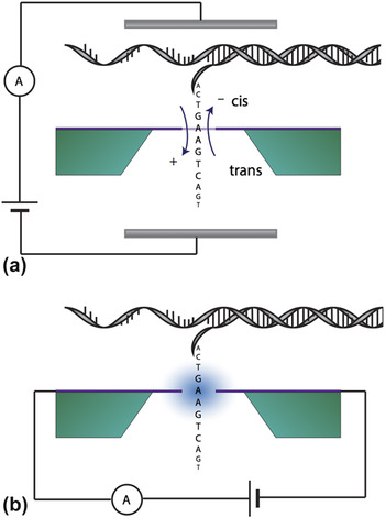
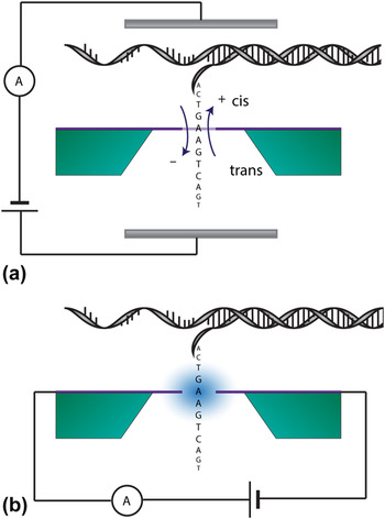
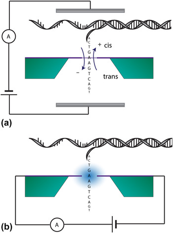
To continue to advance the accuracy, miniaturization, and affordability of these devices a paradigm shift is envisioned from the ionic current blockade to quantum electron tunneling-based sequencing (see Fig. 1). Reference Di Ventra and Taniguchi8 In this configuration, analytes must pass between two electrodes placed just nanometers apart, known as a tunnel junction, allowing electrons to tunnel from one electrode to the other via the analyte molecule through a potential barrier that particles cannot surmount classically. Tunneling sequencing relies on probing the differences in the electronic structure of the bases and other small biomolecules as they move sequentially through the nanogap between the nanoelectrodes. Reference Di Ventra and Taniguchi8
Schematic illustrating the difference between (a) traditional solid-state nanopores sequencing by trans-membrane conductance of an ionic solution and (b) tunneling sequencing where the bases are read via transverse conductance (in-plane current).

Tunneling sequencing of DNA was first investigated using electron tunneling in a scanning tunneling microscope (STM), Reference Tanaka9,Reference Tanaka, Hamai, Kanno and Kawai10 where the analyte was firmly adsorbed onto a Cu(111) surface without any possibility of controlled translocation under the STM tip. Reference Tanaka9 Realization of DNA imaging demanded the development of an aggregation-preventing approach for the deposition of DNA molecules on the metallic surface, and imaging was achieved via cryogenic STM measurements of double-stranded DNA. This pioneering study revealed the existence of a transport gap surrounded by peaks that result from electrons tunneling in a double-barrier system—one tunnel barrier between the STM tip and the DNA molecule and a second barrier between the molecule and the substrate. Reference Shapir, Cohen, Calzolari, Cavazzoni, Ryndyk, Cuniberti, Kotlyar, Di Felice and Porath11
The discovery of a distinct electronic fingerprint of one of the DNA bases (guanine) allowed for its identification in a single-stranded DNA (ssDNA) and the partial sequencing of DNA with a known nucleotide order by STM. Reference Tanaka and Kawai12 Guanine could be distinguished from the other three bases in a topographic measurement at a fixed bias voltage, i.e., without the need for full tunneling spectroscopy, enabling fast sequencing. With additional development, full sequencing of short DNA strands has now also been realized. Reference Huang, He, Chang, Zhang, Liang, Li, Tuchband, Fuhrmann, Ros and Lindsay13
An important conceptual advancement in DNA sequencing was the proposed use of nanopores integrated with tunnel junctions—first described by Zwolak and Di Ventra. Reference Zwolak and Di Ventra14 Tunnel junctions with sufficiently small probing areas should be able to analyze electronic fingerprints of single nucleotides in a DNA strand independent of the influence of the nearest neighboring nucleotides. If nanoscale fabrication challenges can be overcome, a robust integrated nanopore-tunnel junction device would combine advantages of nanopores and tunneling sequencing into a single device. Namely, control over the motion of DNA strands via the applied trans-membrane bias voltage from nanopore sequencing and high accuracy based on the tunneling current variations from the different electronic structure of bases. Reference Agah, Zheng, Pasquali and Kolomeisky15
With this vision in mind, tunnel junctions have been fabricated via a variety of approaches. Tunnel junctions aligned with nanopores were prepared on insulating membranes by means of transmission electron beam ablation lithography Reference Fischbein and Drndic16 and high-resolution focused ion beam (FIB) milling. Reference Maleki, Mohammadi and Ziaie17 In an alternative approach, electrodes embedded in long quasi-one-dimensional nanochannels were used to detect individual DNA strands. Reference Liang and Chou18 Mechanically controllable break junctions have also been used to distinguish between solutions of single nucleotides. Reference Tsutsui, Taniguchi, Yokota and Kawai19 Solutions containing thymine (T), cytosine (C), and guanine (G) give rise to different conductance values corresponding to the HOMO–LUMO gaps of the monophosphates when measured using tunneling sequencing. Reference Tsutsui, Taniguchi, Yokota and Kawai19 Analyses of nucleotide monophosphates and short DNA strands have also been performed using electromigrated tunneling electrodes embedded in nanopores, showing a bimodal current peak distribution for strands consisting of two different bases (guanine and thymine). Reference Tsutsui, Rahong, Iizumi, Okazaki, Taniguchi and Kawai20
A study comparing the ionic and tunneling current recorded simultaneously in a gold-tunnel junction that was co-aligned with a FIB milled nanopore showed that two different classes of events can be detected Reference Ivanov, Instuli, McGilvery, Baldwin, McComb, Albrecht and Edel21 : (i) a short signal (with a mean dwell time of 0.3 ms) corresponding to fast DNA translocation only detected in the ionic current; and (ii) a long signal (4.7 ms) due to a decreased DNA translocation rate and an improved signal-to-noise ratio (SNR) due to nonspecific bonding to the tunnel junction. Mean dwell times recorded for the second class of events by both methods were in good agreement, indicating a common origin of signals detected by tunneling and ionic current, thus demonstrating the efficacy of the approach. Reference Ivanov, Instuli, McGilvery, Baldwin, McComb, Albrecht and Edel21 To achieve an acceptable SNR, it is often necessary to only allow a small portion of the tunnel junction to be exposed to maximize the signal from the analyte of interest. In liquid environments electronic noise in tunnel junctions can be a serious limitation; however, it can be reduced by coating the devices with a few nanometers of a passivation layer such as SiO2 Reference Arima, Tsutsui, Morikawa, Yokota and Taniguchi22,Reference Morikawa, Yokota, Tsutsui and Taniguchi23 or Al2O3. Reference Muthusubramanian, Galan, Maity, Eelkema, Grozema and Van Der Zant24
Alternate experimental configurations of tunnel junctions have been realized in parallel to the development of metal tunnel junctions. These include carbon nanowire tunnel junctions aligned with nanopores and patterned via electron beam induced deposition from organic precursors. Reference Spinney, Collins, Howitt and Smith25,Reference Ivanov, Freedman, Kim, Albrecht and Edel26 This approach, which allows for the fabrication of electrodes aligned with nanopores, is perceived as a straightforward alternative capable of being adapted to multiple geometries. Recently, new approaches are being conceived which build on these foundations. One such approach which utilizes graphene as the ultimately thin version of carbon nanowire has already shown early promises in a variety of biosensing applications.
II. GRAPHENE BIOSENSORS
Due to graphene’s remarkable properties, such as atomic thinness, high conductivity, and chemical stability Reference Geim27 in both air and liquid, it has attracted considerable attention for applications in biosensing. Reference Mohanty and Berry28–Reference Yang, Yang, Zou, Wu, Wan, Cao, Liao, Yuan and Duan30 The ability to synthesize graphene via scalable methods Reference Li, Magnuson, Venugopal, An, Suk, Han, Borysiak, Cai, Velamakanni, Zhu, Fu, Vogel, Voelkl, Colombo and Ruoff31,Reference Bae, Kim, Lee, Xu, Park, Zheng, Balakrishnan, Lei, Kim, Song, Kim, Kim, Özyilmaz, Ahn, Hong and Iijima32 and pattern it with atomic precision has further motivated many researchers to incorporate it as the active component in biomolecular sensors. These sensing approaches span from graphene field-effect transistors Reference Dong, Shi, Huang, Chen and Li33,Reference Ohno, Maehashi and Matsumoto34 to trans-electrode membranes Reference Garaj, Hubbard, Reina, Kong, Branton and Golovchenko29 to tunnel junctions, Reference Malec and Davidović35 all of which exploit unique properties of the material. Among these, configurations with nanogaps and nanoconstrictions offer the impressive capability to probe analytes electronically at the atomic scale with single-molecule sensitivity and high-time resolution.
Many techniques have been developed for fabricating nanoscale features in graphene. These can generally be classified into the paradigmatic categories of top-down and bottom-up fabrication methods, with much of the focus by the research community on the former. Bottom-up approaches include graphene nanoribbons, Reference Cai, Ruffieux, Jaafar, Bieri, Braun, Blankenburg, Muoth, Seitsonen, Saleh, Feng, Mullen and Fasel36 DNA templated synthesis, Reference Sokolov, Yap, Liu, Kim, Ci, Johnson, Wang, Vosgueritchian, Koh, Chen, Park and Bao37 and block co-polymer lithography. Reference Choi, Kuru, Choi, Noh, Hong, Das, Choi and Jin38 Within the bottom-up methods, graphene nanoribbons with the capability to specify edge shape, dimensions, Reference Li, Wang, Zhang, Lee and Dai39 functionalization, Reference Boukhvalov and Katsnelson40 and doping Reference Huang, Yan, Zhou, Wu, Gu, Duan and Liu41 have shown the most promise, particularly when used in conjunction with graphene nanogaps. Top-down approaches include electron beam lithography (EBL) patterning, Reference Cao, Dong, Liu, He, Gan, Yu, Steigerwald, Wu, Liu and Guo42 FIB milling, Reference Zhou, Maguire, Jadwiszczak, Muruganathan, Mizuta and Zhang43 transmission electron microscopy sculpting, Reference Song, Schneider, Xu, Pandraud, Dekker and Zandbergen44 scanning probe methods, Reference Zhang, Fu, Pan, Yu, Liu, Luo, Wang, Yang and Hou45 and feedback-controlled electroburning. Reference Prins, Barreiro, Ruitenberg, Seldenthuis, Aliaga-Alcalde, Vandersypen and Van Der Zant46
Of the top-down fabrication approaches, EBL and feedback-controlled electroburning have been developed the most for graphene tunnel junctions. In particular Cao et al. have demonstrated the successful fabrication of arrays of tunnel junctions in single-layer graphene using EBL followed by oxygen plasma etching. Reference Cao, Dong, Liu, He, Gan, Yu, Steigerwald, Wu, Liu and Guo42 Briefly, a dashed line of 5 nm width was patterned in Poly(methyl methacrylate) (PMMA) using ultrahigh-resolution EBL, which was then developed and patterned via reactive ion etching to transfer the pattern to the graphene resulting in lines of nanoelectrodes ranging in width from 1 to 10 nm. These electrodes were also functionalized by carboxylic acid end groups and terpyridyl ligands to facilitate specific binding interactions. EBL which has been demonstrated for nanoscale patterning of graphene for other nanoscale quantum applications Reference Moser and Bachtold47 offers a possible scalable solution for the fabrication of nanogaps; however, consistency and yield will need to improve for the research to continue to progress.
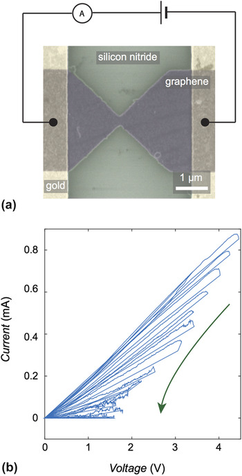
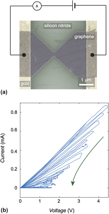
Feedback-controlled electroburning Reference Prins, Barreiro, Ruitenberg, Seldenthuis, Aliaga-Alcalde, Vandersypen and Van Der Zant46 is a technique, which as the name suggests, allows for real-time in situ monitoring of the graphene sculpting on the nanoscale (see Fig. 2). The ingenuity of the feedback mechanism is that the process can be tailored in response to variations in the graphene before the tunnel junction is produced and can respond accordingly as the process rapidly evolves. This provides a mechanism to controllably and consistently produce a high yield of either constrictions or nanogaps with the desired nanoscale dimensions. Reference Moser and Bachtold47,Reference Lau, Mol, Warner and Briggs48 Many groups have demonstrated variations on the technique Reference Lau, Mol, Warner and Briggs48–Reference Lumetti, Candini, Godfrin, Balestro, Wernsdorfer, Klyatskaya, Ruben and Affronte50 showing its efficacy on both single-layer graphene and few-layer graphene, Reference Prins, Barreiro, Ruitenberg, Seldenthuis, Aliaga-Alcalde, Vandersypen and Van Der Zant46 at room temperatures, in ambient conditions, and in vacuum, Reference Nef, Posa, Makk, Fu, Halbritter, Schonenberger and Calame49 with applications from single-molecule devices Reference Lumetti, Candini, Godfrin, Balestro, Wernsdorfer, Klyatskaya, Ruben and Affronte50 to single-electron transistors. Reference Puczkarski, Gehring, Lau, Liu, Ardavan, Warner, Briggs and Mol51 Briefly, to sculpt graphene on the nanoscale via feedback-controlled electroburning, micron-scale ribbons of graphene are formed and notched [see Fig. 2(a)], typically via a lithography process followed by oxygen etching. This notched portion forms a constriction which when a bias voltage is applied between the two ends of the ribbon joule heating will occur at the narrowest point. When sufficiently high current densities are achieved, joule heating can cause graphene at the edges of the constriction to react with oxygen at high temperatures. This causes a gap to open controllably at the edges of the constriction which in turn modifies the conductance across the ribbon. This change in conductance is sensed via the feedback mechanism and the current is abruptly reduced. The bias voltage is then ramped again and the process proceeds until the desired endpoint is achieved. Fabricating features in this manner avoids a catastrophic rupture of the constriction, allowing for a nanometer-scale gap or constriction to be formed, as desired.
(a) False-color scanning electron micrograph of a graphene constriction on a silicon-nitride substrate. The graphene is contacted via gold leads. (b) Typical current–voltage traces recorded during feedback-controlled electroburning of a graphene nanogap.
Initially, graphene was explored as a solid-state analogue to tremendously successful biological nanopores for DNA sequencing Reference Garaj, Hubbard, Reina, Kong, Branton and Golovchenko29 where trans-membrane conductance of ionic current through a nanometer-scale aperture was monitored while the DNA translocated electrophoretically through the pore. This showed feasibility, however, encountered difficulties, such as strong graphene DNA interactions Reference Husale, Sahoo, Radenovic, Traversi, Annibale and Kis52 and challenges in reproducibly creating pores. Reference Goyal, Lee, Darvish, Ahn and Kim53 Recently, several new schemes utilizing graphene nanogaps and nanoconstrictions and transverse (in-plane) measurements have been explored (see Fig. 3). These include monitoring the in-plane current through a constriction with a localized nanopore either within Reference Traversi, Raillon, Benameur, Liu, Khlybov, Tosun, Krasnozhon, Kis and Radenovic54,Reference Girdhar, Sathe, Schulten and Leburton55 or near the pore, Reference Balan, Drndi, Puster and Rodrı56 bilayer graphene constrictions, Reference Sadeghi, Algaragholy, Pope, Bailey, Visontai, Manrique, Ferrer, Garcia-Suarez, Sangtarash and Lambert57–Reference Paulechka, Wassenaar, Kroenlein, Kazakov and Smolyanitsky59 detecting electronic changes from localized strain due to a DNA translocating through a nanopore in a constriction, Reference Paulechka, Wassenaar, Kroenlein, Kazakov and Smolyanitsky59 changes in constriction current due to physisorption of analytes on the graphene surface Reference Le, Kara, Schröder, Hyldgaard and Rahman60 or from a channel below, Reference Min, Kim, Cho and Kim61 and modulations in tunneling current through a graphene nanogap. Reference Postma62 The breadth of configurations demonstrate the versatility of nanoscale features in graphene exploiting quantum phenomena. In many of these device architectures, the graphene electrodes are placed between the two chambers and the DNA is typically translocated through the nanopore via an applied bias across the device. Confinement of the translocating DNA relative to the electrodes ensures that each base is read sequentially. However, the confinement can be achieved through other means, such as channels. Reference Min, Kim, Cho and Kim61
Schematic demonstrating the organization of types of nanopore sequencing starting from the delineation between biological and solid-state, trans-membrane and transverse conductance (see Fig. 2) and finally different geometries for transverse conductance-based sequencing.
Of these approaches graphene tunnel junctions offer the most viable routes to an experimental demonstration. The approach consists of fabricating graphene nanogaps co-aligned with a pore within the substrate material, via a method such as feedback-controlled electroburning. The graphene electrodes, which form a tunnel junction, would then be passivated entirely except for in the vicinity of the nanogap where the DNA is electrophoretically driven through. This is similar, in principle, to how single molecules are probed currently Reference Prins, Barreiro, Ruitenberg, Seldenthuis, Aliaga-Alcalde, Vandersypen and Van Der Zant46,Reference Island, Holovchenko, Koole, Alkemade, Menelaou, Aliaga-Alcalde, Burzurí and van der Zant63,Reference Barreiro, Van Der Zant, Vandersypen and van der Zant64 but done sequentially with the addition of an aperture for translocation.
The predicted advantages of transverse conductance sequencing methods using graphene parallel those of biological nanopores include long read lengths, real-time data, label-free direct molecular analysis, the capability to detect methylation and other epigenetic markers, Reference Shendure and Ji65 portability, and scalability via parallelization. 66 Due to their solid-state nature and unique-material properties, graphene nanoelectrodes also offer further hypothesized benefits, including stability and durability, Reference Xu, Wu, Schneider, Houben, Malladi, Dekker, Yucelen, Dunin-Borkowski and Zandbergen67 tunable gap width, Reference Lau, Mol, Warner and Briggs48 atomic thinness, Reference Novoselov, Geim, Morozov, Jiang, Zhang, Dubonos, Grigorieva and Firsov68 the ability to tune sensitivity via functionalization, Reference Sint, Wang and Kral69,Reference Prasongkit, Grigoriev, Pathak, Ahuja and Scheicher70 being planar and flexible, Reference Reina, Jia, Ho, Nezich, Son, Bulovic, Dresselhaus, Kong, Kim, Zhao, Jang, Lee, Kim, Ahn, Kim, Choi and Hong71 and the promise of leveraging semiconductor manufacturing techniques for further miniaturization and low-cost production of biosensors at wafer scale.
III. FUNCTIONALIZATION
Tuning the width of graphene nanogaps provides a straightforward way to discriminate between species of interest due to differences in size; however, given the range of biomolecules of interest with similar sizes, more refinement is desired. One such method of tailoring the graphene is through functionalization, which ranges from noncovalent Reference Schneider, Xu, Hage, Luik, Spoor, Malladi, Zandbergen and Dekker72 to covalent bonding. Reference Georgakilas, Otyepka, Bourlinos, Chandra, Kim, Kemp, Hobza, Zboril and Kim73 Covalent bonding has several configurations from common graphene edge terminations to the possibility of specifically designed functionalization such as tether molecules Reference Im, Biswas, Liu, Zhao, Sen, Biswas, Ashcroft, Borges, Wang, Lindsay and Zhang74 or nucleobases Reference Prasongkit, Grigoriev, Pathak, Ahuja and Scheicher70 currently used in other systems.
Functionalization of the nanogaps and nanoconstrictions could help to facilitate bonding of various degrees between analytes of interest while also stabilizing edges and promoting preferred orientations of molecules for detection. Noncovalent bonding typically takes the form of π–π interactions, van der Waals forces, and hydrogen bonding. These most commonly occur when the analyte of interest bonds to the graphene such as in physisorption-based detection methods Reference Le, Kara, Schröder, Hyldgaard and Rahman60 or when the fluid containing the analyte interacts with the graphene. Common covalent functionalization includes hydroxylation and hydrogenation, Reference Cohen-Tanugi and Grossman75 which are suspected to occur in many formation methods of graphene nanogaps and nanoconstrictions, to silicon termination. Reference Lee, Yang, Zhou, Pennycook, Pantelides and Chisholm76 Recently, there has been a growing interest in the specific functionalization of the edges of tunnel junctions and graphene nanogaps to permit tailoring of tunnel junctions for molecular recognition.
Molecular recognition using functionalized electrodes Reference He, Lin, Zhang and Lindsay77 relies on the formation of strong bonds between reader molecules (sometimes also called adapter molecules or tethering molecules depending on molecule type and configuration) and the electrodes (e.g., by thiol chemistry for gold or palladium electrodes Reference Chang, Sen, Zhang, Gyarfas, Ashcroft, Lefkowitz, Peng and Lindsay78 ) and weak noncovalent interactions between the reader molecules and the analyte. Reference Lindsay79 The formation of a bond between the reader and analyte molecules gives rise to a current peak with a time duration and amplitude that are characteristic of that specific interaction. Reference Lindsay79 This approach improves DNA reads by decreasing the contact resistance of the junction and by limiting the number of possible molecular orientations of the DNA strand in the gap. Reference Chang, Huang, He, Liang, Zhang, Li, Chen, Sankey and Lindsay80 Slowing down DNA translocation also enhances the SNR by allowing for a longer signal averaging interval. Reference Pedersen, Boynton, Di Ventra, Jauho and Flyvbjerg81 This can be achieved by functionalization of the tunnel junction with a reader molecule that weakly binds to the DNA, acting as a ‘molecular velcro’. Reference Krishnakumar, Gyarfas, Song, Sen, Zhang, Krstić and Lindsay82,Reference Lindsay, He, Sankey, Hapala, Jelinek, Zhang, Chang and Huang83
Molecules that specifically bind to a given DNA base can also increase the accuracy of the DNA sequencing. This can be attained through the use of van der Waals interaction (π–π stacking) with the nucleotides. Reference Biswas, Sen, Im, Biswas, Krstic, Ashcroft, Borges, Zhao, Lindsay and Zhang84 The tunneling current through these interactions depends on the strength of the bonding with the bases, allowing for differentiation between molecules in the gap with either two or three hydrogen bonds. Reference Chang, He, Kibel, Lee, Sankey, Zhang and Lindsay85 The differences in the strength of the bonding can also modulate the dwell time of a base in a junction facilitating detection. This approach subtly differs from solely detecting the change in tunneling current amplitude and can provide additional signals for differentiating the bases. Reference Lindsay, He, Sankey, Hapala, Jelinek, Zhang, Chang and Huang83,Reference Chang, He, Kibel, Lee, Sankey, Zhang and Lindsay85
IV. GENOMICS, PROTEOMICS, AND GLYCOMICS
To date, much of the focus for tunnel junctions has been on DNA sequencing applications Reference Wang, Yang and Wang86 ; however, methylation of DNA, which contributes to differential gene expression, can also be probed. Reference Cortini, Barbi, Caré and Lavelle87 Methylated DNA gives rise to a higher tunneling current than its nonmethylated form in a MCBJ due to a better energetic alignment with the electrodes Fermi level. Reference Tsutsui, Matsubara, Ohshiro, Furuhashi, Taniguchi and Kawai88 The potential for tunnel junctions can also be extended beyond DNA entirely.
In addition to genomics, the fields of glycomics (the detection and analysis of carbohydrates) and proteomics (the detection and analysis of proteins) have prospects for tunnel junctions. Reference Lindsay5 The detection of analytes at the single-molecule level is particularly important in the case of proteins, which—contrary to DNA molecules—cannot be amplified by a polymerase chain reaction. Reference Lindsay5 Moreover, proteins and peptides often do not have any net charge, or the charge they do posses is not evenly distributed over the structure, hindering the possibility of dragging them electrophoretically through tunnel junctions. These issues may be overcome by the functionalization of neutral peptides with short and charged DNA oligomers by click chemistry. Reference Biswas, Song, Borges, Lindsay and Zhang89 Negatively charged oligomers can act as a molecular thread and exert a dragging force on the protein forcing it to translocate through the tunnel junction. The biggest challenge in protein sequencing, however, is in the expected range of variability in the sequencing signal. Whereas ssDNA is a linear chain constructed of only four different bases, proteins are composed of twenty fundamental building-blocks, termed amino acids. To identify proteins it is therefore necessary to develop a method capable of discriminating between these amino acids.
Recently, 8 out of 20 amino acids were detected in a gold MCBJ through which short peptides could be partially sequenced. Reference Ohshiro, Tsutsui, Yokota, Furuhashi, Taniguchi and Kawai90 A higher selectivity, resulting in the identification of 18 out of 20 amino acids was achieved through functionalization with molecules that form weak, noncovalent bonds to the amino acids. Reference Zhao, Ashcroft, Zhang, Liu, Sen, Song, Im, Gyarfas, Manna, Biswas, Borges and Lindsay91 This also enabled the differentiation between various forms of the amino acids, including methylated and nonmethylated forms, L- and D-enantiomers, and isobaric amino acids. Reference Zhao, Ashcroft, Zhang, Liu, Sen, Song, Im, Gyarfas, Manna, Biswas, Borges and Lindsay91
Carbohydrates are characterized by a complex secondary structure that cannot be resolved with simple mass spectrometry and therefore require bulky NMR techniques. While recognition tunneling using functionalized metal tunnel junctions is viable method to identify nanomolar concentrations of some saccharides, Reference Im, Biswas, Liu, Zhao, Sen, Biswas, Ashcroft, Borges, Wang, Lindsay and Zhang74 the identification of the large variety of different carbohydrates is still being developed as it will demand the collection of an entire library of fingerprints for the variety of building blocks found in carbohydrates. Nevertheless, recognition tunneling may provide additional information about the structure of linear polysaccharides on the single-molecule level that cannot be obtained using other techniques. Reference Im, Biswas, Liu, Zhao, Sen, Biswas, Ashcroft, Borges, Wang, Lindsay and Zhang74
V. MACHINE LEARNING
Signals collected in experiments with tunnel junctions are typically analyzed by the characteristic current spikes produced with a particular focus on amplitude and duration. However, the relatively large distribution of values of these parameters makes the analysis cumbersome. Specifically, the overlap between peaks and the convolution of many signals recorded for different DNA bases and biomolecules can hinder their analysis and lower the identification accuracy. One possible solution to this issue is the application of machine learning methods for the analysis of the wide variety of features in the signals from tunnel junctions. Reference Krstic, Ashcroft and Lindsay92 In particular, Support Vector Machines (SVMs) Reference Pedersen, Boynton, Di Ventra, Jauho and Flyvbjerg81 have attracted a growing interest recently. It has been demonstrated that for single nucleotide solutions, the simultaneous analysis of basic parameters, such as duration and amplitude of individual peaks and groups of peaks, with additional analysis of less intuitive parameters obtained by Fourier and wavelet analysis of these signals, can increase the accuracy of identification. Reference Chang, Huang, Liu, Zhang, Liang, Akahori, Li, Gyarfas, Shumway, Ashcroft, He and Lindsay93 These parameters were further analyzed with an SVM to identify the subset of parameters providing the highest accuracy of assignment. The outcome of the SVM analysis is a set of properties that can be used to assign a characteristic signal to the nucleotide with the highest accuracy. Reference Chang, Sen, Zhang, Gyarfas, Ashcroft, Lefkowitz, Peng and Lindsay78 It has also been demonstrated that the clustering of data from a SVM can lead to the increased accuracy of analysis, in particular, by taking into account the parameters describing contextual information (groups of peaks) and not only the single-peak properties. Reference Chang, Huang, Liu, Zhang, Liang, Akahori, Li, Gyarfas, Shumway, Ashcroft, He and Lindsay93
A similar SVM approach has also been used for the analysis of tunneling experiments using mixtures of different amino acids, increasing the accuracy of amino acid assignment. Reference Zhao, Ashcroft, Zhang, Liu, Sen, Song, Im, Gyarfas, Manna, Biswas, Borges and Lindsay91 SVMs trained on amino acids did not recognize them as constituents in peptide sequences, showing that there is a difference between a signal generated by free amino acids and the same amino acids in the short peptide chain. Nevertheless, applying the SVM to short peptides in a separate experiment showed it to be highly accurate even though the peptides were differing by a single amino acid. Reference Chang, Huang, Liu, Zhang, Liang, Akahori, Li, Gyarfas, Shumway, Ashcroft, He and Lindsay93 Machine learning was also used to analyze data from scanning tunneling spectroscopy performed on mononucleotide ssDNA in an STM configuration. Reference Korshoj, Afsari, Khan, Chatterjee and Nagpal94 Parameters used for this analysis contained HOMO and LUMO values and band gap energy, along with transition voltage, tunneling barrier, and electron effective mass, all at positive and negative biases. These parameters were analyzed with a modified naive-Bayes classifier leading to an increase in base calling accuracy compared to an analysis performed taking into account only HOMO and LUMO values. Reference Korshoj, Afsari, Khan, Chatterjee and Nagpal94
VI. OUTLOOK
With the further introduction of machine learning techniques to both the fabrication of graphene biosensors and the interpretation of their signals, progress can be expected to continue at a rapid pace. Tunnel junction devices, due to their high sensitivity and ability to be tailored toward many biological analytes of interest can be expected to have an important role along with a host of additional sensing modalities in biosensors of the future. This is likely to include being utilized in implantable devices, which will be enabled by robust solid-state devices. These sensors will also allow for real-time diagnostics at concentration levels that currently are not possible and drive a shift toward personalized medicine. This capability would allow for the detection of indicators of deteriorating health earlier on, enabling a shift toward proactive healthcare that is tailored to the patient. Along the way toward this vision, the devices would, if successful, offer an alternative approach to achieving the $100 genome Reference Singer95 and usher in an era of ubiquitous DNA sequencing. Reference Erlich96
ACKNOWLEDGMENTS
This work was funded by the U.K. EPSRC (Grants Nos. EP/J015067/1 and EP/N017188/1). JAM is a RAEng Engineering for Development Research Fellow.