INTRODUCTION
Most marine invertebrates display planktonic dispersive phases in their early life stages (McEdward, Reference McEdward1995). In particular, the dispersal ability of sessile or sluggishly motile animals, including bivalves, is mostly restricted during this period. Consequently, the spatial distribution patterns of adult populations are largely dependent on the dispersal of their larvae (Lewin, Reference Lewin1986). Therefore, data on the spatio-temporal patterns of planktonic larvae with satisfactory taxonomic resolution are critically important for investigating early life histories of marine invertebrates.
However, it is quite difficult to identify most marine invertebrate larvae based on their morphology under the optical microscope because of the morphological similarity between closely related species and the phenotypic plasticity observed during their developmental stages and due to environmental variables (Medeiros-Bergen et al., Reference Medeiros-Bergen, Olson, Conroy and Kocher1995). Bivalve larvae have long been morphologically identified by the shape of their shell hinges (Kasyanov, Reference Kasyanov1984), but empirical identification using microscopy was difficult.
Molecular techniques for identifying invertebrate larvae
In recent years, molecular techniques have been applied for identifying marine invertebrate larvae among various phyla (Olson et al., Reference Olson, Runstadler and Kocher1991; Medeiros-Bergen et al., Reference Medeiros-Bergen, Olson, Conroy and Kocher1995). With regard to bivalves, molecular techniques such as PCR-RFLP (Côrte-Real et al., Reference Côrte-Real, Holland and Dixon1994; Toro, Reference Toro1998), multiplex PCR (Hare et al., Reference Hare, Palumbi and Butman2000; Larsen et al., Reference Larsen, Frischer, Rasmussen and Hansen2005; Taris et al., Reference Taris, Baron, Sharbel, Sauvage and Boudry2005), and DNA probes (Bell & Grassle, Reference Bell and Grassle1998; Frischer et al., Reference Frischer, Danforth, Tyner, Leverone, Marelli, Arnold and Blake2000) or immunological techniques (Ikegami et al., Reference Ikegami, Mitsuno, Kataoka, Yajima and Komatsu1991; Paugam et al., Reference Paugam, Le Pennec, Marhic and Andre-Fontaine2003) were applied for the identification of bivalve larvae. Consequently, these molecular methods have clarified a previously unrecognizable phase of their lives (Bierne et al., Reference Bierne, Launey, Naciri-Graven and Bonhomme1998; Toro et al., Reference Toro, Innes and Thompson2004).
Cryopreservation of samples
Most of these reports deal with living or deep-frozen (preserved at –80°C) marine invertebrate larvae, including bivalves (Côrte-Real et al., Reference Côrte-Real, Holland and Dixon1994; Medeiros-Bergen et al., Reference Medeiros-Bergen, Olson, Conroy and Kocher1995; Baldwin et al., Reference Baldwin, Black, Sanjur, Gustafson, Lutz and Vrijenhoek1996; Bell & Grassle, Reference Bell and Grassle1998; Frischer et al., Reference Frischer, Hansen, Wyllie, Wimbush, Murray and Nierzwick-Bauer2002; Lopez-Pinon et al., Reference Lopez-Pinon, Insua and Mendez2002; Passamonti et al., Reference Passamonti, Boore and Scali2003; Bendezu et al., Reference Bendezu, Slater and Carney2005; Livi et al., Reference Livi, Cordisco, Damiani, Romanelli and Crosetti2006), which are also referred to in many other reports. However, cryopreservation is a cumbersome method to use in field work because it requires cumbersome equipment like liquid nitrogen or bulky electric freezers. Furthermore, careful handling is required to reduce or avoid the thawing frequency to as much as possible in order to prevent the degradation of larval DNA by hydrolytic or oxidative endogenous nucleases (Dessauer et al., Reference Dessauer, Cole, Hafner, Hillis and Moritz1996).
Using fixatives to preserve larvae
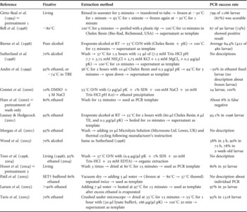
Fixatives were originally used to keep figures or tissues of animals for long term storage. In particular, alcohol-based fixatives (like ethanol) are effective to preserve DNAs of animals (Dessauer et al., Reference Dessauer, Cole, Hafner, Hillis and Moritz1996; Fukatsu, Reference Fukatsu1999). Such alcohol-based fixatives are thought to denature and inactivate tissue nucleases by dehydration, and the dehydration will inhibit hydrolysis of phosphodiester bonds of DNA (Fukatsu, Reference Fukatsu1999). Alcohol-based fixatives have also been used for fixing marine invertebrates (Dawson et al., Reference Dawson, Raskoff and Jacobs1998). Therefore, storage of bivalve larvae in alcohol-based fixatives is believed to be a practical method instead of cryopreservation. Previously, a few studies have applied molecular techniques for this type of larval fixation (Table 1).
Review of the previous methods of bivalve larval PCR on fixed samples, containing the methods of this study.

Wash, washed with filtrated seawater; pK, proteinase K; O/N, over night reaction; RT, room temperature.
† SET buffer: 0.375 M NaCl, 2.5 mM EDTA, 40 mM Tris–HCl, pH 7.8; ‡lysate buffer: 670 mM Tris–HCl pH 8.8, 166 mM ammonium sulphate, 0.1% Tween-20, proteinase K (1 mg/ml).
However, most of these reports have employed expensive commercial kits or redundant pretreatment processes such as the use of chelating resin in order to extract DNA from bivalve larvae; other studies have reported the use of a standard DNA extraction method of SDS-proteinase K digestion and phenol/chloroform extraction (Sambrook & Russell, Reference Sambrook and Russell2001). These troublesome steps were used to extract the DNA from bivalve larval tissues to purify their DNA because the cell lysis contains various inhibitors of PCR, whereas these extraction steps require a considerable number of handling steps that lead to the loss of template larvae or contamination of other DNAs at a high frequency. Furthermore, the amount of DNA in individual larvae is so small that extraction steps may diminish the yield of larval DNA (Bierne et al., Reference Bierne, Launey, Naciri-Graven and Bonhomme1998; Sutherland et al., Reference Sutherland, Stewart, Kenchington and Zouros1998; Morgan & Rogers, Reference Morgan and Rogers2001; Frischer et al., Reference Frischer, Hansen, Wyllie, Wimbush, Murray and Nierzwick-Bauer2002; Deagle et al., Reference Deagle, Bax and Patil2003; Patil et al., Reference Patil, Gunasekera, Deagle and Bax2005). In addition, some of these reports indicated that PCR amplification of fixed larvae frequently failed due to unknown mechanisms (Andre et al., Reference Andre, Lindegarth, Jonsson and Sundberg1999; Wood et al., Reference Wood, Beaumont, Skibinski and Turner2003; Larsen et al., Reference Larsen, Frischer, Rasmussen and Hansen2005).
For these reasons, simple and successful pretreatment methods are urgently required for fixed samples. However, each of these previous reports had adopted different methods for this pretreatment step, and no information is available regarding the most robust pretreatment and the most adequate fixative for bivalve larvae.
Planning of this study
The purpose of this study is to find the most practical method that enables PCR amplifications of bivalve larvae DNA preserved in fixatives. To develop a practical method for field researches, it is important that: (1) the method require only a few handling processes; (2) each process does not require expensive commercial kits; (3) the total processing time is short; and (4) the result is successful. Among these, (1) and (2) are more essential because larval identification is usually applied to a large number of field samples (Medeiros-Bergen et al., Reference Medeiros-Bergen, Olson, Conroy and Kocher1995; Bierne et al., Reference Bierne, Launey, Naciri-Graven and Bonhomme1998; Launey & Hedgecock, Reference Launey and Hedgecock2001; Toro et al., Reference Toro, Innes and Thompson2004; Taris et al., Reference Taris, Baron, Sharbel, Sauvage and Boudry2005).
In this study, we compared several pretreatment methods from previous reports to determine the most practical method. Based on the results we decided that the pretreatment modified from the method of Taris (Taris et al., Reference Taris, Baron, Sharbel, Sauvage and Boudry2005) was the most appropriate one for the fixation of larval samples.
MATERIALS AND METHODS
Larval samples
Larvae were obtained from spawning adult mussels according to the protocol of Beaumont (Beaumont & Abdulmatin, Reference Beaumont and Abdulmatin1994). The Mediterranean blue mussel Mytilus galloprovincialis was commercially purchased and maintained in 3.0% artificial seawater (ASW) at 10°C for at least 10 days. To induce spawning, they were exposed to thermal stimuli ranging from 10 to 20°C repeatedly until they spawned. After the spawned eggs and sperms were mixed in ASW, they were washed carefully by pouring them into a 30-μm mesh sieve and transferred to clean, filtered, natural seawater at 15°C. Fertilized eggs were reared without feeding and aeration until they developed into the D-type veliger stage larvae within 48 hours. The seawater including D-type larvae was filtrated through a 60-μm mesh to remove debris. The larvae were resuspended in ASW, and centrifuged at 700 g for 3 minutes. After discarding the supernatant, the larvae were resuspended again in ASW.
Fixation processes for the larvae
To fix the bivalve larvae, numerous larvae were picked up and transferred onto a watch glass. Using a 0.5–10 µl micropipette (Gilson, Middleton, WI, USA), they were washed and rehydrated in distilled water for 5 minutes under a light microscope. This washing step was repeated 3 times by replacing the distilled water each time in order to eliminate contaminants such as NaCl, which inhibit PCR amplification. Each larva was then transferred into a 1.5-ml microcentrifuge tube and 1 ml of the fixative (100% ethanol, 70% ethanol, 70% acetone and 10% neutralized formalin/water) was added. All the samples were stored at 25°C (RT) for 30 minutes, and then 1 week, 1 month and 1 year under shaded conditions in order to avoid exposure to UV rays (Dessauer, Reference Dessauer, Cole, Hafner, Hillis and Moritz1996). After fixation, the fixed samples were picked up from the tubes and washed 3 times for 5 minutes in distilled water to eliminate the fixatives from larval tissue. After this step, the following pretreatment processes were applied to the larvae.
Comparison of the PCR pretreatments
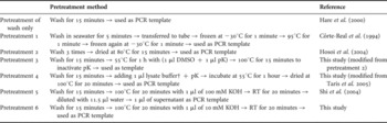
To identify the most effective PCR pretreatment method, we compared the performance of the pretreatments by using 3 procedures reported in previous literature that employed fixed larvae and 3 other procedures modified from original procedures. The details of each procedure are described in Table 2. The previous pretreatments were those of Hare (Hare et al., Reference Hare, Palumbi and Butman2000), Côrte-Real (Côrte-Real et al., Reference Côrte-Real, Holland and Dixon1994), Hosoi (Hosoi et al., Reference Hosoi, Hosoi-Tanabe, Sawada, Ueno, Toyohara and Hayashi2004) and Shi (Shi et al., Reference Shi, Datar, Liu, Wu, Zhang, Cote and Taylor2004). We have selected these pretreatments based on the criteria noted in the introduction. They do not employ organic extraction, chelating resins, or commercial kits because the use of these methods increases the number of handling steps and is more expensive.
The details of 7 pretreatments on bivalve larvae tested in this study.

Wash, washed with distilled water to induce hypotonic lysis, and/or to eliminate contaminants in each larval body; pK, proteinase K (0.2 mg/ml).
† lysate buffer: 670 mM Tris–HCl pH 8.8, 166 mM ammonium sulphate, 0.1% Tween-20, proteinase K (1 mg/ml).
PCR analysis
After performing the above-mentioned pretreatment processes, PCR tests were performed for 4 individual larvae by using each combination of pretreatment and fixative for the appropriate fixation duration. Each PCR test was triplicated. In total, 12 larvae were tested. In this study, we used a partial region of the nuclear large subunit (LSU) ribosomal gene as the target gene because the mitochondrial genes that have been widely used as molecular markers for species identification show heteroplasmic inheritance in several bivalves, including the mussel M. galloprovincialis (Baldwin et al., Reference Baldwin, Black, Sanjur, Gustafson, Lutz and Vrijenhoek1996; Burzynski et al., Reference Burzynski, Zbawicka, Skibinski and Wenne2003; Larsen et al., Reference Larsen, Frischer, Rasmussen and Hansen2005). PCR of the nuclear LSU ribosomal gene was performed as follows: forward primer bvLSUD1f, 5′-GGGTGGTAAACTCCAYCTAARGC-3′ and reverse primer bvLSUD3r, 5′-CTRCGGACCTCCATCAGAGTTTCC-3′ (PCR primers described prior to this research (Hosoi et al., Reference Hosoi, Hosoi-Tanabe, Sawada, Ueno, Toyohara and Hayashi2004) were used). In order to validate the pretreatment of larval DNA for PCR, the most appropriate pretreatment was applied to another nuclear gene of an adhesive protein, which codes for a mussel byssus protein and has been used as a reliable molecular marker for analysing the mussel population structure (Inoue et al., Reference Inoue, Waite, Matsuoka, Odo and Harayama1995; Wood et al., Reference Wood, Beaumont, Skibinski and Turner2003). The adhesive protein gene was amplified using the primer set Me 15 and Me 16 (Inoue et al., Reference Inoue, Waite, Matsuoka, Odo and Harayama1995). All PCR amplifications were performed in 20 µl of reaction mixture containing 1.0 U Taq (New England Biolabs, Beverley, MA, USA), 0.2 mM dNTPs, 1× standard Taq buffer, and 0.5 µM primers. Although some previous studies recommend the use of hotstart Taq polymerase or PCR additives that could improve larval PCR amplification (Côrte-Real et al., Reference Côrte-Real, Holland and Dixon1994; Hare et al., Reference Hare, Palumbi and Butman2000; Patil et al., Reference Patil, Gunasekera, Deagle and Bax2005; Larsen et al., Reference Larsen, Frischer, Rasmussen and Hansen2005), we used only a standard recombinant Taq polymerase in all the examinations to compare the effectiveness of the pretreatments under normal conditions; however, for pretreatment 2, 10% DMSO was used as a facilitator, according to our previous study. Thermocycling was performed as follows: the preheat step for 5 minutes at 94°C, followed by 35 cycles of 1 minute at 94°C, 1.5 minutes at 55°C, and 2.5 minutes at 72°C, and a final elongation step of 10 minutes at 72°C. As a positive control for the PCR amplifications, the nuclear LSU ribosomal RNA gene fragment of M. galloprovincialis cloned into the pGEM-T Easy vector (Promega Corporation, Madison, WI, USA) was amplified using the primers bvLSUD1f and bvLSUD3r. The fragment was used as the PCR template. As a negative control, PCR with no template was performed for each experiment. The success rate of PCR was defined as the rate of individual larvae successfully amplified per total analysed individual larvae. Successful PCR was judged based on the amount of PCR products available for use in the following analyses (e.g. PCR-RFLP). The PCR products were evaluated using electrophoresis in 1.2% ethidium bromide-stained agarose gel in Tris-acetate-EDTA (TAE) buffer and observed under a UV transilluminator.
Statistical analysis
The significance of difference in the PCR success rates was tested between: (1) wash only pretreatment as a negative control and the other combinations involving different pretreatments along with fixatives for the appropriate fixation duration; and (2) 30 minute-fixed larvae and 1-week-, 1-month- or 1-year-fixed larvae for each pretreatment. The significance of difference in the success rates was evaluated using Fisher's exact test (Sokal & Rohlf, Reference Sokal and Rohlf1994).
RESULTS
The most effective pretreatment for living larvae
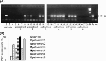
First, PCR targeting the nuclear LSU ribosomal RNA gene was performed for specimens of living larvae for each pretreatment method (Figure 1). The expected 791-bp sized bands of M. galloprovincialis larvae were amplified. The success rates of the 4 pretreatments based on the PCR products (1, 3, 4 and 6 described in Table 2) were significantly greater than that for the wash only pretreatment (Figure 1B; Fisher's exact test, P < 0.05 for pretreatment 1 and P < 0.01 for pretreatments 3, 4 and 6).
(A) Representative results of pretreated PCR amplification performed on Mytilus galloprovincialis living larvae using LSU ribosomal RNA gene targeted primers. Each of pretreatments is described in Materials and Methods and Table 2. Lanes 1–4, wash only pretreatment; lanes 5–8, pretreatment 1; lanes 9–12, pretreatment 2; lanes 13–16, pretreatment 3; lanes 17–20, pretreatment 4; lanes 21–24, pretreatment 5; lanes 25–28, pretreatment 6; lane M, molecular size marker (HindIII-digested lambda DNA); lane Pc, positive control (M. galloprovincialis LSU ribosomal RNA gene fragment constructed plasmid used as a template); lane Nc, negative control (no templates). Arrows on the right hand side denote the band of 791 bp; (B) the success rate of PCR amplification of 12 living individual bivalve larvae with each pretreatment. Significance of each pretreatment in contrast to wash only pretreatment as a negative control is indicated above the column with *(P < 0.05), **(P < 0.01).

Application of the pretreatment processes to fixed larvae
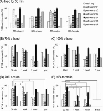
Next, we examined the effects of the 4 fixatives and fixation duration on the larvae coupled with each pretreatment. First, to confirm the effectiveness of each pretreatment with regard to enhancing the PCR success rate for larvae fixed using various fixatives, we compared the success rate of each pretreatment for the 30 minute-fixed larvae (Figure 2A). With regard to the 70% ethanol-fixed larvae, the PCR success rate was improved in the cases of pretreatments 1 and 4, which were found to be superior to the wash only pretreatment. In the 100% ethanol-fixed larvae, improvement in the success rate was confirmed only for pretreatment 4. In the 70% acetone-fixed larvae, pretreatments 1, 3, and 4 showed higher success rates than the wash only pretreatment. In the 10% formalin-fixed larvae, improvement was observed only for pretreatment 4.
The success rate of pretreated PCR amplification of bivalve larvae. (A) Fixed for 30 minutes with each of fixatives of 70% ethanol, 100% ethanol, 70% acetone and 10% formalin. (B), (C), (D) and (E) represents the results of the effects of fixation duration and pretreatment of wash only, 1, 3, 4 and 6. Significance of each pretreatment in contrast to wash only pretreatment as a negative control is indicated above the columns with *(P < 0.05), **(P < 0.01). Significance of the time-course decrease of PCR success rate compared to 30 minutes was indicated under the solid line with †(P < 0.05), ††(P < 0.01).

When the success rates of PCR amplification were tested over time with regard to pretreatments 1, 3, 4 and 6, which were effective for the 30-minute fixed larvae, some pretreatments on fixed larvae tended to show a time-course decrease in the success rate, but the temporal pattern was not obvious through 1 year in 70% and 100% ethanol-fixed larvae. On the other hand, some pretreatments on acetone- and formalin-fixed larvae showed a significant decrease over time (Figure 2D & E; P < 0.05, 0.01). The most notable difference was observed in the larvae that were processed using pretreatment 4. The DNA of fixed larvae that underwent pretreatment 4 showed good amplification and significantly higher success rates than those that underwent the wash only pretreatment in 70% ethanol, 100% ethanol and 10% formalin (Fisher's exact test, P < 0.05).
DNA amplification of the adhesive protein gene
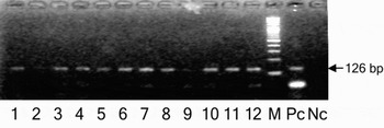
PCR amplification using the Me 15 and Me 16 primers was performed. These primers amplified the expected 126-bp sized bands of M. galloprovincialis larvae processed by pretreatment 4 after a 1-month fixation in 70% ethanol (Figure 3). The results demonstrated the success rate of PCR to be 90% (18/20) for pretreatment 4, and it was not significantly different from that of the LSU rRNA gene (Fisher's exact test, P > 0.05).
Gel electrophoresis of PCR amplicons of Mytilus galloprovincialis adhesive protein gene with Me 15/16 primers of each of 70% ethanol preserved larvae. Lanes 1–12, PCR products; lane M, 2-log DNA ladder (New England Biolabs, Beverley, MA); lane Pc, positive control (M. galloprovincialis ribosomal LSU fragment constructed plasmid as a template); lane Nc, negative control (no template).

DISCUSSION
Even though many studies applied PCR for species identification of bivalve larvae, the reason for PCR inhibition of bivalve larvae has not been explained by any systematic study thus far (Table 1). This is because the factors that cause PCR inhibition are very diversified in the same way as the studies of holothurian larvae (Medeiros-Bergen et al., Reference Medeiros-Bergen, Olson, Conroy and Kocher1995; Evans et al., Reference Evans, White and Ward1998). Most of those factors which inhibit PCR have not been identified, researchers have to treat their zooplankton samples to avoid these indefinite factors just after sampling (Bucklin, Reference Bucklin, Harris, Wiebe, Lenz, Skjoldal and Huntley2000). DNA degradation or modification of samples which influence PCR success rate were probably caused by those factors; for example, storage conditions of the larvae after sampling (storage buffer, time, temperature and light), washing time and washing stringency of the larvae before PCR, the existence of tightly closed shells, the difference of species of larvae, size of the larvae (large individuals contain a considerable number of endogenous inhibitors), and the presence of heating or evaporation steps during the pretreatment. Altering even one of these factors would dramatically change the final outcome, i.e. the PCR success rate. This study focused on the pretreatment steps immediately before PCR, while keeping the other factors constant for each larval sample.
The most effective pretreatment
In this study, PCR following the wash only pretreatment could not consistently amplify the DNA of living bivalve larvae that was used as the PCR template (Figure 1B). In addition, fixed larvae, too, could not be stably amplified by this method (Figure 2). Inconsistency in the PCR success rates for fixed larvae has not been reported, except for a few papers on ethanol fixation (Medeiros-Bergen et al., Reference Medeiros-Bergen, Olson, Conroy and Kocher1995; Andre et al., Reference Andre, Lindegarth, Jonsson and Sundberg1999; Wood et al., Reference Wood, Beaumont, Skibinski and Turner2003; Larsen et al., Reference Larsen, Frischer, Rasmussen and Hansen2005). The present study scrutinized the difficulty of conducting PCR on the DNA of bivalve larvae. However, the success rates of 3 pretreatments (wash only and pretreatments 1 and 2) would be less than that of the original articles because we used only the standard Taq polymerase in all the experiments in order to evaluate the effectiveness of each pretreatment under the simplest condition. If other efficient Taqs were used according to the original descriptions of the 3 pretreatments, the PCR success rates would be as high as that of the original studies.
Some effective pretreatments for fixed bivalve larvae were screened via a comparative experiment involving 6 pretreatments. Initially, pretreatment 4 was more effective for living larvae in contrast to the wash only pretreatment (Figure 1B; Fisher's exact test, P < 0.01), and this pretreatment was also effective for larvae fixed with 70% ethanol, 100% ethanol and 10% formalin for 1 year. Pretreatment 4 involved the use of ammonium sulphate and Tween 20 and proteinase K digestion. Because proteinase K was also used in pretreatment 3, this improvement may be caused by the presence of ammonium sulphate and/or Tween 20. Ammonium sulphate was used to detect the presence of virus in the urine directly by PCR (Olive et al., Reference Olive, Simsek and Al-Mufti1989). However, it was less popular as a PCR facilitator in contrast to DMSO, BSA, Tween 20 and glycerol (Rolfs, Reference Rolfs1992; Griffin & Griffin, Reference Griffin and Griffin1994; Abu Al-Soud & Radstrom, Reference Abu Al-Soud and Radstrom2000). Ammonium sulphate and/or Tween 20 would actually be suitable for the pretreatment of bivalve larvae prior to PCR, although the underlying mechanism has not yet been determined.
Alkaline treatments, for example, the addition of KOH or NaOH solutions have been popularly used for DNA extraction in order to save effort and time (Rolfs, Reference Rolfs1992; Sambrook & Russell, Reference Sambrook and Russell2001). Recently, this method was applied for retrieving DNA from formalin-fixed paraffin-embedded tissues in order to use them as PCR templates without phenol–chloroform extraction (Shi et al., Reference Shi, Cote, Wu, Liu, Datar, Shi, Liu, Lim and Taylor2002, Reference Shi, Datar, Liu, Wu, Zhang, Cote and Taylor2004). Therefore, we attempted to use this alkaline treatment with 2 different concentrations of KOH (pretreatments 5 and 6). The PCR success rate of pretreatment 6 was significantly improved in experiments using living larvae in contrast to the wash only pretreatment (Fisher's exact test, P < 0.01). Pretreatment 6 showed a relatively high PCR success rate in some fixed larvae; however, its effectiveness was not significant in contrast to the wash only pretreatment.
Fixation of bivalve larvae in formalin
As shown in Figure 2B–E, extension of fixation time of bivalve larvae in formalin or acetone caused a temporal decrease in the PCR success rates for some pretreatments. After the PCR, we confirmed that the soft body of each larva in PCR mixture was enough digested. Therefore, the decrease of PCR success rate was not due to the rigid cell membranes but the other factors. It is most likely that these decreases in the PCR success rates are due to DNA degradation. In formalin-fixed tissues, PCR inhibition was caused by not only DNA degradation (e.g. DNA cleavage) but also DNA modification (e.g. formation of crosslinks across DNA–protein or DNA–DNA bonds) or the presence of inhibitor(s) for polymerase activity in the PCR buffer (Wilson, Reference Wilson1997; Srinivasan et al., Reference Srinivasan, Sedmak and Jewell2002; Inadome & Noguchi, Reference Inadome and Noguchi2003). Larval DNA cleavage critically lowered the PCR success rates, but the success rate for pretreatment 4 by using formalin-fixed larvae was as high as those obtained with the other fixatives (Figure 2E). Based on these results, it may be suggested that pretreatment 4 also facilitates PCR reactions of modified DNA or antagonizes the effect of polymerase inhibitor(s) to some extent.
Ethanol fixation of bivalve larvae
Figure 2B & C suggest that the significant time-dependent decrease in PCR success rates had not occurred in ethanol-fixed larvae. However, the PCR success rate for these ethanol-fixed larvae was too low (below 50%) to be practically applied, unless pretreatment 4 was employed. These low success rates observed with regard to ethanol-fixed tissues may be observed under similar conditions in formalin-fixed larvae; however, the mechanism of DNA damage caused by ethanol is not known in contrast to that by formalin (Bancroft & Stevens, Reference Bancroft and Stevens2002). On the other hand, many studies used alcoholic fixatives or acetone to preserve adult invertebrate tissues for long periods (over years), and they showed that DNA extracted from fixed samples by the standard phenol–chloroform extraction method can be used as PCR templates (Dawson et al., Reference Dawson, Raskoff and Jacobs1998; Fukatsu, Reference Fukatsu1999). This implies that ethanol and acetone can at least prevent the sample DNA from undergoing complete degradation and that organic extraction of the sample DNA makes it available as PCR templates. However, this organic extraction method is impractical for larvae because of its laboriousness. Alternatively, pretreatment 4 will provide a forcible method for PCR. It may be concluded that: (1) PCR inhibition due to DNA degradation or modification is found in fixed bivalve tissues; however, (2) pretreatment 4, in comparison to other pretreatments or the absence of any pretreatment, can facilitate PCR amplification in the presence of such damaged DNA as efficiently as organic extraction. Based on the success of pretreatment 4, the importance of careful choice of a DNA amplification method for fixed samples is brought to light.
Specific factors in bivalve larvae
While fixed bivalve larvae contain similar factors as fixed human tissues, some characteristic features of bivalve larvae also exist. For example, the presence of the shell may induce instability in the PCR amplification (Bell & Grassle, Reference Bell and Grassle1998; Taris et al., Reference Taris, Baron, Sharbel, Sauvage and Boudry2005). When their shells were decalcified by acids like hydrochloride, strong inhibition of amplification was observed (data not shown). This was probably caused by the increased concentration of calcium ions in the PCR reaction buffer, or hydrolysis of DNA which was teared into shreds in the acidic solution. On the other hand, their tightly closed shells may prevent the DNA of lysed cells from eluting out of their shells. However, crushing the shells using needle or plastic tips (Bell & Grassle, Reference Bell and Grassle1998; Taris et al., Reference Taris, Baron, Sharbel, Sauvage and Boudry2005) was difficult and time consuming. In this study, the effectiveness of pretreatment 4 may make the crushing of bivalve shells unnecessary.
Our results showed that pretreatment 4, i.e. the ammonium sulphate method, was robust enough to other various fixatives even after 1-year fixation, and this method was feasible with regard to other primer pairs (Figure 2). This study revealed 2 solutions to the problems involved in the study of bivalve larvae. First, ethanol preservation could be used at the time of sampling bivalve larvae in the field. The availability of convenient fixatives for the preservation of larvae would increase the number of opportunities for field sampling in various situations hereafter. Second, the extraction steps before PCR can be abbreviated since this method does not require the use of expensive commercial kits or standard SDS–phenol–chloroform extraction. Thus, it enables the analysis of a large number of samples in the course of high-throughput studies. Studies on ecology, evolution, and population genetics, which are conducted in the open ocean or the deep sea, that ordinarily involve long-term preservation of samples on board or in other sampling fields where enough laboratory equipments are lacking will be greatly benefited by this method involving ethanol fixation and subsequent pretreatments.
ACKNOWLEDGEMENTS
We would like to thank the numerous colleagues and collaborators of the laboratories in Kyoto and Ibaraki for their technical advice and help on rearing larvae and laboratory experiments. We are especially grateful to C. Shinzato and K. Sakamoto for their comments on an early version of the manuscript draft. Two anonymous referees made important contributions to this manuscript.