




No CrossRef data available.
Article contents
Left and right basal ganglia and frontal activity during language generation: Contributions to lexical, semantic, and phonological processes
Published online by Cambridge University Press: 12 February 2004
Abstract
fMRI was used to determine the frontal, basal ganglia, and thalamic structures engaged by three facets of language generation: lexical status of generated items, the use of semantic vs. phonological information during language generation, and rate of generation. During fMRI, 21 neurologically normal subjects performed four tasks: generation of nonsense syllables given beginning and ending consonant blends, generation of words given a rhyming word, generation of words given a semantic category at a fast rate (matched to the rate of nonsense syllable generation), and generation of words given a semantic category at a slow rate (matched to the rate of generating of rhyming words). Components of a left pre-SMA–dorsal caudate nucleus–ventral anterior thalamic loop were active during word generation from rhyming or category cues but not during nonsense syllable generation. Findings indicate that this loop is involved in retrieving words from pre-existing lexical stores. Relatively diffuse activity in the right basal ganglia (caudate nucleus and putamen) also was found during word-generation tasks but not during nonsense syllable generation. Given the relative absence of right frontal activity during the word generation tasks, we suggest that the right basal ganglia activity serves to suppress right frontal activity, preventing right frontal structures from interfering with language production. Current findings establish roles for the left and the right basal ganglia in word generation. Hypotheses are discussed for future research to help refine our understanding of basal ganglia functions in language generation. (JINS, 2003, 9, 1061–1077.)
Keywords
Information
- Type
- THEMATIC ARTICLES
- Information
- Journal of the International Neuropsychological Society , Volume 9 , Issue 7 , November 2003 , pp. 1061 - 1077
- Copyright
- © 2003 The International Neuropsychological Society
INTRODUCTION
Although investigators have speculated about the role of the basal ganglia in language since the end of the 19th century (Broadbent, 1872; Marie, 1906; Wernicke, 1874), little consensus has been reached regarding the involvement, not to mention the role, of the basal ganglia in language. Suggested functions of the basal ganglia in movement and language have included: facilitation/initiation of desired actions and suppression of undesired actions (Gerfen, 1992; Penney & Young, 1986), selection of actions from competing alternatives (Jueptner & Weiller, 1998; Mink, 1996), and facilitation of controlled, as opposed to automatic processing (Copland et al., 2000a). Although aphasia is common after dominant basal ganglia lesion, evidence currently suggests that the more florid language symptoms in such cases are related to concomitant cortical dysfunction (Nadeau & Crosson, 1997; Weiller et al., 1993). Nonetheless, Mega and Alexander (1994) showed that the basal ganglia play a more subtle role in the generation of words, and more recent evidence indicates that the basal ganglia have a broader role in complex aspects of language, that is, executive language functions (Copland et al., 2000b). This latter role is consistent with the anatomic position of basal ganglia structures in multiple frontal–basal-ganglia–thalamic loops (Middleton & Strick, 2000), indicating that the function of these loops must be considered to understand the role of the basal ganglia in language.
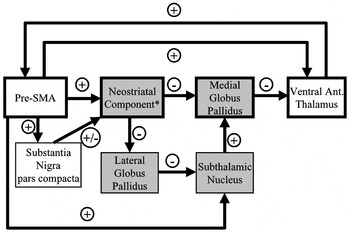
The phylogenetically older structures of these loops, deep within the cerebral hemispheres, typically have not been considered to possess the information storage capacity of the cerebral cortex. The connections of the basal ganglia (Figure 1) are well known and give some clues regarding their function. These structures are situated in multiple anatomically segregated closed loops; each loop includes a distinct region of the cerebral cortex (Middleton & Strick, 2000). At the subcortical level, the loops are not interconnected, and access to the various subcortical structures is limited to a finite set of brain structures. The caudate nucleus and putamen comprise the neostriatum. Much of the putamen receives input from motor and premotor cortex, while components of the caudate nucleus receive input largely from various prefrontal structures (Hoover & Strick, 1999; Parent, 1990). The corticostriatal neurotransmitter is glutamate, an excitatory neurotransmitter (Gerfen, 1992). In turn, the neostriatal components of these loops each project to a distinct segment of the medial globus pallidus, which is known to be the output organ of the basal ganglia. The major striatopallidal neurotransmitter is gamma-aminobutyric acid (GABA), an inhibitory neurotransmitter, though neuropeptides that may act as neurotransmitters also are found in these projections (Gerfen, 1992). Each medial pallidal segment projects to specific thalamic nuclei, each of which projects back primarily to the cortical region which contributes fibers to the neostriatal component of its particular loop. Thalamocortical projections are reciprocated by corticothalamic projections. The pallidothalamic connections employ the inhibitory neurotransmitter GABA, while the thalamocortical and corticothalamic neurotransmitter is glutamate (Gerfen, 1992). For a complete discussion of neurotransmitters within cortical–basal ganglia loops, the reader is referred to Gerfen (1992). Although most basal ganglia loops originate in and target frontal cortical regions, temporal and parietal loops also exist (Middleton & Strick, 2000). Thus, the connections of the basal ganglia indicate that they influence cortical processing. Other basal ganglia connections that influence the loops are shown in Figure 1 (Gerfen, 1992; Nambu et al., 2000). Finally, while this loop structure has largely been conceptualized as a unilateral system, cortical projections, especially from medial frontal cortex, to the contralateral neostriatum also exist (Buchanan et al., 1994; Cowan & Wilson, 1994; Inase et al., 1999; Morino et al., 1994; Wang & Pickel, 1998). How contralateral projections influence cortical–basal ganglia loops has not been extensively explored.

Schematic diagram of pre–SMA–basal ganglia loop. Components of the “direct loop” (discussed in Introduction) are shown in boxes with heavy black lines. There is also an “indirect loop” which, due to space considerations, was not detailed in the Introduction. Components of the “indirect subloop” are shown with shaded boxes. The neostriatal component* of the loop probably consists of striatal gray bridges, but also may include the caudate nucleus or putamen on either side of the capsule (Inase et al., 1999). Direction of pathways between structures is indicated by the arrows. Reciprocated connections are indicated by separate arrows. +'s in circles indicate excitatory pathways; −'s in circles indicate inhibitory pathways. Note that dopaminergic projections from the substantia nigra pars compacta to the neostriatum have both excitatory and inhibitory effects, depending upon the target neurons (Gerfen, 1992).
While functional neuroimaging offers promise as a tool to unravel the role of the basal ganglia in language, results of functional imaging studies to this point have been less than definitive (Cabeza & Nyberg, 2000). In particular, word generation studies have shown consistent activation of medial frontal cortex, usually near the boundary of pre-SMA and the rostral cingulate zone (e.g., Crosson et al., 1999, 2001; Petersen et al., 1988; Warburton et al., 1996). SMA (supplementary motor area) is the posterior portion of medial Brodmann's area 6, while pre-SMA is the anterior portion of medial Brodmann's area 6. SMA has reciprocal connections with motor and pre-motor cortices, while pre-SMA has reciprocal connections with prefrontal cortex (Matsuzaka et al., 1992; Picard & Strick, 1996). Anatomic studies clearly show pre-SMA projections to striatal gray matter spanning the internal capsule and to the caudate nucleus and putamen on either side of the capsule (Inase et al., 1999). Further, the ventral anterior and dorsal medial thalamus are connected to pre-SMA (Wiesendanger & Wiesendanger, 1985), and Akkal et al. (2002) recently identified the pallidal segment of the pre-SMA–basal ganglia loop. While such anatomic studies highlight the striatal, pallidal, and thalamic components of this loop, these components have not shown consistent activity in functional imaging studies of word generation. Limitations in numbers of subjects, number of trials during functional neuroimaging, and experimental design contribute to inconsistency in activation of basal ganglia and thalamic structures during word generation. Indeed, even work within single laboratories, using identical tasks, has proven inconsistent in this regard. For example, Warburton et al. (1996) compared verb generation to a resting state using positron emission tomography (PET) in three separate studies performed in their laboratory. Medial frontal activity appeared in all three studies. However, this comparison yielded activity in the left and right heads of the caudate nuclei in only one of the three experiments. While thalamic activity was present in two of three studies, the location varied considerably between the two studies. Numbers of subjects in these experiments varied between 4 and 9. In a separate set of experiments that used varying imaging modalities, overt versus covert word production, and different rates of production, word-stem completion and visual fixation were compared (Ojemann et al., 1998; Palmer et al., 2001; Rosen et al., 2000). Although medial frontal cortex was active in all comparisons of these studies, activity in the left and right putamen was inconsistent between studies, and no activity was found in the caudate nucleus. Thalamic activity was more consistent and was located in thalamic centers related to motor functions. Numbers of subjects in these studies varied between 5 and 10. With such inconsistencies in basal ganglia activity during word generation and the probable methodological issues contributing to these inconsistencies, few attempts have been made to delineate the conditions under which basal ganglia activity is present or absent.
However, in a recent verbal working memory study, Crosson et al. (1999) showed pre-SMA and anterior thalamic activity in verbal working memory tasks involving lexical items but not in a verbal working memory task in which lexical items were not attended to. On the other hand, caudate nucleus activity appeared only for a semantic working memory task but not for a working memory task relying upon rhyming words. These findings raise two questions: First, is retrieval or manipulation of pre-existing lexical items what determines whether pre-SMA and its basal ganglia and thalamic targets become active? And, second, is such activity unique to working memory or does it apply to other verbal tasks, such as word generation?
The purpose of this study was to determine which aspects of language generation engage pre-SMA and related structures in the basal ganglia and thalamus. The most general question was whether pre-SMA and its related subcortical structures would show similar or dissimilar patterns of activity when language generation involved lexical versus non-lexical processes, phonological versus semantic processes, and fast versus slow generation. To the degree that cognitive operations of working memory and word generation overlap, the verbal working memory study of Crosson et al. (1999) suggested that pre-SMA and the ventral anterior thalamus would be activated by lexical as opposed to non-lexical processes. However, the findings of Crosson et al. also suggested that the caudate nucleus would be engaged by semantic as opposed to phonological processes. The word-stem completion findings of Palmer et al. (2001) also indicate that faster rates of word generation could dampen medial frontal activity though rate of generation does not appear to impact thalamic activity.
Thus, we endeavored to design a study of word generation to determine factors that engage pre-SMA and related basal ganglia and thalamic structures. A series of language generation tasks was developed that systematically varied lexical versus nonlexical processing, phonological versus semantic processing, and high versus low density of response. Given that basal ganglia and thalamic activity could not be consistently replicated in previous studies, even using identical word generation tasks within the same laboratories, we used several methodological devices to ensure optimal sensitivity within these subcortical structures. First, a relatively large number of subjects and trials was used to enhance reliability of analyses and sensitivity to activity in deep brain structures. Second, a blocked trial format (i.e., multiple responses to a single stimulus) was used. Pilot data from our laboratory demonstrated that the extended activity generated by this format for word generation produces a greater amplitude of signal change in medial frontal cortex than a single-event format. Thus, the blocked trial format enhances signal-to-noise ratio, and therefore provides for greater sensitivity than single-event paradigms. Third, a resting state was used as a control state because initiation of other activities might involve basal ganglia functions (Heilman et al., 2003), and because recent evidence has indicated that a resting baseline enhances activity in language-related structures in comparison to using other language tasks as a baseline state (Newman et al., 2001). Finally, a relatively stringent statistical threshold for activity was employed so that cluster size thresholds could be decreased, thereby allowing us to visualize activity in relatively small subcortical structures.
Competing hypotheses were as follows: (1) in a study with adequate sensitivity to subcortical activity, consistent activity will be seen in pre-SMA and related basal ganglia and thalamic structures when language generation involves retrieval of lexical items but not when it involves sub-lexical processes; (2) alternatively, pre-SMA and related subcortical structures will show different patterns of activity during word generation, reflecting varying roles in language generation. Specifically, using Crosson et al.'s (1999) working memory findings as a template, pre-SMA and its thalamic target will be active when word generation involves retrieval of lexical items, but the caudate nucleus will be active only when word generation is based on retrieval of semantic information.
METHODS
Research Participants
Twenty-one students, faculty, or staff at the University of Florida and/or residents of Gainesville, Florida (12 male, 9 female) participated. Ages ranged from 18 to 38 years; education ranged from 13 to 20 years (M = 16.5, SD = 2.0). All participants were native English speakers, were right hand dominant (Edinburgh Handedness Inventory: Oldfield, 1971), and gave written informed consent in accordance with a protocol approved by the Health Center Institutional Review Board at the University of Florida.
Word Production Tasks
All participants performed four language generation tasks silently during scanning sessions:
- In nonsense syllable generation participants heard beginning and ending consonant blends and generated as many nonsense syllables as possible (e.g., for “sk … lf” they might respond “skalf, skilf, skoolf …”).
- In rhyme generation, participants heard a stimulus word and generated as many rhyming words as possible (e.g., for “cat,” they might respond “bat, hat, rat …”).
- In fast category member generation participants heard a category and generated as many members of the category as possible (e.g., for “birds,” they might respond “sparrow, wren, hawk …”). On the basis of pilot data, categories were chosen for which the generation rate was equivalent to nonsense syllable generation.
- In slow category member generation participants heard a category and generated as many category members as possible. On the basis of pilot data, categories were chosen for which the generation rate was equivalent to that of rhyme generation.
In separate imaging runs, 17.4 s blocks of each language production task were alternated with 17.4 s blocks of rest during which participants were discouraged from thinking any words to themselves. During word generation blocks and the resting control state, subjects were instructed to focus on a fixation point (cross) located outside the scanner, which they could see through a mirror attached to the head coil. Imaging runs began with a block of rest and were 6.4 cycles long. Subjects performed two imaging runs of each task. Three lists of stimuli (i.e., categories, rhyming words, beginning and ending consonant blends) were developed for each task. In each task for each subject, two lists were chosen in a pseudorandom fashion for use in imaging runs. The third list for each task was used to obtain oral generation rates after the scanning session.
Stimulus Presentation and Task Performance
All word production was accomplished silently to avoid image artifacts created when participants speak during scanning and to avoid activation elicited when participants hear their own voice. Silent production has proved reliable for imaging cortical regions such as pre-SMA in our own (e.g., Crosson et al., 1999, 2001) and other (e.g., Chao et al., 1999; Warburton et al., 1996) laboratories. Order of presentation of the language production tasks was pseudorandomized. Categories and cues were presented via an IBM 380ED notebook computer using a Kenwood KR-A4070 amplifier, a Realistic 31-2005 Ten Band Stereo Frequency Equalizer, a JBL 2446J 16 ohm speaker attached to an air conduction transducer, and foam insert ear phones. Output was biased toward the high end of the frequency spectrum to compensate for the loss of amplitude in higher frequencies through the air conduction apparatus. Before beginning experiments, individual thresholds for word recognition during scanning were determined, and stimuli were delivered at 30 to 35 dB above threshold.
Image Acquisition
Functional and structural images were acquired on a GE 1.5T Signa scanner using a dome-shaped quadrature radio frequency head coil. T1-weighted axial scout scans were acquired to place sagittal functional images in the brain, and head alignment in the coil was adjusted, if necessary, such that the interhemispheric fissure was within two degrees of vertical. Twenty-two functional image slices (6.4–6.9 mm thick) covered the whole brain. The plane between slices 11 and 12 was placed in the interhemispheric fissure. A series of 64 functional images (10 images for each rest–task cycle) was acquired for each of the 22 sagittal slices using a gradient echo spiral scan acquisition (King et al., 1995; Macovski, 1985; Noll et al., 1995), TE = 40 ms, TR = 1740 ms, FA = 60°, FOV = 18 cm, 2 spirals. Images were reconstructed into a 128 × 128 matrix. Subsequent to functional images, T1-weighted structural images were acquired for 124 × 1.3 mm thick sagittal slices, using a 3D spoiled GRASS volume acquisition (TE = 7 ms; TR = 27 ms; NEX = 1; FOV = 24 cm; matrix size = 256 × 192).
Image Analyses
Functional images were analyzed with the Analysis of Functional Neuroimaging (AFNI) program (Cox, 1996). Functional images for each imaging run were spatially registered to a base image using a 3-dimensional registration algorithm, visually inspected for gross artifact, and viewed in a cine loop to detect residual motion. For each task, images from a participant were discarded due to significant artifacts or motion on either of the two runs. This procedure left data for analysis from 19 subjects in nonsense syllable generation, 21 subjects in rhyme generation, and 20 subjects for each category-member generation task. Mean signal intensities for individual images in the slice-time matrix were normalized to the group mean, and voxels for which the standard deviation of the signal in the time series exceeded 5% of the mean signal were set to zero to attenuate large vessel effects and residual motion artifacts. Linear drift in the time series was removed using Gram-Schmidt orthogonalization. The acquired functional time series in each voxel was regressed against sinusoidal reference waveforms, time-locked to the alternating cycles of word generation and rest (Bandettini et al., 1993). Nine phase-shifted reference waveforms were used to compensate for temporal differences in acquisition of each spiral within the 22 slices. The waveform generating the highest correlation was used for each voxel. Use of this regression technique assumes that the form of the hemodynamic response will be similar between cortical and subcortical structures. A composite functional image for each run for each subject was generated using magnitude of least squares fit (MLSF), an additive function that contains information about the temporal correlation of the acquired time series with the selected reference wave form, as well as the amplitude of signal changes in the acquired time series. Functional images derived for the 2 runs of each task were averaged. Subsequently, anatomic and averaged functional images were linearly interpolated to 1 mm3 voxels, co-registered, and converted to stereotactic coordinates (Talaraich & Tournoux, 1988). Functional image volumes were smoothed (3 mm FWHM Gaussian filter) to compensate for intersubject variability in structural and functional anatomy. This relatively small degree of smoothing was used to prevent reduction of signal in small subcortical regions by volume averaging with larger surrounding regions of inactivity.
Voxel by voxel analyses comparing each task to the resting state were found to be more sensitive than other analysis schemes to basal ganglia and thalamic changes, consistent with the findings of Newman et al. (2001) that language task–rest comparisons have greater sensitivity than language task–language task comparisons. Thus, given the focus of this paper on basal ganglia and thalamic contributions to word and nonsense syllable production, we present these analyses. Student's t-tests were conducted across participants on a voxel-by-voxel basis comparing alternations between each of the 4 tasks and rest to a null hypothesis of no change in activity from rest to language production. Both a statistical probability threshold applied on a voxel by voxel basis and a cluster-size threshold of contiguous voxels to identify regions of significant activity were used. Clusters of activity were defined using a statistical probability threshold for voxel-wise t test of p < .00001. When regression analyses with nine random reference waveforms were performed on the current data at this probability level, no significant clusters were found. (This approach to defining cluster size and probability thresholds was derived from the work of Bullmore et al., 1996, and Forman et al., 1995.) Thus, the ability to confidently reject false positive activity at this probability level allows us to use a relatively small contiguity threshold of volume greater than 50 μl to define significant activity, thereby increasing sensitivity to small regions of activity in the basal ganglia and thalamus.
RESULTS
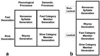
Figure 2a shows that generation of nonsense syllables and generation of rhyming words both emphasized phonological as opposed to semantic processing, while fast and slow category-member generation emphasized semantic as opposed to phonological processing. Generation of nonwords and fast category-member generation were matched by pilot data for their relatively high density of response generation, while generation of rhyming words and slow category-member generation were matched by pilot data for their relatively low density of response generation. Figure 2b indicates that tasks also could be divided according to whether words (i.e., lexical items) or nonsense syllables (i.e., nonlexical items) are generated; words are generated on the rhyme generation, fast category-member, and slow category-member generation tasks, while nonwords are generated on the nonsense syllable generation task. These divisions of task characteristics indicate what variables determine the participation of different frontal, basal ganglia, and thalamic structures in language generation.

Experimental task characteristics. The four word generation tasks can be classified as requiring phonological versus semantic processing or fast versus slow generation (a), but also can be classified by whether they require nonlexical versus lexical processing (b).
For each task, oral generation rates were obtained for one of three lists outside the scanner. Mean rates of item generation for each task are displayed in Table 1. Two-tailed t tests (p < .05) indicate that fast category-member generation and nonsense syllable generation do not differ from one another, and slow category-member generation and rhyme generation do not differ from one another, as expected for both comparisons. The rate of item generation for nonsense syllables was significantly greater than the rate for both rhyme generation and slow category-member generation. Fast category-member generation showed a significantly faster rate than slow category-member generation. However, fast category member generation did not differ from rhyme generation, as expected. In summary, oral generation rates outside the scanner were generally confirmatory of pilot data, with the exception that the rate of fast category-member generation did not differ significantly from the rate of rhyme generation.
Experimental subjects' mean production rates for each task (per 17.4 s period)

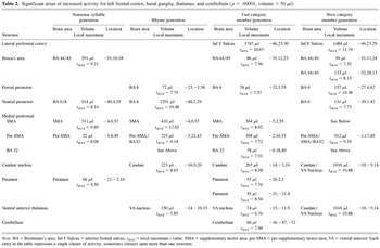
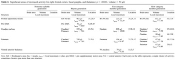
In voxel-by-voxel statistical analyses for each task, t tests were used to compare task–rest activity cycles to a null hypothesis of no change between rest and task, using a statistical probability threshold of p < .00001 with a cluster size threshold of volume greater than 50 μl. Activity in structures of the frontal lobe, basal ganglia, thalamus, and cerebellum is presented in Table 2 for the left hemisphere and Table 3 for the right hemisphere. The major frontal cortical regions activated in the left hemisphere on one or more tasks include: cortex along the inferior frontal sulcus, cortex along the precentral sulcus, pre-SMA and cortex in BA 32 (collectively referred to as the pre-SMA in text), and SMA.
Significant areas of increased activity for left frontal cortex, basal ganglia, thalamus, and cerebellum (p < .00001, volume > 50 μl)

Significant areas of increased activity for right frontal cortex, basal ganglia, and thalamus (p < .00001, volume > 50 μl)

Activity in the Left Pre-SMA Loop, SMA, and the Putamen
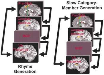
Pre-SMA is the anterior portion of medial Brodmann's area 6 (Matsuzaka et al., 1992; Paus, 2001; Picard & Strick, 1996, 2001). In our work, we have found that a coronal plane 4 mm anterior to the posterior margin of the anterior commissure best separates pre-SMA from SMA, which occupies the posterior portion of medial Brodmann's area 6 (Crosson et al., 2001). Robust activity in left pre-SMA was seen in three tasks compared to resting baseline (Table 2): rhyme generation (Figure 3), fast category-member generation, and slow category-member generation (Figure 3). This activity borders on and includes the most superior portion of Brodmann's area 32. In the fourth task, nonsense syllable generation, almost no activity was seen in pre-SMA. What little activity appeared in pre-SMA (less than one seventh of the volume in other tasks) was posterior to activity in the other three tasks. If a relaxed statistical threshold of p < .0001 was applied to ascertain the extent of activity in pre-SMA, the activity became contiguous with SMA activity, and, therefore, it most likely is an extension of that SMA activity. Thus, the left pre-SMA showed a volume of activity in tasks involving word generation, and only a very small volume of activity in the task involving generation of nonwords, that is, nonsense syllables. Activity in two other left-hemisphere structures, the dorsal left caudate nucleus plus the adjacent capsule and the left ventral anterior thalamus, followed the same pattern (Figure 3; Table 2). Even when the statistical threshold was lowered to p < .001, there was no activity in either the dorsal caudate nucleus or the ventral anterior thalamus during the nonsense syllable generation task. Given the known connections of pre-SMA with the caudate nucleus and adjacent capsular gray matter (Inase et al., 1999) and with the ventral anterior thalamus (Wiesendanger & Wiesendanger, 1985), activity in pre-SMA, the dorsal lateral caudate nucleus, and the ventral anterior thalamus in all likelihood represents involvement of a pre-SMA–dorsal caudate–ventral anterior thalamic loop in generation of words. The only component of the loop that was not activated by word generation tasks was the medial globus pallidus; the probable reasons for a lack of pallidal activity are briefly discussed below.

Activation of left pre-sma–dorsal caudate–ventral anterior thalamic loop during rhyme and slow category member generation. The left pre-SMA–dorsal caudate–ventral anterior thalamic loop was activated by tasks requiring lexical processing. Rhyme generation (left), slow category member generation (right), and fast category member generation (not shown) all activated the same components of this loop. In each of these tasks, left pre-SMA, the left dorsal caudate nucleus extending into the internal capsule, and the left ventral anterior (VA) thalamus showed a significant increase in activity relative to a resting baseline. In the medial globus pallidus (MGP), significant activity changes were absent, probably because of alternating increases and decreases in activity during generation of word sequences (see text). For nonsense syllable generation, neither the dorsal caudate nucleus nor the ventral anterior thalamus showed significant activity increases, and the pre-SMA activity increase was minimal compared to other tasks. Significant clusters of activity (volume > 50 μl) were determined by a statistical probability of p < .00001 (yellow). To visualize the extent of clusters, activity also is shown at a statistical probability of p < .0001 (red). The vertical green line on the pre-SMA slices represents the dividing line between SMA and pre-SMA.
All tasks activated left SMA to some degree. SMA is known to project to the putamen (Inase et al., 1999), but the left putamen showed significant activity only for nonsense syllable and fast category-member generation (Table 2). Thus, the left putamen was active for tasks that involved a faster rate of language generation, irrespective of whether subjects generated words or nonwords. For this reason, the case for involvement of the entire left SMA-putamen-thalamic loop is not as strong as it is for the pre-SMA loop. Elements of this loop other than SMA and the putamen (i.e., globus pallidus or ventral lateral thalamus) did not show significant activity, and SMA was active in all tasks while the putamen was active only in selected tasks.
Activity in Other Left Frontal Structures
Two other areas of left frontal cortex showed patterns of activity for tasks that paralleled elements of the experimental design. First, cortex along the inferior frontal sulcus was active only for the fast and slow semantic generation tasks (Table 2), indicating that this cortex is involved in the semantic aspects of word generation. In a previous study (Crosson et al., 2001), this area was even more active when word generation was guided by external semantic cues. Second, lateral premotor cortex along the precentral sulcus was active to some degree in all tasks. However, the most robust activity was seen during the nonsense syllable and the rhyme generation tasks (Table 2). Indeed, the maximum activity for these two tasks was in a ventral position along the sulcus, within 6 mm or less in every plane for these two tasks. The maxima of activity for the smaller volumes in the semantic generation tasks were far removed from maxima in the syllable and rhyme generation tasks. This finding indicates there is an area in the left lateral premotor cortex that is involved in processing phonological information during language generation.
The fact that Broca's area demonstrated significant activity in three of the four tasks deserves mention, even though the pattern does not parallel any of the experimental design elements. Broca's area demonstrated activity for the nonsense syllable, slow category-member, and fast category-member generation tasks. The fact that activity patterns in Broca's area did not parallel any of our design elements suggests determinants of activity in Broca's area other than those covered by the design elements of the current study. It was recruited both in semantic tasks and in a task where novel syllables were produced.
Activity in Right Basal Ganglia and Frontal Structures
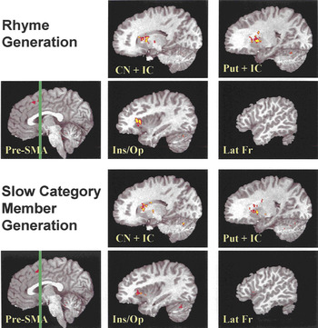
As in the left pre-SMA, left dorsal caudate nucleus, and left ventral anterior thalamus, the right basal ganglia demonstrated significant activity increases relative to rest for tasks requiring generation of words but not for nonsense syllable generation; however, the increased activity was more wide-spread in the right than in the left basal ganglia. For the rhyme, slow category-member, and fast category-member generation tasks, activity occurred in both the right putamen and the lateral portion of the right dorsal caudate nucleus (Figure 4; Table 3). In all cases this activity extended into the anterior limb of the internal capsule, between the putamen and the dorsal caudate nucleus. Although activity clusters from the left pre-SMA region extend minimally into the right hemisphere for these tasks, one small independent area of significant activity in the right pre-SMA was evident only on the rhyme generation task. The substantial activity in the right putamen, right caudate nucleus, and intervening capsular region, in the absence of any consistent activity in the right pre-SMA region across lexical tasks suggests that the source of the right-hemisphere basal ganglia activity may be input from the left pre-SMA. In the macaque, this region of the right basal ganglia, including the striatal gray bridges spanning the capsule, is a projection area of the left pre-SMA (Inase et al., 1999), suggesting that the left pre-SMA could be the source for this right neostriatal activity.

Right basal ganglia and frontal activity for rhyme and slow category member generation. For language generation tasks requiring lexical processing, right basal ganglia activity (a, b) is more diffuse than in the left hemisphere, and with the exception of the right frontal opercular-insular junction (d), there is little medial or lateral frontal activity. Significant activity during rhyme generation is shown in the top two rows, and significant activity during slow category member generation is shown in the bottom two rows. For both of these tasks, activity increases occurred in the right dorsal caudate nucleus [CN] and anterior limb of the internal capsule [IC] (a), in the right putamen [Put] and IC (b), and at the junction of the right frontal operculum [Op] and insula [Ins] (d). Little or no activity occurred in the right medial frontal cortex [pre-SMA] (c) or lateral frontal [Lat Fr] (e) cortex for either task. Fast category member generation (not shown) demonstrated a similar activity pattern to the other tasks with a lexical component. Nonsense syllable generation did not evoke significant activity in the right basal ganglia or frontal cortex. Significant clusters of activity (volume > 50 μl) were determined by a statistical probability of p < .00001 (yellow). To visualize the extent of clusters, activity also is shown at a statistical probability of p < .0001 (red). The vertical green line on the pre-SMA slices represents the dividing line between SMA and pre-SMA.
Only one right frontal area was active across the lexical tasks (Table 3). This area was at the juncture of the pars opercularis and the insula. This region is close to cortex that could be considered a right-hemisphere homologue to Broca's area. The only other area of right-sided activity in the frontal cortex or basal ganglia loops was limited to an area of activity in the right ventral anterior thalamus in the fast category member generation task. Thus, when compared to left cortical and subcortical activity, activity of the right basal ganglia was more widespread but occurred within the context of limited right frontal activity.
Activity Decreases
Some areas of activity decrease in the various tasks relative to rest occurred (Table 4). One area of decreased activity was found in a frontal region for rhyme generation; it was in right BA 32 anterior to the genu of the corpus callosum. No other area of decreased activity for any task-rest comparison was found in the frontal lobes, basal ganglia, or thalamus. Some areas of decreased activity were found in the posterior cingulate or temporoparietal regions for the lexical tasks. One area of activity decrease occurred in the left parahippocampal gyrus for nonsense syllable generation.
Significant areas of decreased activity (p < .00001, volume > 50 μl)

DISCUSSION
The current experiment was designed to distinguish contributions of the pre-SMA and related basal ganglia and thalamic structures to lexical, semantic, and phonological aspects of language generation. In previous studies, thalamic nuclei and particularly the basal ganglia have shown inconsistent activity during word generation tasks, even when identical tasks were imaged in the same laboratories (Ojemann et al., 1998; Palmer et al., 2001; Rosen et al., 2000; Warburton et al., 1996). Given such inconsistencies, we took steps to improve sensitivity to basal ganglia and thalamic activity: We used a relatively large number of subjects, a blocked trial format, two experimental runs for each task, rest (visual fixation) as our control state, and a stringent statistical probability with a small cluster size which allowed us to image small subcortical structures. With improved sensitivity to subcortical activity, simple competing hypotheses could be explored. These hypotheses postulated either that activity in pre-SMA and connected subcortical structures would occur consistently whenever language generation involved lexical as opposed to non-lexical processes, or that pre-SMA and connected subcortical structures would show varying patterns of activity across the different language generation tasks reflecting different functional profiles. Findings clearly favor consistent activity of pre-SMA, the dorsal caudate nucleus, and ventral anterior thalamus across language generation tasks involving lexical retrieval. The following discussion of these findings will deal with a number of issues related to this finding: a possible role for the left pre-SMA–dorsal caudate–ventral anterior thalamic loop in lexical retrieval, alternatives this explanation of our findings, development of testable hypotheses about the precise nature of the role of this loop in lexical retrieval, and the absence of activity in one constituent of this loop in the current study (i.e., the globus pallidus). In addition we will discuss four other issues: (1) the role of the right basal ganglia in the generation of words; (2) differential involvement of lateral frontal structures in phonological versus semantic processes; (3) areas of decreased activity for tasks compared to the resting baseline; and (4) limitations of the current study.
The Left Pre-SMA–Dorsal Caudate Nucleus–Ventral Anterior Thalamic Loop in Lexical Retrieval
In the current study, left pre-SMA and adjacent Brodmann's area 32, the left dorsal caudate nucleus and adjacent capsule, and the ventral anterior thalamus all were activated on tasks involving retrieval of pre-existing lexical items, whether based on semantic or phonological processes. Extension of dorsal caudate activity into the capsule is assumed to represent the grey bridges between the caudate nucleus and putamen to which pre-SMA projects (Inase et al., 1999). While a very small portion of pre-SMA is activated on nonsense syllable generation, this activity is posterior to pre-SMA activity in other tasks and appears to be merely an extension of the larger SMA activity. While portions of dorsal premotor cortex are active on all of these tasks, there are significant differences in locations indicating that these are different foci of activity. No other left-hemisphere regions showed exclusive activity on all of these lexical tasks. Even in studies of the same word generation tasks within the same laboratory (Ojemann et al., 1998; Palmer et al., 2001; Rosen et al., 2000; Warburton et al., 1996), previous studies have not shown such consistent activity in the basal ganglia or thalamus. For the current study, we attribute the consistency of activity in these structures to the measures that we took to ensure sensitivity to subcortical activity. The most obvious conclusion is that these structures (left pre-SMA, left dorsal caudate nucleus, left ventral anterior thalamus) are linked to facilitate retrieval of lexical items from pre-existing stores during language generation. This conclusion is bolstered by the known connections between pre-SMA and this general region of the striatum (Inase et al., 1999), between pre-SMA and the ventral anterior thalamus (Wiesendanger & Wiesendanger, 1985), and between pre-SMA and the globus pallidus via the thalamus (Akkal et al., 2002). Such connections are a prerequisite for these structures to function as a unified system. Nonetheless, other potential explanations for the similar pattern of activity between these structures should be addressed.
Alternative Explanations to a Role for the Left Pre-SMA Loop in Lexical Retrieval
As just noted, left pre-SMA, the left dorsal caudate nucleus, and the left ventral anterior thalamus consistently showed increased activity in the tasks requiring generation of a lexical item. Put most simply, alternative explanations to involvement of the pre-SMA loop in lexical retrieval would involve hypothesizing an uncoupling of the activity in at least one of these structures from activity in the others during generation of lexical items. These explanations clearly violate the law of parsimony (Occam's razor). The most parsimonious conclusion regarding activity in pre-SMA, the dorsal caudate nucleus and adjacent capsule, and ventral anterior thalamus during lexical generation is that these structures participate in a system that contributes to retrieval of lexical items from pre-existing stores during word generation. In the absence of compelling evidence for some alternative explanation, we will accept for now the most parsimonious one. As just noted, the anatomical connections between these structures bolster the idea that they are involved in a unified system. While the current data alone cannot determine how the pre-SMA–dorsal caudate–ventral anterior thalamic loop contributes to lexical generation, data from previous studies can be used to generate hypotheses that may lead to future research.
The Nature of the Role of the Pre-SMA Loop in Lexical Retrieval
Several lines of evidence bear upon the role of this pre-SMA loop in word generation. First, previous findings from our laboratory indicate that the left pre-SMA region is involved in semantically driven word generation but not in word repetition (Crosson et al., 2001). When considered with the current results, that finding suggests that it is not the mere use of a lexical item in task performance, but the retrieval of that lexical item from pre-existing stores that engages the pre-SMA loop. Second, recent evidence has accumulated that medial frontal cortex around the paracingulate sulcus, in a location similar to that of our study, shows greater activity at times when multiple responses are possible and subjects must choose between the competing responses (Barch et al., 2000; Botvinick et al., 1999; Carter et al., 2000; Milham et al., 2001; van Veen et al., 2001)1
When one plots Talairach coordinates (Talaraich & Tournoux, 1988) from many previous studies requiring monitoring of response competition, activity peaks are generally in the paracingulate sulcus (Barch et al., 2000; Botvinick et al., 1999; Carter et al., 2000; Milham et al., 2001; van Veen et al., 2001). The paracingulate sulcus is the dividing line between the rostral cingulate zone (BA 32) and pre-SMA (the anterior portion of medial BA 6; Paus et al., 1996). Generally, investigators in these studies have referred to the cortex in this area as anterior cingulate cortex, favoring the BA 32 side of the paracingulate sulcus. However, we have chosen the designation of pre-SMA for two reasons: The first is simplicity of presentation. The second is that we performed a study of word generation in which activity on the upper versus the lower bank of the paracingulate sulcus was compared in individual participants (Crosson et al., 1999). Significantly more activity occurred on the upper bank of the sulcus than the lower bank, indicating that more activity occurred in pre-SMA than the rostral cingulate zone. No matter what designation is used for this area, it should be kept in mind that it includes portions of both pre-SMA and the rostral cingulate zone. It also should be noted that pre-SMA and the rostral cingulate zone connect with similar areas of prefrontal cortex (Picard & Strick, 1996).
Absence of Globus Pallidus Activity in Current Findings
Perhaps the greatest limitation regarding our interpretation that the dominant pre-SMA–dorsal caudate–ventral anterior thalamic loop plays a role in retrieving lexical forms during word generation is the absence of any activity changes in the medial globus pallidus. As in similar cortical-basal ganglia loops, the medial globus pallidus must be situated between the dorsal caudate nucleus and the ventral anterior thalamus in the pre-SMA loop (Akkal et al., 2002). Why were no activity changes seen in the medial pallidal segment of the loop? The development of a testable hypothesis would be useful for future research.
Figure 1 shows that in addition to direct connections from the striatum to the medial globus pallidus, there is an indirect subloop from the striatum to the lateral globus pallidus to the subthalamic nucleus to the medial globus pallidus (Gerfen, 1992) and connections from pre-SMA to the subthalamic nucleus to the medial globus pallidus (Nambu et al., 2000). It has been proposed that inhibitory inputs from the striatum to the medial globus pallidus (direct loop) serve to inhibit pallidal neurons, which reduces their inhibition of thalamic neurons. The effect is to enhance thalamocortical excitation allowing desired actions to be performed. The excitatory inputs from the subthalamic nucleus to the medial globus pallidus, via subsequent pallidothalamic projections, inhibit surrounding thalamocortical activity that represents competing behaviors. Output of the subthalamic nucleus can be driven either by the indirect subloop or by direct connections from pre-SMA to the subthalamic nucleus (Gerfen, 1992; Inase et al., 1999; Mink, 1996; Nambu et al., 2000; Redgrave et al., 1999).
Evidence strongly indicates that synaptic input drives the hemodynamic response (Lauritzen, 2001). One possible reason for a lack of change in medial pallidal activity in the current tasks is the following: Our word generation tasks required generation of a sequence of words (either from a category or a rhyming cue). Competing responses that must be inhibited during production of the first response must be disinhibited to allow for later responses representing different category members or rhyming words, and responses given earlier in a sequence must be inhibited so that different words can be generated later in the sequence. One way this might happen is that both the response facilitation input to the medial pallidum from the direct loop and the response suppression input to the medial pallidum from the subthalamic nucleus show alternating cycles of increased and decreased activity which result in no net change in synaptic activity across a word generation block. These alternating cycles of increased and decreased pallidal input would allow for switching from one word to another as the response sequence develops. Future experiments should be developed to test this hypothesis. One challenge in doing so would be to develop imaging techniques sensitive to activity changes in the relatively small portion of the subthalamic nucleus connected to pre-SMA. Finally, based purely on input to the ventral anterior thalamus from the medial globus pallidus, one would expect activity in the ventral anterior thalamus to mirror that of the medial globus pallidus (i.e., in this hypothesis, no net change across blocks of word generation). However, it must be remembered that the connections of ventral anterior thalamus with pre-SMA are reciprocal in nature. Thus, corticothalamic input could account for ventral anterior thalamic activity even if cycles of increased and decreased input from the globus pallidus tend to cancel each other across word generation blocks.
Role of the Right Basal Ganglia in Word Generation
We must now turn our attention to the activity in the right basal ganglia on the tasks involving the retrieval of lexical items, the same tasks that engaged the left pre-SMA–dorsal caudate–ventral anterior thalamic loop. In comparison to left-hemisphere activity, right hemisphere basal ganglia activity occupied more widespread segments of the neostriatum, including the lateral portion of the dorsal caudate nucleus, the putamen, and the capsular region between them. In the presence of this relatively widespread engagement of the right neostriatum, only a single significant region of right frontal activity was present across these tasks, the region at the junction of the frontal operculum and the insula. One possible explanation for the absence of greater frontal activity in the presence of relatively widespread neostriatal activity in the right hemisphere is that the neostriatal activity somehow acts to suppress right frontal activity to keep it from interfering with word generation processes in the left hemisphere. The lateral portion of the right caudate nucleus, right putamen, and gray bridges of the intervening capsular region are known to receive afferents from the left pre-SMA region (Inase et al., 1999). Thus, we hypothesize that input from left pre-SMA to the right striatum plays a role in suppressing right frontal activity. This hypothesis regarding suppression of right frontal mechanisms has profound implications for lateralization of language production and should be tested in future experiments.
Lateral Frontal Structures in Phonological Versus Semantic Processes
It should be noted that other areas of activity in left lateral frontal cortex showed patterns that corresponded to experimental manipulations. First, activity along the inferior frontal sulcus was active only for tasks involving semantic processing in word generation (i.e., both category-member generation tasks). This result is consistent with previous findings from our laboratory (Crosson et al., 2001) indicating that this area is active for tasks involving category-member generation but not for word repetition, since word repetition can be done on the basis of lexical knowledge without reference to semantic information conveyed by the words. Second, a specific region of premotor cortex along the precentral sulcus was active primarily during language generation tasks involving phonological components. In electrical stimulation studies, Ojemann (1983) has shown that motor and phonological processes are closely linked, which would explain why premotor cortex becomes active in tasks requiring phonological processing.
Activity Decreases in Tasks Versus the Resting Baseline
Activity decreases occurred in the posterior cingulate and temporoparietal regions for tasks involving generation of lexical items. We attribute this decreased activity to a decreased focus on external stimuli during word generation. In other words, cortices associated with attentional processes (Heilman et al., 2003) show decreased activity in the absence of overt demands for attention to external stimuli. We have noticed the opposite effect when subjects focus upon an external visual stimulus in the absence of overt responses, i.e., a decrease in frontal activity in the presence of increased activity in primary visual and visual association cortices (Crosson et al., 1994; Nadeau et al., 1997). Such findings have important implications for intentional vs. attentional systems (see Heilman et al., 2003 for discussion of these systems).
Limitations in Current Study
One additional limitation in the current study should be noted. We are dependent on task-rest comparisons rather than direct task to task comparisons for interpretation of our findings; direct task to task comparisons were not sensitive to changes in subcortical activity. For example, we did a region of interest analysis with direct task to task comparisons. This analysis confirmed greater involvement of left pre-SMA in lexical as opposed to nonlexical processing, greater involvement of cortex in the left IFS in semantic as opposed to phonological processing, and greater involvement of cortex around the precentral sulcus in phonological as opposed to semantic activity. However, this analysis was not sensitive to activity differences in subcortical structures. In part, this lack of difference appears to be due to significantly increased variability of activity in subcortical structures during nonsense syllable generation, especially in the left dorsal caudate nucleus. However, Newman et al. (2001) found that a resting baseline enhances activity in language-related structures in comparison to using other language tasks as a baseline state. In other words, in subcortical structures where sensitivity of fMRI measures is at issue, comparison of language activity to resting activity will yield greater sensitivity than comparison of activity from one language task to activity from a second language task. Finally, it should be remembered that activity in the left dorsal caudate nucleus, the left ventral anterior thalamus, and the right basal ganglia did not reach significance during nonsense syllable generation even when the statistical threshold was relaxed from the p < .00001 level used to identify significant clusters of activity to the p < .0001 level. Thus, while we can conclude that significant activity occurs in the subcortical structures during lexical generation tasks but not during nonsense syllable generation, we cannot conclude that activity levels are significantly different between these tasks. Increased variability as well as reduced amplitude of activity in subcortical structures contributes to a lack of significant activity during nonsense syllable generation.
Summary
Based upon our data and the primate literature, we have inferred the existence of a left pre-SMA–dorsal caudate–ventral anterior thalamic loop involved in lexical retrieval. We have shown that all components of this loop, except the medial globus pallidus, demonstrated increased activity during generation of words but not during generation of nonsense syllables. It does not matter whether retrieved words were category members or rhyming words. Drawing upon previous literature as well as our findings, we hypothesized that activity in this loop was related to maintaining a bias toward the retrieval of one lexical item versus competing alternatives for each response during word generation blocks. Relatively widespread activity in the right basal ganglia during the generation of lexical items may be driven by input from the left pre-SMA region and may be responsible for keeping right frontal structures from interfering with language generation. Studies to confirm or reject hypotheses generated by the current findings should be conducted. In particular, some attention should be given to discerning the specific role of the basal ganglia and thalamic components of the loop. Do the subcortical structures merely enhance functioning of the cortical structure to which they are connected, or do they add a specific cognitive function beyond that of the cortex. Findings of Copland and colleagues (Copland, 2003; Copland et al., 2000a) hint that the subcortical components may be involved in maintaining response biases across time that are set by the cortical components of the loop. However, in a lesion model, cases of damage to the left pre-SMA region would have to be included to answer this question. Manipulation of the response bias in functional neuroimaging studies and, at the same time, using strategies to enhance subcortical activity may be a more practical way to assess this hypothesis.
ACKNOWLEDGMENTS
This work was supported by Grant DC03455 from the National Institute on Deafness and Other Communication Disorders.
References
REFERENCES
Akkal, D.,
Dum, R.P., &
Strick, P.L.
(2002).
Cerebellar and basal ganglia inputs to the pre-supplementary area
(Pre-SMA).
Society for Neuroscience Abstract Viewer/Itinerary Planner,
Online, Program No. 462.
14.
Bandettini, P.A.,
Jesmanowicz, A.,
Wong, E.C., &
Hyde, J.S.
(1993).
Processing strategies for time-course data sets in functional MRI
of the human brain.
Magnetic Resonance in Medicine,
30,
161–173.Google Scholar
Barch, D.M.,
Braver, T.S.,
Saab, F.W., &
Noll, D.C.
(2000).
Anterior cingulate and the monitoring of response conflict:
Evidence from an fMRI study of overt verb generation.
Journal of Cognitive Neuroscience,
12,
298–309.Google Scholar
Botvinick, M.,
Nystrom, L.E.,
Fissell, K.,
Carter, C.S., &
Cohen, J.D.
(1999).
Conflict monitoring versus selection for action in anterior
cingulate cortex.
Nature,
402,
179–181.Google Scholar
Broadbent, G.
(1872).
On the cerebral mechanisms of speech and thought.
London.
Buchanan, S.L.,
Thompson, R.H.,
Maxwell, B.L., &
Powell, D.A.
(1994).
Efferent connections of the medial prefrontal cortex in the rabbit.
Experimental Brain Research,
100,
469–483.Google Scholar
Bullmore, E.,
Brammer, M.,
Williams, S.C.R.,
Rabe-Hesketh, S.,
Janot, N.,
David, A.,
Mellers, J.,
Howard, R., &
Sham, P.
(1996).
Statistical methods of estimation and inference for functional MR
image analysis.
Magnetic Resonance in Medicine,
35,
261–277.Google Scholar
Cabeza, R. &
Nyberg, L.
(2000).
Imaging cognition II: An empirical review of 275 PET and fMRI
studies.
Journal of Cognitive Neuroscience,
12,
1–47.Google Scholar
Carter, C.S.,
Macdonald, A.M.,
Botvinick, M.,
Ross, L.L.,
Stenger, A.,
Noll, D., &
Cohen, J.D.
(2000).
Parsing executive processes: Strategic vs. evaluative functions of the
anterior cingulate cortex.
Proceedings of the National Academy of Sciences, U.S.A.,
97,
1944–1948.Google Scholar
Chao, L.L.,
Haxby, J.V., &
Martin, A.
(1999).
Attribute-based neural substrates in temporal cortex for perceiving
and knowing about objects.
Nature Neuroscience,
2,
913–919.Google Scholar
Copland, D.A.,
Chenery, H.J., &
Murdoch, B.E.
(2000a).
Processing lexical ambiguities in word triplets: Evidence of
lexical–semantic deficits following dominant nonthalamic
subcortical lesions.
Neuropsychology,
14,
379–390.Google Scholar
Copland, D.A.,
Chenery, H.J., &
Murdoch, B.E.
(2000b).
Persistent deficits in complex language function following dominant
nonthalamic subcortical lesions.
Journal of Medical Speech-Language Pathology,
8,
1–15.Google Scholar
Cowan, R.L. &
Wilson, C.J.
(1994).
Spontaneous firing patterns and axonal projections of single
corticostriatal neurons in the rat medial agranular cortex.
Journal of Neurophysiology,
71,
17–32.Google Scholar
Cox, R.W.
(1996).
AFNI: Software for analysis and visualization of functional
magnetic resonance images.
Computers in Biomedical Research,
29,
162–173.Google Scholar
Crosson, B.,
Sadek, J.R.,
Maron, L.,
Gökçay, D.,
Mohr, C.M.,
Auerbach, E.J.,
Freeman, A.J.,
Leonard, C.M.,
Briggs, R.W.
(2001).
Relative shift in activity from medial to lateral frontal cortex
during internally versus externally guided word generation.
Journal of Cognitive Neuroscience,
13,
272–283.Google Scholar
Crosson, B.,
Sadek, J.R.,
Bobholz, J.A.,
Gökçay, D.,
Mohr, C.M.,
Leonard, C.M.,
Maron, L.,
Auerbach, E.J.,
Browd, S.R.,
Freeman, A.J., &
Briggs, R.W.
(1999).
Activity in the paracingulate and cingulate sulci during word
generation: An fMRI study of functional anatomy.
Cerebral Cortex,
9,
307–316.Google Scholar
Crosson, B.,
Williamson, D.J.G.,
Shukla, S.S.,
Honeyman, J.C., &
Nadeau, S.E.
(1994).
A technique for localizing activation in the human brain with
[99mTc]-HMPAO SPECT: A validation study using
visual stimulation.
Journal of Nuclear Medicine,
35,
755–763.Google Scholar
Forman, S.D.,
Cohen, J.D.,
Fitzgerald, M.,
Eddy, W.F.,
Mintun, M.A., &
Noll, D.C.
(1995).
Improved assessment of significant activation in functional
magnetic resonance imaging (fMRI): Use of a cluster-size threshold.
Magnetic Resonance in Medicine,
33,
636–647.Google Scholar
Gerfen, C.R.
(1992).
The neostriatal mosaic: Multiple levels of compartmental
organization in the basal ganglia.
Annual Review of Neuroscience,
15,
285–320.Google Scholar
Heilman, K.M.,
Watson, R.T., &
Valenstein, E.
(2003).
Neglect and related disorders. In
K.M. Heilman &
E. Valenstein (Eds.),
Clinical neuropsychology
(4th ed., pp.
296–346).
New York:
Oxford University Press.
Hoover, J.E. &
Strick, P.L.
(1999).
The organization of cerebellar and basal ganglia outputs to primary
motor cortex as revealed by retrograde transneuronal transport of
herpes simplex virus type 1.
Journal of Neuroscience,
19,
1446–1463.Google Scholar
Inase, M.,
Tokuno, H.,
Nambu, A.,
Akazawa, T., &
Takada, M.
(1999).
Corticostriatal and corticosubthalamic input zones from the
presupplementary motor area in the macaque monkey: Comparison with the
input zones from the supplementary motor area.
Brain Research,
833,
191–201.Google Scholar
Jueptner, M. &
Weiller, C.
(1998).
A review of differences between basal ganglia and cerebellar
control of movements as revealed by functional imaging studies.
Brain,
121,
1437–1449.Google Scholar
King, K.F.,
Foo, T.K.F., &
Crawford, C.R.
(1995).
Optimized gradient waveforms for spiral scanning.
Magnnetic Resonance in Medicine,
34,
156–160.Google Scholar
Lauritzen, M.
(2001).
Relationship of spikes, synaptic activity, and local changes of
cerebral blood flow.
Journal of Cerebral Blood Flow and Metabolism,
21,
1367–1383.Google Scholar
Macovski, A.
(1985).
Volumetric NMR imaging with time-varying gradients.
Magnetic Resonance in Medicine,
2,
29–40.Google Scholar
Marie, P.
(1906).
Revision de la question de l'aphasie: Que faut-il penser des
aphasies sous-corticales (aphasies pures)? [Review of the question
of aphasia: What to think about subcortical aphasias (pure
aphasias)?]
La Semaine Medicale,
42.Google Scholar
Matsuzaka, Y.,
Aizawa, H.,
Tanji, J.
(1992).
A motor area rostral to the supplementary motor area
(presupplementary motor area) in the monkey: Neuronal activity during a
learned motor task.
Journal of Neurophysiology,
68,
653–662.Google Scholar
Mega, M.S. &
Alexander, M.P.
(1994).
Subcortical aphasia: The core profile of capsulostriatal infarction.
Neurology,
44,
1824–1829.Google Scholar
Middleton, F.A. &
Strick, P.L.
(2000).
Basal ganglia and cerebellar loops: Motor and cognitive circuits.
Brain Research Review,
31,
236–250.Google Scholar
Milham, M.P.,
Banich, M.T.,
Webb, A.,
Barad, V.,
Cohen, N.J.,
Wszalek, T., &
Kramer, A.F.
(2001).
The relative involvement of anterior cingulate and prefrontal
cortex in attentional control depends on nature of conflict.
Brain Research: Cognitive Brain Research,
12,
467–473.Google Scholar
Mink, J.W.
(1996).
The basal ganglia: Focused selection and inhibition of competing
motor programs.
Progress in Neurobiology,
50,
381–425.Google Scholar
Morino, P.,
Mascangni, F.,
McDonald, A., &
Hökfelt, T.
(1994).
Cholecystokinin corticostriatal pathway in the rat: Evidence for
bilateral origin from medial prefrontal cortical areas.
Neuroscience,
59,
939–952.Google Scholar
Nadeau, S.E. &
Crosson, B.
(1997).
Subcortical aphasia.
Brain and Language,
58,
355–402.Google Scholar
Nadeau, S.E.,
Hammond, E.,
Williamson, D.J., &
Crosson, B.
(1997).
Resting and stimulated states in functional imaging studies:
Evidence of differences in attentional and intentional set.
Neuropsychiatry, Neuropsychology, and Behavioral Neurology,
10,
162–163.Google Scholar
Nambu, A.,
Tokuna, H.,
Hamada, I.,
Kita, H.,
Imanishi, M.,
Akazawa, T.,
Ikeuchi, Y., &
Hasegawa, N.
(2000).
Excitatory cortical inputs to pallidal neurons via the subthalamic
nucleus in the monkey.
Journal of Neurophysiology,
84,
289–300.Google Scholar
Newman, S.D.,
Twieg, D.B., &
Carpenter, P.A.
(2001).
Baseline conditions and subtractive logic in neuroimaging.
Human Brain Mapping,
14,
228–235.Google Scholar
Noll, D.C.,
Cohen, J.D.,
Meyer, C.H., &
Schneider, W.J.
(1995).
Spiral k-space MR imaging of cortical activation.
Magnetic Resonance Imaging,
5,
49–56.Google Scholar
Ojemann, G.A.
(1983).
Brain organization for language from the perspective of electrical
stimulation mapping.
Behavioral and Brain Sciences,
2,
189–230.Google Scholar
Ojemann, J.G.,
Buckner, R.L.,
Akbudak, E.,
Snyder, A.Z.,
Ollinger, J.M.,
McKinstry, R.C.,
Rosen, B.R.,
Petersen, S.E.,
Raichle, M.E., &
Conturo, T.E.
(1998).
Functional MRI studies of word-stem completion: Reliability across
laboratories and comparison to blood flow imaging with PET.
Human Brain Mapping,
6,
203–215.Google Scholar
Oldfield, R.C.
(1971).
The assessment and analysis of handedness: The Edinburgh Inventory.
Neuropsychologia,
9,
97–113.Google Scholar
Palmer, E.D.,
Rosen, H.J.,
Ojemann, J.G.,
Buckner, R.L.,
Kelley, W.M., &
Petersen, S.E.
(2001).
An event-related fMRI study of overt and covert word stem
completion.
NeuroImage,
14,
182–193.Google Scholar
Parent, A.
(1990).
Extrinsic connections of the basal ganglia.
Trends in Neuroscience,
13,
254–258.Google Scholar
Parent, A. &
Hazrati, L.-N.
(1993).
Anatomical aspects of information processing in primate basal
ganglia.
Trends in Neuroscience,
16,
111–116.Google Scholar
Paus, T.
(2001).
Primate anterior cingulate cortex: Where motor control, drive and
cognition interface.
Nature Reviews Neuroscience,
2,
417–424.Google Scholar
Paus, T.,
Tomaiuolo, F.,
Otaky, N.,
MacDonald, D.,
Petrides, M.,
Atlas, J.,
Morris, R., &
Evans, A.C.
(1996).
Human cingulate and paracingulate sulci: Pattern, variability,
asymmetry, and probabilistic map.
Cerebral Cortex,
6,
207–214.Google Scholar
Penney, J.B. &
Young, A.B.
(1986).
Striatal inhomogeneities and basal ganglia function.
Movement Disorders,
1,
3–15.Google Scholar
Petersen, S.E.,
Fox, P.T.,
Posner, M.I.,
Mintun, M., &
Raichle, M.E.
(1988).
Positron emission tomographic studies of the cortical anatomy of
single-word processing.
Nature,
331,
585–589.Google Scholar
Picard, N. &
Strick, P.L.
(1996).
Motor areas of the medial wall: A review of their location and
functional activation.
Cerebral Cortex,
6,
342–353.Google Scholar
Picard, N. &
Strick, P.L.
(2001).
Imaging the premotor areas.
Current Opinion in Neurobiology,
11,
263–272.Google Scholar
Redgrave, P.,
Prescott, T.J., &
Gurney, K.
(1999).
The basal ganglia: A vertebrate solution to the selection problem?
Neuroscience,
89,
1009–1023.Google Scholar
Rosen, H.J.,
Ojemann, J.G.,
Ollinger, J.M., &
Petersen, S.E.
(2000).
Comparison of brain activation during word retrieval done silently
and aloud using fMRI.
Brain and Cognition,
42,
201–217.Google Scholar
Strick, P.L.,
Dum, R.P, &
Picard, N.
(1995).
Macro-organization of the circuits connecting the basal ganglia
with the cortical motor areas. In
J.C. Houk,
J.L. Davis, &
D.G. Beiser (Eds.),
Models of information processing in the basal ganglia (pp.
117–130).
Cambridge, MA:
MIT Press.
Talaraich, J. &
Tournoux, P.
(1988).
Co-planar stereotaxic atlas of the human brain. 3-dimensional
proportional system: An Approach to cerebral imaging.
New York:
Thieme Medical Publishers.
van Veen, V.,
Cohen, J.D.,
Botvinick, M.M.,
Stenger, V.A., &
Carter, C.S.
(2001).
Anterior cingulate cortex, conflict monitoring, and levels of
processing.
NeuroImage,
14,
1302–1308.Google Scholar
Wang, H. &
Pickel, V.M.
(1998).
Dendritic spines containing μ-opioid receptors in rat striatal
patches receive asymmetric synapses from prefrontal corticostriatal
afferents.
Journal of Comparative Neurology,
396,
223–237.Google Scholar
Warburton, E.,
Wise, R.J.S.,
Price, C.J.,
Weiller, C.,
Hadar, U.,
Ramsay, S., &
Frackowiak, R.J.S.
(1996).
Noun and verb retrieval by normal subjects: Studies with PET.
Brain,
119,
159–179.CrossRefGoogle Scholar
Weiller, C.,
Willmes, K.,
Reiche, W.,
Thron, A.,
Insensee, C.,
Buell, U., &
Ringelstein, E.B.
(1993).
The case of aphasia or neglect after striatocapsular infarction.
Brain,
116,
1509–1525.CrossRefGoogle Scholar
Wernicke, C.
(1874).
Der aphasische symptomencomplex [The complex of symptoms in
aphasia].
Breslau, Germany:
Cohn and Weigert.
Wiesendanger, R. &
Wiesendanger, M.
(1985).
Thalamic connections with medial area 6 (supplementary motor
cortex) in the monkey (Macaca fascicularis).
Experimental Brain Research,
59,
91–104.Google Scholar
Schematic diagram of pre–SMA–basal ganglia loop. Components of the “direct loop” (discussed in Introduction) are shown in boxes with heavy black lines. There is also an “indirect loop” which, due to space considerations, was not detailed in the Introduction. Components of the “indirect subloop” are shown with shaded boxes. The neostriatal component* of the loop probably consists of striatal gray bridges, but also may include the caudate nucleus or putamen on either side of the capsule (Inase et al., 1999). Direction of pathways between structures is indicated by the arrows. Reciprocated connections are indicated by separate arrows. +'s in circles indicate excitatory pathways; −'s in circles indicate inhibitory pathways. Note that dopaminergic projections from the substantia nigra pars compacta to the neostriatum have both excitatory and inhibitory effects, depending upon the target neurons (Gerfen, 1992).
Experimental task characteristics. The four word generation tasks can be classified as requiring phonological versus semantic processing or fast versus slow generation (a), but also can be classified by whether they require nonlexical versus lexical processing (b).

Experimental subjects' mean production rates for each task (per 17.4 s period)
Significant areas of increased activity for left frontal cortex, basal ganglia, thalamus, and cerebellum (p < .00001, volume > 50 μl)
Significant areas of increased activity for right frontal cortex, basal ganglia, and thalamus (p < .00001, volume > 50 μl)
Activation of left pre-sma–dorsal caudate–ventral anterior thalamic loop during rhyme and slow category member generation. The left pre-SMA–dorsal caudate–ventral anterior thalamic loop was activated by tasks requiring lexical processing. Rhyme generation (left), slow category member generation (right), and fast category member generation (not shown) all activated the same components of this loop. In each of these tasks, left pre-SMA, the left dorsal caudate nucleus extending into the internal capsule, and the left ventral anterior (VA) thalamus showed a significant increase in activity relative to a resting baseline. In the medial globus pallidus (MGP), significant activity changes were absent, probably because of alternating increases and decreases in activity during generation of word sequences (see text). For nonsense syllable generation, neither the dorsal caudate nucleus nor the ventral anterior thalamus showed significant activity increases, and the pre-SMA activity increase was minimal compared to other tasks. Significant clusters of activity (volume > 50 μl) were determined by a statistical probability of p < .00001 (yellow). To visualize the extent of clusters, activity also is shown at a statistical probability of p < .0001 (red). The vertical green line on the pre-SMA slices represents the dividing line between SMA and pre-SMA.
Right basal ganglia and frontal activity for rhyme and slow category member generation. For language generation tasks requiring lexical processing, right basal ganglia activity (a, b) is more diffuse than in the left hemisphere, and with the exception of the right frontal opercular-insular junction (d), there is little medial or lateral frontal activity. Significant activity during rhyme generation is shown in the top two rows, and significant activity during slow category member generation is shown in the bottom two rows. For both of these tasks, activity increases occurred in the right dorsal caudate nucleus [CN] and anterior limb of the internal capsule [IC] (a), in the right putamen [Put] and IC (b), and at the junction of the right frontal operculum [Op] and insula [Ins] (d). Little or no activity occurred in the right medial frontal cortex [pre-SMA] (c) or lateral frontal [Lat Fr] (e) cortex for either task. Fast category member generation (not shown) demonstrated a similar activity pattern to the other tasks with a lexical component. Nonsense syllable generation did not evoke significant activity in the right basal ganglia or frontal cortex. Significant clusters of activity (volume > 50 μl) were determined by a statistical probability of p < .00001 (yellow). To visualize the extent of clusters, activity also is shown at a statistical probability of p < .0001 (red). The vertical green line on the pre-SMA slices represents the dividing line between SMA and pre-SMA.

Significant areas of decreased activity (p < .00001, volume > 50 μl)
 You have
Access
- 162
- Cited by