INTRODUCTION
Vermetids are sessile gastropods, occurring in intertidal to subtidal zones of warm-water seas (Keen, Reference Keen1961; Safriel, Reference Safriel1966; Ekman, Reference Ekman1953). Some species are solitary, whereas others form aggregations with up to 60,000 individuals m−2 (Miloslavich & Penchaszadeh, Reference Miloslavich and Penchaszadeh1992). The resulting biogenic reefs protect the shoreline from wave erosion and support a highly diverse community (Lipkin & Safriel, Reference Lipkin and Safriel1971; Ben-Eliahu, Reference Ben-Elihau1976; Fishelson & Haran, Reference Fishelson and Haran1987; Goren & Galil, Reference Goren and Galil2001).
Dendropoma petraeum (Monterosato, 1884) is a gregarious reef-building vermetid, endemic to the Mediterranean Sea. The species is included in Appendix II (Strictly Protected Fauna Species) of the Bern Convention, and in Annex II (Endangered or Threatened Species) of the Protocol for Specially Protected Areas and Biodiversity in the Mediterranean (SPABIM Protocol of the Barcelona Convention). The reefs have been listed as threatened biostructures in the Mediterranean Red Data Book (UNEP/IUCN/GIS POSIDONIE, 1990).
The Dendropoma reefs extend along the Mediterranean shores from Spain to the Levant (Safriel, Reference Safriel1966; Calvo et al., Reference Calvo, Templado and Penchaszadeh1998, Reference Calvo, Templado, Oliverio and Machordom2009). A recent study on the genetic diversity of D. petraeum populations throughout their habitat range revealed the existence of four geographically-separated phylogroups within the species (Calvo et al., 2009). One of these groups comprised Lebanese and Israeli populations. This finding provided the genetic support to the differences in reproductive characteristics among populations described in Usvyatsov (Reference Usvyatsov2007) and Calvo et al. (Reference Calvo, Templado, Oliverio and Machordom2009). Since there is evidence that each population is a separate species, thorough research of the ecology of each is needed for full understanding of the differences among them.
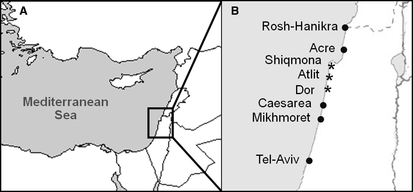
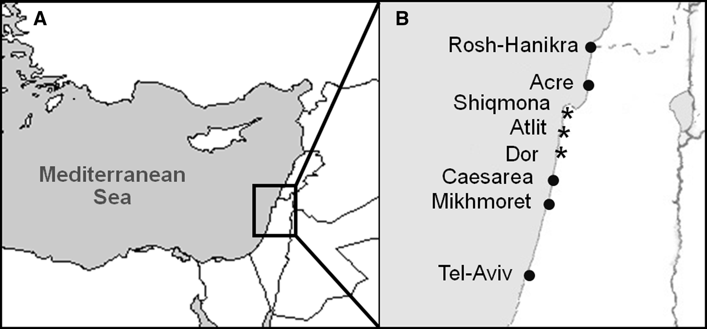
Off the Israeli coast vermetid reefs were recorded from Rosh-Hanikra to Mikhmoret (Safriel, Reference Safriel1966; see Figure 1 for map). The reefs consist of shallow flat-bottom basins encircled by raised overhanging rims. Dendropoma forms the reef's seaward ledges, where it is constantly swept by the surf, and exposed to air at low tide (Safriel, Reference Safriel1974). The high turbulence at the reef's edge is an advantage to this filter-feeding species (Hughes, Reference Hughes1978), while its thick shell and tightly-sealed operculum protect it from predators, desiccation and waves (Safriel, Reference Safriel1974).
(A) The Levant basin; (B) Israeli Mediterranean coast. Sampling sites are marked by asterisks.
During the 1960s and 1970s living Dendropoma covered 3% of the area of the raised rims (Lipkin & Safriel, Reference Lipkin and Safriel1971); a recent estimate puts it at 1% for most of the sites (Klerman et al., Reference Klerman, Fine and Galil2004). Much of the deterioration resulted from anthropogenic activity: the Acre reefs were lost to urban development, and the ones off Akhziv-Rosh Hanikra had been covered by dense algal growth due to a high organic load from an industrial outfall nearby (Klerman et al., Reference Klerman, Fine and Galil2004).
The western Mediterranean Dendropoma clade was found to be gonochoristic (Calvo et al., Reference Calvo, Templado and Penchaszadeh1998). The gonad, spread lengthwise throughout the visceral mass, becomes visible at the start of the reproductive period and degenerates at its conclusion (Calvo et al., Reference Calvo, Templado and Penchaszadeh1998). The males are aphallic, releasing sperm in the form of pelagic spermatophores (Hadfield & Hopper, Reference Hadfield and Hopper1980; Calvo & Templado, Reference Calvo and Templado2005). The females capture the spermatophores and store them until the eggs mature and fertilization is possible. Fertilized ova are encapsulated in a transparent envelope and released into the mantle cavity. The egg capsules are brooded lying free in the cavity (Calvo et al., Reference Calvo, Templado and Penchaszadeh1998). Egg capsules may contain several embryos in different developmental stages. The hatchlings are at the crawling juvenile stage, and subsist a few hours to find a suitable substrate; once settled, they remain sedentary.
The characteristics and reproductive cycle of Dendropoma were previously studied off south-east Spain (Calvo et al., Reference Calvo, Templado and Penchaszadeh1998; Calvo & Templado, Reference Calvo and Templado2005), and the male reproductive cycle was described for the Italian population (D'Ancona Lunetta & Damiani, Reference D'Ancona Lunetta and Damiani2002). Calvo et al. (Reference Calvo, Templado, Oliverio and Machordom2009) reported not only genetic differences among populations, but also some developmental characteristics, such as protoconch morphology and numbers of egg capsules brooded by females. However, little information exists on differences in reproductive activity between populations, and few histological images are available in the literature.
Here we provide a detailed account of these issues in the Levantine population of the D. petraeum-complex, extending our preliminary description of differences in reproductive activity (Usvyatsov, Reference Usvyatsov2007). Our objectives were to examine the occurrence and the reproductive cycle of Dendropoma off the northern Mediterranean coast of Israel, and compare them with those of the population off south-east Spain, reported by Calvo et al. (Reference Calvo, Templado and Penchaszadeh1998). Thus, our findings provide information on reproductive differences between Dendropoma clades, supplementing the genetic analysis performed by Calvo et al. (Reference Calvo, Templado, Oliverio and Machordom2009).
MATERIALS AND METHODS
Aggregation density
Specimens of Dendropoma were collected at Atlit, Dor and Shiqmona (Figure 1) from January to June 2003. Chunks of biogenic crusts were removed from the reef, the tube-like shells broken and their contents removed.
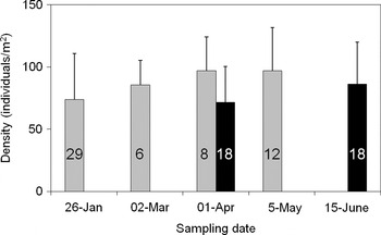
Density of live specimens was examined at Atlit and Shiqmona (see Figure 2 for dates and number of samples). Samples, each with a surface area of 12.5 cm2, were examined for the presence of live specimens, and their number extrapolated to 1 m2.
Average aggregation density (individuals per m2) of live specimens of Dendropoma petraeum at Shiqmona (grey) and Atlit (black), as sampled in 2003. Error bars are standard deviations. Numbers indicated are the number of 12.5 cm2 samples taken.

Histology
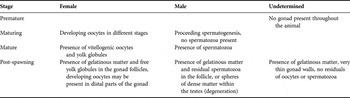
Samples were collected from January 2003 to June 2004. The specimens were fixed in Bouin, embedded in paraffin, sectioned at 5 µm and stained with haematoxylin and eosin. Stained specimens were classified according to their reproductive stage (Table 1). If they lacked a visible gonad or were identified as post-reproductive with no sex cells or products, they were classified as ‘undetermined sex’. Specimens showing both male and female gonads were classified as hermaphrodites. Both undetermined and hermaphrodite specimens were subtracted from the total before calculating percentage of females. Histological specimens were deposited at the National Collections of Natural History at Tel Aviv University (catalogue number MO 71543-71557-TAU).
Classification of gonad development stages in Dendropoma petraeum-complex.

Egg capsules
Brooding females were collected on 30 March 2003 and 15 June 2003 at Atlit and on 1 April 2003 and 8 June 2003 at Shiqmona. The mantle cavity of the females was slit open, and egg capsules were removed and counted. The capsules' size was measured to 0.06 mm using an ocular micrometer in a Leica MZ12 binocular microscope. The transparent capsule wall allowed examination of the number and developmental stage of the eggs/embryos contained within.
Aggregation density, capsule size, number of capsules per female and number of eggs/embryos per capsule were tested for normality using the Shapiro–Wilk test (alpha set to 0.05). A log10 transformation was applied when necessary to achieve normality. Variables were then tested using the t-distribution for differences between sampling times or sites. A Bonferroni correction was applied, if data were used for more than one test.
The capsules and the histological sections were photographed using a Nikon Eclipse E200 microscope with a Nikon Colorview II digital camera.
RESULTS
Aggregation density
Aggregation densities of Dendropoma, calculated per 1 m2, are shown in Figure 2. An increase in density with time was observed at both sites, however it was not statistically significant (t-test, P > 0.05). No significant differences in densities between the two sites were observed using data pooled over time (t-test, P > 0.05).
Histology
A total of 549 specimens were collected from the three sites. The division of specimens by site and sex as well as percentage of females, are given in Table 2.
The number of histologically treated specimens, divided according to site and sex. Percentage F is the percentage of females at each site, omitting hermaphrodites and unidentified specimens.

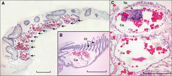
A female specimen with ovaries containing oocytes at different stages of development is shown in Figure 3. Eggs within the mantle cavity were observed in a few female specimens (Figure 3B).
(Colour online). (A) A female specimen with ripe gonad follicles, marked by arrows (magnification ×40, scale bar is 50 µm); (B) an egg capsule (Ca) showing yolk granules (red), brooded in the pallial cavity. Ctenidium is marked as ‘Ct’ (magnification ×40, scale bar is 50 µm); (C) spermatozoa cluster (Sp) having entered an egg capsule (Ca) (magnification × 100, scale bar is 20 µm).

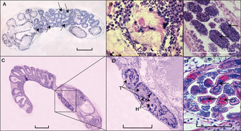
Male specimens with mature, degenerating and parasitized testes are illustrated in Figure 4. Spermatogenesis resulted in spheres of spermatozoa at the centre of the testes (Figure 4A, B), expelled through the gonoduct (Figure 4C, D). The unexpelled spermatozoa degenerated and formed dense spheres within the testes (Figure 4E). These were apparently more prone to parasitism (Figure 4F), as parasites were encountered only in post-reproductive males with degenerated spermatozoa (6 of 21 at Shiqmona, 3 of 9 at Dor and 2 of 6 at Atlit).
(Colour online). (A) A mature male with ripe gonad follicles, marked by arrows (magnification × 40, scale bar is 50 µm); (B) a ripe follicle; maturing sex cells are at the perimeter; the mature spermatozoa are at the centre (magnification × 400, scale bar is 10 µm); (C) a mature male, showing spermatozoa spheres in the gonoduct (marked by square) (magnification ×40, scale bar is 50 µm); (D) enlarged from (C); spheres of spermatozoa, showing the heads (H, blue) and tails (T, pink) of spermatozoa (magnification ×100, scale bar is 30 µm); (E) degenerating sex cells within the gonad follicle (magnification × 400, scale bar is 10 µm); (F) parasitic infestation on degenerating male gonads; the parasites are marked by arrows (magnification ×400, scale bar is 10 µm).

No spermatophores were observed within males. However, clusters of spermatozoa were observed in some histological sections of females, probably from captured and disrupted spermatophores (Figure 3C).
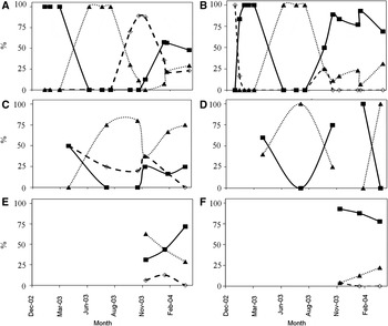
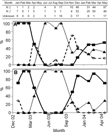
As the developmental stages of the gonads did not differ among sites (Figure 5), the data were pooled for ease of presentation and analysis. The pooled developmental stages are presented in Figure 6. Both sexes were reproductively active between January 2003 and June 2003, and September 2003–May 2004, when sampling ended. Reproductive activity probably began in October, with males maturing faster than females. Half of the males examined had mature testes in November, whereas 50% of the females reached maturity only in February. Few immature testes were observed throughout the sampling period; their percentage peaked at the start of the reproductive season and decreased to zero by early December. However, immature ovaries were observed in most samples. Between June and September 2003 all specimens were reproductively inactive, except for a single maturing female, collected in August 2003 at Atlit. Post-reproductive specimens of both sexes were recorded throughout the year. However, the percentage of post-reproductive females was higher than that of the males (Figure 6). Unidentified post-reproductive specimens were observed throughout the sampling period.
Reproductive activity of female (A, C, E) and male (B, D, F) Dendropoma collected at Shiqmona (A, B), Atlit (C, D) and Dor (E, F). Activity is shown as percentage of the 3 reproductive stages: premature (open diamond, dashed line); mature (solid square, solid line); and post-spawning (solid triangle, dotted line). Unidentified specimens are not shown.

Reproductive activity of female (A) and male (B) Dendropoma specimens throughout 2003–2004. Data are from pooled samples from Shiqmona, Atlit and Dor. Activity is shown as percentage of the 3 reproductive stages: premature (open diamond, dashed line); mature (solid square, solid line); and post-spawning (solid triangle, dotted line). Unidentified specimens are not shown. Numbers of female, male and undetermined-sex specimens collected each month are detailed in the table, aligned with the x-axis of the figure.

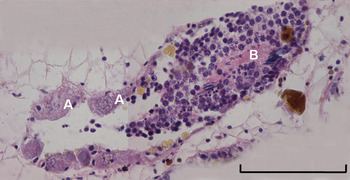
Seven hermaphroditic specimens were recorded (see Table 2 for collection sites). One specimen had mature or maturing cells of both sexes (Figure 7), others had maturing cells of one sex, and degenerating cells of the other (not shown).
(Colour online) A gonad from a hermaphroditic specimen collected at Shiqmona in December 2003 with maturing oocytes (A) and spermatozoa (B) (magnification × 40). The scale bar is10 µm in length.

Egg capsules
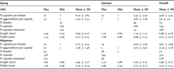
A total of 332 capsules from Shiqmona and 324 from Atlit were extracted from the pallial cavities of 72 and 74 females, respectively. Of these, the sizes of 178 and 168 capsules (from Shiqmona and Atlit, respectively) were measured (Table 3). Capsule length did not differ significantly between sites or sampling dates (t-test, P > 0.05). The width of capsules collected in Atlit increased significantly between April and June (one-tailed t-test, P < 0.001); there was no such increase in capsules collected in Shiqmona. The numbers of capsules per female and eggs/embryos per capsule were recorded (Table 3). These values decreased significantly between spring and summer samples at both sites (one-tailed t-tests, P < 0.05 for both).
Number of capsules per female, capsule size and number of eggs/embryos per capsule, observed in spring 2003 (30 March at Atlit and 1 April at Shiqmona) and in summer 2003 (8 June at Atlit and 15 June at Shiqmona). The maximum (Max), minimum (Min), mean and standard deviation (SD) values are given. N, number.

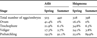
The distribution of embryonic stages at both sites is given in Table 4. In each site the percentage of ova dropped to zero in summer, while the percentage of pre-hatchlings increased to ~90%. This is in accordance with the reproductive stages observed (Figure 6), as all individuals collected in June were in post-reproductive stage.
Distribution of embryonic development stages, observed in spring 2003 (30 March at Atlit and 1 April at Shiqmona) and in summer 2003 (8 June at Atlit and 15 June at Shiqmona).
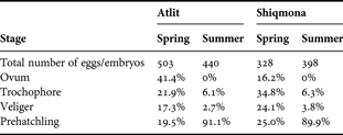
Spring samples in Shiqmona had a more even distribution of developmental stages than Atlit samples (Table 4). Thus while Atlit samples contained 63% ova and trochophores, in Shiqmona these stages constituted only 50% of embryos. According to the distribution of embryonic developmental stages (Table 4) hatching probably ended by late June–early July.
DISCUSSION
Aggregation
The calculated value of aggregation density is similar to previously reported values (Miloslavich & Penchaszadeh, Reference Miloslavich and Penchaszadeh1992). However, due to the extreme patchiness of Dendropoma aggregation, our estimate, based on an average of small samples, may be inaccurate. Moreover, the basins chosen for sampling, in both Atlit and Shiqmona, were those containing live Dendropoma. Therefore, the calculated aggregation numbers represent the maximal aggregation observed, rather than the mean aggregation of a particular reef. The insignificant increase of density over the sampling months at both sites could result from newly settled juveniles, since in our study pre-hatchlings were seen as early as March.
Reproductive cycle
The histological results reveal that the period of reproductive activity for both sexes lasted from October–November to May–June at the three sampling sites. Thus, the reproductive period of the Israeli population is similar in length to the one described by Calvo et al. (Reference Calvo, Templado and Penchaszadeh1998), but has a later start (October instead of August–September). However, brooding seems to start earlier or proceed faster in the Israeli population. Calvo et al. (Reference Calvo, Templado and Penchaszadeh1998) state that in March and April 48.5% of capsules contained early stages of embryos (ova and trochopores); a similar percentage was recorded in Shiqmona at the same season, despite a much later start in gonad maturation.
Annual fluctuation in the onset of gametogenesis is likely. Comparison of our data from 2003 and 2004 suggests that in 2003 spawning ended later than in 2004. However, this may be due to lower sampling effort in 2003. A longer-span study would be necessary to establish year-to-year variability in reproductive activity.
The lower percentage of post-reproductive males versus females encountered from December 2003 to May 2004 could suggest that either: (1) more males than females commence gametogenesis due to the lower energetic cost of spermatogenesis; or (2) assuming that male sexual products degenerate faster than the female ones, it is possible that most or all of the unknown-sex specimens are males. Overall, males matured earlier than females and maintained a higher percentage of activity throughout the reproductive period.
Post-reproductive specimens of both known and unknown sex were recorded throughout the year. This could suggest that: (1) a part of the population does not commence gametogenesis, possibly recovering from the previous year's spawning; or (2) an individual's reproductive period is short in comparison to the population's reproductive period, and not fully synchronized with other individuals.
Calvo et al. (Reference Calvo, Templado and Penchaszadeh1998) described an entirely gonochoristic population. Also, the percentage of females in the Israeli population, although variable, was lower than reported by Calvo et al. (Reference Calvo, Templado and Penchaszadeh1998) for the population off south-east Spain. These findings may suggest a higher anthropogenic stress on the vermetid reefs in Israel or a different reproductive strategy by the Levantine species. The overall differences in commencement of reproductive and brooding periods could reflect a difference in water temperature regime between the two regions. The warmer water of the eastern Mediterranean could induce earlier reproduction.
Egg capsules
Egg capsules collected at Atlit and Shiqmona differed greatly from those reported by Calvo et al. (Reference Calvo, Templado and Penchaszadeh1998) for south-east Spain. The overall mean number of capsules per female was lower than reported by Calvo et al. (Reference Calvo, Templado and Penchaszadeh1998), whereas the overall number of eggs/embryos per capsule was significantly higher (Figure 8). The number of eggs/embryos per capsule was significantly different between the Spanish population and both Israeli sites at both sampling dates (one-tailed t-tests, P ≪0.001 for both). The number of capsules per female was not compared between the Spanish and Israeli populations, as Calvo et al. (Reference Calvo, Templado and Penchaszadeh1998) provided only the mean values.
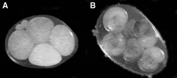
Egg capsules with eggs (A) and veligers (B) (photograph by A. Klerman).

Pooled egg capsule length for each site was significantly larger than reported by Calvo et al. (Reference Calvo, Templado and Penchaszadeh1998) (one-tailed t-test, P < 0.01 at both sites). The width of the egg capsules at Shiqmona and Atlit was significantly higher than in Spain (one-tailed t-test, P < 0.001).
The increase in capsule size from March/April to June at both sites was probably due to the development of the embryos within the capsules. While Calvo et al. (Reference Calvo, Templado and Penchaszadeh1998) describe the increase in capsule size as being due to increase in length, in the Israeli populations the length increase was insignificant. However, the Atlit capsules grew significantly in width, while the Shiqmona ones did not. The difference in width increase between the sites was probably due to different rates of development. The embryonic development at Atlit was slower than at Shiqmona. This observation was unexpected, since our histological results suggested similar gonad maturation at both sites. As shown in Table 4, more developed embryos were collected at Shiqmona than in Atlit. Therefore spring-time capsule size in Shiqmona could be initially higher, hence decreasing the spring-to-summer difference in width.
The decrease in number of brooded capsules per female with time was probably due to hatching. However, the reason for the significant decrease in the number of eggs/embryos per capsule at both Atlit and Shiqmona is unknown.
Our results show differences between the Spanish and the Israeli populations of D. petraeum-complex in reproductive period, presence of hermaphrodites, percentage of females, capsule size, number of capsules per female and number of eggs/embryos per capsule. These could be due to differences in temperature regimes, water pollution levels or genetic differences. If the latter is true, our results provide the phenotypic data, supplementing the known genetic differences between the Mediterranean populations of D. petraeum-complex. If the Levantine population is recognized as a separate species, its protection is vital. It is then endemic not only to the Mediterranean, but to the increasingly diminishing patches along the northern Israeli, Lebanese and Syrian coasts, which experience a significant anthropogenic pressure. The decrease in habitat range means immediate changes to this endangered species' conservation potential.
ACKNOWLEDGEMENTS
We would like to thank Dr Elisaveta Moiseeva for her considerable help in preparation and interpretation of histological materials; Dr Marta Calvo for assistance with identification of parasitized samples and Dr Alexander Klerman for collecting the specimens and examining the egg capsules. This research received no specific grant from any funding agency, commercial or not-for-profit sectors.