Acinetobacter baumannii has emerged as one of the most important nosocomial pathogens in intensive care units (ICUs) worldwide.Reference Munoz-Price and Weinstein 1 , 2 It can survive for extended periods of time on multiple environmental surfaces; thus, the hospital environment serves as a major reservoir for this organism.Reference Munoz-Price, Namias and Cleary 3 , Reference Hess, Shardell and Johnson 4 The capacity of this organism to contaminate hospital surfaces for long periods is associated with nosocomial outbreaks.Reference Rosa, Arheart and Depascale 5 , Reference Latibeaudiere, Rosa, Laowansiri, Arheart, Namias and Munoz-Price 6
A. baumannii has diverse mechanisms of antimicrobial resistance that impact therapeutic options when infection develops. Therefore, prevention of horizontal transmission among inpatients is essential in reducing morbidity and mortality associated with this organism.Reference Latibeaudiere, Rosa, Laowansiri, Arheart, Namias and Munoz-Price 6 , Reference Munoz-Price 7
At our institution, carbapenem-resistant A. baumannii (hereafter A. baumannii) has been endemic for the past 20 years.Reference Munoz-Price, Arheart and Nordmann 8 , Reference Munoz-Price, Carling and Cleary 9 As part of a multistep approach by the infection control department to contain this pathogen, a point-prevalence surveillance study was performed in a single ICU. In this study, 23% of the air samples from rooms occupied by patients that were either colonized or infected with this organism were positive for A. baumannii.Reference Munoz-Price, Fajardo-Aquino and Arheart 10 More recently, we demonstrated an association between anatomic sources of colonization with A. baumannii and air contamination with this pathogen.Reference Shimose, Doi and Bonomo 11 The air tested positive for A. baumannii in 26% of the samples from rooms of rectally colonized patients, compared to only 11% of samples from rooms of patients with respiratory tract colonization.Reference Shimose, Doi and Bonomo 11 Furthermore, environmental surfaces were also more commonly contaminated with A. baumannii in rooms of rectally colonized patients as opposed to those with respiratory tract colonization.Reference Rosa, Depascale, Cleary, Fajardo-Aquino, Kett and Munoz-Price 12
Building upon these previous data, we designed the present study to prospectively determine the rates of concomitant air and environmental contamination in ICU rooms of A. baumannii–colonized patients based on their anatomic source of colonization. Additionally, we evaluated the impact of ICU layout in relation to these associations.
METHODS
Study Settings
This study was performed in 6 adult ICUs at a 1,500- bed hospital in Miami, Florida; 3 of these ICUs have an open layout and 3 have single-occupancy rooms. Units with open layouts (multiple beds within a same room) included the Trauma ICU (TICU), which has one large 8-bed pod and four 4-bed pods; the Surgical ICU A (SICU-A), with five 4-bed pods; and the Medical ICU A (MICU-A), with four 4-bed pods and two 2-bed pods. Single-occupancy units included Medical ICU B (MICU-B), with 8 rooms; Surgical ICU B (SICU-B), with 20 rooms; and the Neurosurgical ICU (NSICU), with 24 single rooms.
Since 2009, this institution has conducted surveillance cultures to detect A. baumannii as part of an infection control intervention.Reference Munoz-Price 7 At this institution, surveillance cultures are obtained from every patient on the day of admission to any adult ICU and then weekly thereafter throughout the ICU stay. Culture swabs are obtained from the rectum and, if the patient is intubated, from the respiratory tract. An A. baumannii–positive patient was defined as a patient with carbapenem-resistant A. baumannii from at least 1 surveillance culture. Electronic medical records were not accessed; data collection, specifically surveillance cultures results, was based solely on microbiology results received by the infection control department. This study was approved by the Institutional Review Board at the University of Miami.
Microbiology
This study was performed on all consecutive A. baumannii–positive patients admitted to our ICUs between October 2013 and February 2014. Once an A. baumannii–positive patient was identified, air and environmental samples from these patients’ rooms were obtained for up to 10 consecutive days or until the patient expired or was discharged from the ICU. Air sampling was conducted using open blood agar plates (BBL, Becton Dickinson, Franklin Lakes, NJ) placed at a height of approximately 2 feet from the headboards and 2 feet from the roof tiles.Reference Shimose, Doi and Bonomo 11 These plates were exchanged every 24 hours until the end of the observation period. Upon collection, plates were thoroughly swabbed, regardless of appearance, using pre-moistened sterile swabs. Swabs were incubated for 48 hours at 37°C on trypticase soy broth (TSB).
Environmental surfaces sampled included bed rails, bedside tables, ventilator panels, and intravenous pumps.Reference Huslage, Rutala, Sickbert-Bennett and Weber 13 Sterile Q-tips pre-moistened with broth were used to swab environmental surfaces then were immediately placed on TSB and incubated for 48 hours at 37°C. After the incubation period, both air and environmental samples were resuspended and streaked on MacConkey agar plates (Becton Dickinson). A. baumannii was detected based on colony color and morphology. Final identification was performed using Vitek II (bioMerieux, Hazelwood, MO). Antibiotic susceptibility for meropenem was performed using the disk diffusion method, and the results were interpreted based on the Clinical and Laboratory Standards Institute (CLSI) criteria. 14
Molecular Typing
The relatedness among clinical isolates from patients, and air and environmental isolates from the ICU room of the patient was determined by pulsed-field gel electrophoresis (PFGE) using ApaI as the restriction enzyme and were processed using a CHEF DR III instrument as described previously.Reference Qureshi, Hittle and O’Hara 15 The similarities among the clinical, air, and environmental isolates were determined using BioNumerics software v6.0. Isolates with >85% similarity were considered to be closely related.Reference Towner, Levi and Vlassiadi 16 – Reference Vali, Dashti, Opazo-Capurro, Dashti, Al Obaid and Evans 18
Statistics
Proportions of positive air and environmental samples based on patient’s anatomic colonization site were analyzed using χ2 or Student t test as appropriate.
RESULTS
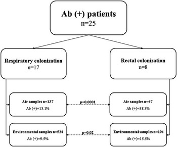
During the 5-month study period, ambient air and environmental surfaces were cultured daily and concomitantly in the ICU rooms of 25 consecutive A. baumannii–positive patients. Of these 25 patients, 17 (68%) were colonized in the respiratory tract and 8 (32%) were rectally colonized. No patient had overlapping sources of colonization, and none had had A. baumannii–positive clinical cultures before any samples were collected.
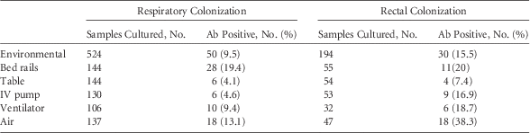
A total of 184 air samples were obtained; among these, 36 (19.5%) were positive for A. baumannii (Table 1). From the rooms of rectally colonized patients, 38.3% of air samples were A. baumannii–positive versus 13.1% from rooms of patients with respiratory colonization (P=.0001) (Table 1). Regarding the environmental samples, 15.5% of surface samples from the rooms of rectally colonized patients were A. baumannii–positive compared with 9.5% from rooms of patients with respiratory colonization (P=.02) (Figure 1). The percentage of rectally colonized patients with at least 1 positive air culture was 75% compared with 64% of patients with respiratory colonization (P=.6). The percentage of rectally colonized patients with at least 1 positive environmental culture was 87% versus 64% for patients with respiratory colonization (P=.2). Concomitant contamination within the same day of both air and environmental surfaces occurred in 17% of samples from rooms of rectally colonized patients but in only 5.1% of samples from rooms of patients with respiratory colonization (P=.02).
Percentage of positive air and environmental samples based on the body source of colonization. Percentage of positive days individual patients were detected to have carbapenem-resistant A. baumannii [Ab (+)] in their corresponding air and environmental samples. Patients were analyzed based on the body source of colonization, either rectally colonized patients or patients with respiratory colonization.

Cumulative Data for Air and Environmental Samples for Rectally Colonized Patients and Patients With Respiratory Colonization

NOTE. Ab, carbapenem-resistant A. baumannii sample; table, bedside table; IV, intravenous.
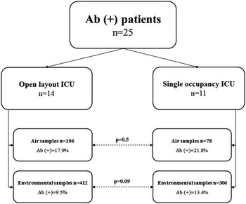
With regard to ICU layout, 14 patients (56%) were admitted to open-layout ICUs and 11 patients (44%) were admitted to single-occupancy ICUs. The rate of air contamination in the open-layout and single-occupancy ICUs was 17.9% and 21.8%, respectively (P=.5). Environmental surfaces were A. baumannii–positive in 9.5% of the samples in the open-layout ICUs versus 13.4% in the single-occupancy ICUs (P=.09) (Figure 2).
Percentage of positive air and environmental samples based on the type of ICU layout. Percentage of positive days individual patients were detected to have carbapenem-resistant A. baumannii [Ab (+)] in their corresponding air and environmental samples. Patients were analyzed based on the type of ICU they were admitted to, either in open-layout or single-occupancy ICUs.

Molecular Typing
We analyzed the corresponding clinical, air, and environmental isolates from 8 different patients; these included 8 clinical isolates, 22 air isolates, and 43 environmental isolates. According to our PFGE results, 13 air isolates (59%) were closely related to their corresponding clinical isolates, and 33 environmental isolates (77%) were closely related to their corresponding clinical isolates. In the open-layout ICUs, 50% of air isolates were closely related to their corresponding clinical isolates; in the single-occupancy ICUs, 70% of air isolates were closely related to their corresponding clinical isolates (P=.9). Environmental isolates were closely related to their corresponding clinical isolates in 68% of open-layout ICU rooms and in 95% of single-occupancy ICU rooms (P=.008) (see Supplementary Material).
DISCUSSION
In this longitudinal and prospective study, concomitant contamination of air and environmental surfaces were tested for up to 10 days in the ICU rooms of 25 consecutive patients with either rectal or respiratory A. baumannii colonization. Rectally colonized patients had more frequent A. baumannii contamination of their ambient air and environment than patients with respiratory colonization. Rectally colonized patients had more concomitant contamination of air and room surfaces. Interestingly, ICU layout did not impact the degree of contamination.
During the past several decades, several reports have been published describing outbreak investigations due to Acinetobacter spp. in which air samples were obtained. In 1987, Gerner-Smidt et alReference Gerner-Smidt 19 found positive air samples with Acinetobacter calcoaceticus serovar anitratus during a 2-year outbreak in a Danish ICU, but unfortunately, the method used to obtain the air samples was not reported.Reference Gerner-Smidt 19 In the same year, an outbreak investigation caused by Acinetobacter anitratus in 2 hospitals in the United Kingdom reported positive Acinetobacter samples from air and environmental surfaces; however, molecular typing of the isolates was not performed.Reference Allen and Green 20 A 1989 study conducted in the Netherlands found Acinetobacter in 12 of 104 air samples (11%) and in 20 of 164 environmental samples (12%); nevertheless, the molecular typing of the air samples in this study did not match with the clinical isolates.Reference Crombach, Dijkshoorn, van Noort-Klaassen, Niessen and van Knippenberg-Gordebeke 21
More recently, in a study published by a group from the University of Maryland, only 1 of 12 air samples belonging to rooms occupied by A. baumannii–positive patients was positive for this organism.Reference Rock, Harris, Johnson, Bischoff and Thom 22 Another study from Thailand also evaluated the presence of air contamination in closed-circuit mechanically ventilated ICUs housing A. baumannii–positive patients. Their air sampling technique consisted of placing 2 settle plates next to each patient twice per week; none of the air samples were positive for A. baumannii.Reference Apisarnthanarak, Tantajina, Laovachirasuwan and Singh 23 Given these discordances, other factors may have influenced the results, such as the method used for sampling the air, the type of ICU, or the frequency and timing of sampling. In our study, air tested positive for A. baumannii in 19.5% of the cultured samples. Our data might differ from the aforementioned studies due to the difference in sampling techniques. We left settle plates open continuously for 24 hours during the entire surveillance period (up to 10 days per patient), which allowed us to detect intermittent contamination of patient rooms. Our preliminary data showed that the presence of A. baumannii in the air varies throughout the day and might be associated with activities occurring inside the patient’s room (eg, diaper or bed linen changes).Reference Shimose, Greissman, Berbel-Caban, DePascale, Cleary and Munoz-Price 24 The fact that patients with rectal colonization had higher ambient air and environmental surface contamination than patients with respiratory colonization could be explained by the presence of a closed-circuit ventilation, which is designed to prevent the spread of such pathogens. Although the rectum might not be a more efficient spreader of A. baumannii than the respiratory tract, rectal contamination seems to be relevant to the degree of bacterial burden in the environment. Additionally, air filtration might have played a role in the results.Reference Munoz-Price 25
The molecular typing results showed that the majority of A. baumannii isolates from patients, air, and environment were closely related, but there was a difference between the numbers of closely related isolates based on the ICU layout. The presence of different strains of A. baumannii in the immediate environment of patients admitted to open-layout ICUs could be explained by a higher degree of horizontal transmission between neighboring patients and by the presence of this organism in the common areas in the ICU, ie, not only contaminating environmental surfaces but also present in the air.
Limitations of this project include its single-center study design and the relatively small sample size. The air sampling method we used was qualitative, with possible low sensitivity that might not be optimal, especially for colonized patients (ie, “low bacterial loads”). In addition, not all study isolates were available for genetic relatedness testing. Moreover, clinical or demographic data were not collected (eg, diarrhea, mechanical ventilation, use of antibiotics, comorbidities), which could have played a role in the results.
Even though our sampling techniques were potentially insensitive, we continued to find different contamination rates of air and environment in the rooms occupied by A. baumannii–positive patients based on colonization source, even when these tests were performed concomitantly. In this study, our findings have been substantiated by molecular typing, and they support the results of our previous studies. The significance of these results remains uncertain, especially with regard to the role of air contamination. However, it is not unreasonable to postulate that air and environmental contamination are directly associated with each other. The relationship between and implications of air and environmental contamination should to be explored in different settings and for other clinically significant pathogens.
ACKNOWLEDGMENTS
Financial support: This investigation was performed with internal funds. Molecular typing of A. baumannii was supported in part by research grants from the National Institutes of Health (grant nos. R01AI104895 and R21AI107302).
Conflicts of interest: Y.D. has served on an advisory board for Shionogi, Meiji, Tetraphase. He has consulted for Melinta and has received research support from Merck and The Medicines Company for studies unrelated to this work. L.S.M.P. has served as a speaker for ECOLAB and as an advisor for Xenex. The remaining authors report no conflicts of interest relevant to this article.
SUPPLEMENTARY MATERIAL
To view supplementary material for this article, please visit http://dx.doi.org/10.1017/ice.2016.69