Introduction
Leishmania infantum is the main causative species of the zoonotic visceral leishmaniasis (VL), with domestic dogs representing the most important parasite reservoirs. Disease control, based on the treatment of human cases, is one of the main measures used to avoid the dissemination of the disease. However, there are only a limited number of drugs available on the market today, and these can cause several side-effects in patients (Sundar and Chakravarty, Reference Sundar and Chakravarty2013). In addition, lipid-based formulations, although effective and less toxic, are expensive (Mohamed-Ahmed et al. Reference Mohamed-Ahmed, Brocchini and Croft2012). In this context, since life-long immunity has been reached in infected and vaccinated mammalian models, prophylactic vaccination to prevent disease has been encouraged (Duarte et al. Reference Duarte, Lage, Martins, Costa, Carvalho, Ludolf, Santos, Vale, Roatt, Menezes-Souza, Fernandes, Tavares and Coelho2017). The immune response associated with the effective protection against VL in humans and dogs has been largely attributed to the development of a predominant T helper (Th)1 immunity, characterized by the production of cytokines, such as interferon-γ (IFN-γ) and interleukin (IL)-12, which subsequently mediates macrophage activation, nitric oxide (NO) production and parasite killing (Miahipour et al. Reference Miahipour, Haji-Fatahaliha, Keshavarz, Gharavi, Mohamadi, Babaloo, Rafati, Younesi, Hosseini and Yousefi2016; Rodrigues et al. Reference Rodrigues, Cordeiro-da-Silva, Laforge, Silvestre and Estaquier2016).
The identification of Leishmania antigens containing the major histocompatibility complex class I- and/or class II-restricted epitopes is an important step towards selecting immunogens, since protection is based on the polarized and long-lasting CD4+ and CD8+ T-cell-dominated immune response against parasites (Agallou et al. Reference Agallou, Margaroni, Athanasiou, Toubanaki, Kontonikola, Karidi, Kammona, Kiparissides and Karagouni2017). As a consequence, Leishmania proteins have been evaluated as recombinant molecules to protect against disease (Iborra et al. Reference Iborra, Parody, Abanades, Bonay, Prates, Novais, Barral-Netto, Alonso and Soto2008; Srivastava et al. Reference Srivastava, Singh, Mishra, Kumar, Gour, Bajpai, Singh, Pandey and Singh2012; Agallou et al. Reference Agallou, Athanasiou, Samiotaki, Panayotou and Karagouni2016). Major advantages have been associated with the use of defined immunogens, as well as by their high purity and production yield; however, these antigens present variable degrees of success, which depend on the animal model used for testing, the parasite species causing the disease, the number and concentration of the administered doses, among other factors (Rafati et al. Reference Rafati, Kariminia, Seyde-Eslami, Narimani, Taheri and Lebbatard2002; Joshi and Kaur, Reference Joshi and Kaur2014; Duarte et al. Reference Duarte, Lage, Martins, Costa, Lage, Carvalho, Ludolf, Santos, Roatt, Menezes-Souza, Fernandes, Tavares and Coelho2016).
As an additional strategy used to select new candidates able to elicit protective immunity against VL, immunoproteomic approaches have been performed in Leishmania promastigotes and/or amastigotes using human and canine sera samples, where antileishmanial antibodies recognize antigenic proteins in parasite extracts (Kumari et al. Reference Kumari, Kumar, Samant, Sundar, Singh and Dube2008; Costa et al. Reference Costa, Andrade, Bartholomeu, Freitas, Pires, Chapeaurouge, Perales, Ferreira, Giusta, Melo and Gazzinelli2011; Abanadés et al. Reference Abanadés, Arruda, Arruda, Pinto, Palma, Aquino, Caldas, Soto, Barral and Barral-Netto2012; Alcolea et al. Reference Alcolea, Tuñón, Alonso, García-Tabares, Ciordia, Mena, Campos, Almeida and Larraga2016). These proteomic screenings have revealed a number of Leishmania-specific and/or conserved proteins that are present in this genus, some of which are annotated as hypothetical in Leishmania genome databases (Agallou et al. Reference Agallou, Athanasiou, Samiotaki, Panayotou and Karagouni2016). One of these candidates, namely LiHyS (LinJ_30_2800), was recognized by antibodies in VL dog's sera, but it was not identified by antibodies in sera from healthy dogs living in an endemic area of disease (Coelho et al. Reference Coelho, Oliveira, Valadares, Chávez-Fumagalli, Duarte, Lage, Soto, Santoro, Tavares, Fernandes and Coelho2012). The present work evaluated the antigenicity of this protein in a recombinant version (rLiHyS) for the canine and human VL, as well as its immunogenicity and protective efficacy against L. infantum infection.
Materials and methods
Ethics statement
Experiments were performed in compliance with the Committee on the Ethical Handling of Research Animals (CEUA) of the Federal University of Minas Gerais (UFMG), Belo Horizonte, Minas Gerais, Brazil (protocol number 333/2015). The present study was also approved by the Ethics Committee of UFMG (protocol number CAAE–32343114.9.0000.5149). A written informed consent was obtained from all subjects, who received an individual copy of the study policy, which was analysed by an independent reviewer.
Mice and parasite
Female BALB/c mice (8 weeks age) were obtained from the breeding facilities of the Department of Biochemistry and Immunology, Institute of Biological Sciences (ICB), UFMG, and were maintained under specific pathogen-free conditions. L. infantum (MHOM/BR/1970/BH46) was used, and parasites were grown at 24 °C in complete Schneider's medium (Sigma-Aldrich, St. Louis, MO, USA), which consisted of Schneider's medium plus 20% heat-inactivated fetal bovine serum (FBS, Sigma-Aldrich, USA), 20 mm L-glutamine, 200 U mL−1 penicillin and 100 µg mL−1 streptomycin, at pH 7·4. The soluble Leishmania antigenic extract (SLA) was prepared from 2 × 10⁸ stationary-phase promastigotes of parasites, as described by Martins et al. (Reference Martins, Duarte, Lage, Costa, Carvalho, Mendes, Roatt, Menezes-Souza, Soto and Coelho2017).
Production of the recombinant antigens
The LiHyS (LinJ_30_2800) protein codifying gene was cloned from L. infantum genomic DNA using specific primers: 5′-TCTCGGATCCATGCGCCAGCGAAAGCAC-3′ (forward) and 5′-TGAAAAGCTTCCATGCGATCCAGTAGATG-3′ (reverse), and BamHI and HindIII as restriction enzymes. The DNA fragment was purified and linked into a pGEM®-T vector system (Promega, USA), and the recombinant plasmid was used to transform Escherichia coli XL1-Blue competent cells. DNA fragments obtained from digestion of pGEM-LiHyS plasmids were ligated into a pET28a-TEV vector, and BL21 cells were transformed into the recombinant plasmid. Sequencing was performed to confirm the identity of the insert by using a MegaBace 1000 automatic sequencer apparatus (Amersham Biosciences, USA). For the purification of rLiHyS, cells were induced with 1·0 µ m isopropyl-β-D-thiogalactopyranoside (IPTG, Promega, Montreal, Canada), and cultures were incubated for 4 h at 37 °C, at which time these were ruptured by six cycles of ultrasound, in cycles of 30 s each (38 MHz), followed by six cycles of freezing (liquid nitrogen, −196 °C) and thawing (+37 °C). Then, the cell debris were removed by centrifugation, and rLiHyS (21·3 kDa) was purified onto a HisTrap HP affinity column (GE Healthcare Life Sciences, NJ, USA) connected to an AKTA system. The eluted fractions were concentrated in Amicon® ultra-15 centrifugal filters, with a 10 000 nominal molecular weight limit (Millipore, Germany), and further purified on a Superdex™ 200 gel-filtration column (GE Healthcare, USA). The A2 recombinant (rA2) protein was produced as described by Zhang et al. (Reference Zhang, Charest, Ghedin and Matlashewski1996). Briefly, pET16b-A2 plasmid, which was kindly provided by Dr Greg Matlashewski (Microbiology and Immunology Department, McGill University, Montreal, Quebec, Canada), was used to express and purify the recombinant protein (53 kDa). For this, transformed E. coli DH5α cells were grown in the presence of 1 mm of IPTG for 3 h (Promega, Montreal, Canada) and later disrupted by five cycles of freezing and thawing, followed by mild ultrasound treatment (five cycles of 30 s each with an ultrasound processor), in which they were centrifuged at 13·000 g for 30 min at 4 °C. After expression, the recombinant protein was purified by a HisTrap HP affinity column connected to an AKTA system. Next, rLiHyS and rA2 proteins were passed through a polymyxin-agarose column (Sigma-Aldrich, USA) to remove any residual endotoxin content (<10 ng of LPS per 1 mg of recombinant protein, measured by the Quantitative Chromogenic Limulus Amebocyte Assay QCL-1000, BioWhittaker, MD, USA).
Affinity purification of the anti-rLiHyS IgG antibody
The specific antibodies against the rLiHyS protein were affinity-purified from a pool of positive anti-rLiHyS sera on an antigen column. For this, BALB/c mice (n = 6) were vaccinated subcutaneously in their left hind footpad with 20 µg of rLiHyS associated with 20 µg of saponin (Quillaja saponaria bark saponin, Sigma). Two doses were administered at 14-day intervals, and 7 days after the last immunization, blood samples were collected and sera were separated. Next, 1 mg of the rLiHyS protein was covalently bound to a cyanogen bromide-activated Sepharose 4B column (GE Healthcare, USA), which was previously equilibrated with 20 mm phosphate buffer, pH 7·4. Coupling and blocking were carried out according to manufacturer instructions. Two millilitres of the sera pool were passed through the antigen column. After washing, the specific antibodies were eluted from the column with 0·1 m glycine, pH 2·8. Finally, the antibody preparation was equilibrated to pH 7·5 with 1 m Tris/HCl buffer. The solution of the antibody was restored to the original volume of the pooled sera (2 mL). An enzyme-linked immunosorbent assay (ELISA) experiment and a sodium dodecyl sulfate-12% polyacrylamide gel (SDS-12% PAGE) with rLiHyS were performed to verify the reactivity and quality of the purified antibody (data not shown).
Sera collection
Sera samples of VL dogs (n = 20) were used. The disease was confirmed by means of the identification of L. infantum kinetoplastid DNA (kDNA) in bone marrow (BM) samples by a polymerase chain reaction (PCR) technique, as well as by positive serological results, which were evaluated by IFAT-LVC® and EIE-LVC® commercial kits (BioManguinhos®, Rio de Janeiro, Brazil). Symptomatic dogs (n = 14) showed three or more of the following symptoms: weight loss; alopecia; adenopathy; onychogryposis; hepatomegaly; conjunctivitis; exfoliative dermatitis on the nose, tail and ear tips; splenomegaly; lymphadenomegaly; and renal azotemia, whereas asymptomatic animals (n = 6) showed positive parasitological and serological results but presented no clinical sign of disease. In addition, sera from healthy animals living in an endemic area of disease (n = 20) were employed. These animals were free of any clinical sign of disease and presented negative serological results. Samples from Leish-Tec®-vaccinated dogs (n = 15), as well as from those experimentally infected with Ehrlichia canis (n = 15) or Babesia canis (n = 10), were used in the assays. Regarding human sera, samples were obtained from VL patients (n = 20, including 12 males and eight females, with ages ranging from 25 to 59 years), which were collected from an endemic area of disease (Belo Horizonte, Brazil). Patients were diagnosed by means of clinical evaluation and demonstration of L. infantum kDNA in BM aspirates, when evaluated by PCR assay. Samples were also obtained from healthy individuals living in an endemic area of disease (n = 20, including 11 males and nine females, with ages ranging from 21 to 53 years, Belo Horizonte, Brazil), which showed no clinical sign of leishmaniasis, in addition to negative serological results from the Kalazar Detect™ Test (InBios® International, USA). Moreover, sera from Chagas disease patients (n = 15, including nine males and six females, with ages ranging from 20 to 55 years) were used, with the infection confirmed by haemoculture by means of the Chagatest® recombinant ELISA v.4·0 kit or the Chagatest® haemagglutination inhibition kit (Wiener lab., Rosario, Argentina).
Immunoblottings and ELISA assays
For the immunoblottings, rLiHyS (10 µg) was submitted to a SDS-12% PAGE and blotted onto a nitrocellulose membrane (0·2 µm pore size, Sigma, St. Louis, USA). Next, membranes were blocked with a solution composed by phosphate buffer saline 1× plus Tween 20 0·05% (PBS-T) added with 5% albumin solution and incubated for 1 h at 37 °C before undergoing the first incubation with human or canine sera pools (1 : 400 and 1 : 200 diluted in PBS-T, respectively). Membranes were washed with PBS-T and anti-human or anti-dog IgG horseradish peroxidase-conjugated antibodies (diluted 1:10 000 and 1:5000 in PBS-T, respectively; SAB 3701282 and A6792 catalogues, respectively, Sigma-Aldrich, USA) were added, at which time a new incubation was developed for 1 h at 37 °C. Reactions were developed by adding 12·5 mg chloronaphthol, 25·0 mg diaminobenzidine and 20 µL H2O2 30 vol., and stopped by adding 10 mL distilled water. The anti-rLiHyS polyclonal antibody was used as a positive control. For the ELISA assays, previous titration curves were performed to determine the most appropriate antigen concentration and antibody dilution to be used. Flexible microtitre immunoassay plates (Jetbiofil®, Belo Horizonte) were coated with rLiHyS, rA2, or SLA (0·5, 1·0 and 1·0 µg per well, respectively), which was diluted in 100 µL of coating buffer (50 mm carbonate buffer, pH 9·6), for 18 h at 4 °C. Next, free binding sites were blocked using 250 µL of PBS-T plus 5% casein, for 1 h at 37 °C. After washing the plates seven times with PBS-T, these were incubated with 100 µL of the individual human or canine sera (1:400 or 1:200, respectively; both diluted in PBS-T), for 1 h at 37 °C. The plates were then washed seven times in PBS-T and incubated with anti-human or anti-dog IgG horseradish peroxidase-conjugated antibodies (diluted 1:15 000 and 1:10 000 in PBS-T, respectively), for 1 h at 37 °C. After washing the plates seven times with PBS-T, reactions were developed by incubation with a solution (100 µL per well) consisting of 2 mg ortho-phenylenediamine, 2 µL H2O2 30 vol. and 10 mL citrate-phosphate buffer, at a pH 5·0, for 30 min in the dark. Reactions were stopped by adding 25 µL 2 N H2SO4, and the optical density was read in an ELISA microplate spectrophotometer (Molecular Devices, Spectra Max Plus, Canada) at 492 nanometres.
PBMCs culture and in vitro lymphoproliferation index
PBMCs were separated from the blood samples of healthy subjects (n = 10) and VL patients (n = 8), as described by Martins et al. (Reference Martins, Duarte, Lage, Costa, Carvalho, Mendes, Roatt, Menezes-Souza, Soto and Coelho2017). For proliferation assays, cells (1 × 10⁷ cells) were labelled with carboxyfluorescein diacetate succinimidyl ester (CFSE). For this, after having been collected and washed, PBMCs were resuspended in pre-warmed PBS 1× containing 0·1% bovine serum albumin, at a final concentration of 1 × 10⁷ cells mL−1, and 2 µL of 5 mm stock Cell Trace CFSE solution (Molecular Probes, USA) was added per 1 mL of cells (at a working concentration of 10 mm). Then, the cells were incubated for 10 min at 37 °C, and the staining was quenched by adding 5 mL of RPMI 1640 plus 10% FBS for 5 min in an ice bath. The cells were then centrifuged three times for 7 min, and 50 µL were collected and analysed on a flow cytometry, aiming to evaluate the percentage of cell labelling. PBMCs were cultured in 48-well flat-bottomed tissue culture plates (Costar, Cambridge, MA, USA), with each well containing 800 µL of RPMI 1640 medium. Next, cells (1 × 10⁶) were added in triplicate to the wells containing 100 µL of RPMI (control) and incubated alone (medium, control background) or stimulated with rLiHyS or rA2 (10 µg mL−1, each). The incubation was carried out in a 5% CO2 atmosphere for 5 days at 37 °C, after which the PBMCs were collected, washed twice in fluorescence-activated cell sorter (FACS) buffer, and fixed using a FACS fixing solution (10 g L−1 paraformaldehyde, 10·2 g L−1 sodium cacodylate and 6·63 g L−1 sodium chloride, pH 7·4), which is also used in the lyses of red blood cells but not of white blood cells. The PBMCs were then stored at 4 °C, prior to flow cytometric acquisition and analysis, which were performed using a FACScalibur® instrument (Becton Dickson – BD, USA). The Cell-Quest™ software package (Franklin Lakes, NJ, USA) was used for data acquisition and analysis based on 30 000 events per sample.
Vaccination and evaluation of the parasite load
BALB/c mice (n = 16, per group) were either vaccinated subcutaneously in their left hind footpad with 20 µg of rLiHyS, associated or not with saponin (20 µg), or they received saline or saponin. Three doses were administered at 14-day intervals. Thirty days after the last immunization, animals (n = 8, per group) were infected subcutaneously in the right hind footpad with 1 × 10⁷ stationary-phase promastigotes of L. infantum; 60 days later they were euthanized, and the liver, spleen, BM) and paws’ draining lymph nodes (dLNs) were collected to evaluate the parasite burden through a limiting-dilution technique, as described by Martins et al. (Reference Martins, Duarte, Lage, Costa, Carvalho, Mendes, Roatt, Menezes-Souza, Soto and Coelho2017). Briefly, organs were homogenized using a glass tissue grinder in sterile saline, and tissue debris were removed by centrifugation at 150 g. Cells were concentrated by centrifugation at 2000 g, and pellets were resuspended in 1 mL of Schneider's insect medium supplemented with 20% FBS. Next, 220 µL was plated onto 96-well flat-bottom microtitre plates (Nunc, Nunclon®, Roskilde, Denmark) and diluted in log-fold serial dilutions in Schneider's medium with a 10−1–10−12 dilution. Pipette tips were discarded after each dilution to avoid carrying adhered parasites from one well to another. Each sample was plated in triplicate, at 24 °C, and read 7 days after the beginning of the culture. Results were expressed as the negative log of the titre adjusted per milligram of organ. The parasite load was also evaluated by a quantitative PCR (qPCR) technique in the spleen of the animals, as described by Duarte et al. (Reference Duarte, Lage, Martins, Costa, Lage, Carvalho, Ludolf, Santos, Roatt, Menezes-Souza, Fernandes, Tavares and Coelho2016), and the results were expressed as the number of parasites per 1000 nucleated cells.
Cytokine assay by capture ELISA and flow cytometry
Thirty days after the last immunization (Before) and 60 days post-infection (After), BALB/c mice (n = 8 per group) were euthanized and their spleen were collected, at which time splenocytes were cultured (5 × 10⁶ cells mL−1) in 24-well plates (Nunc). Cells were incubated in RPMI medium (control) together with 20% FBS, 20 mm L-glutamine, 200 U mL−1 penicillin and 100 µg mL−1 streptomycin, at pH 7·4; or stimulated with rLiHyS or L. infantum SLA (10 and 25 µg mL−1, respectively) for 48 h at 37 °C in 5% CO2. IFN-γ, IL-4, IL-10, IL-12p70 and GM-CSF levels were assessed in the supernatants by an ELISA capture (all obtained from BD OptEIA TM set mouse, Pharmingen®, San Diego, CA, USA), following manufacturer instructions. The involvement of CD4+ and CD8+ T cells in IFN-γ production was also evaluated in the spleen cells, in which cells stimulated with rLiHyS or SLA (10 and 25 µg mL−1, respectively) were incubated in the presence of monoclonal antibodies against mouse IL-12 (C17·8), CD4 (GK 1·5), or CD8 (53-6·7), all in a concentration of 5 µg mL−1. Appropriate isotype-matched controls (no azide/low endotoxin™), rat IgG2a (R35-95) and rat IgG2b (95-1) were used (Pharmingen®, USA). A flow cytometry assay was also performed in these spleen cells, aiming to evaluate the IFN-γ-, TNF-α- and IL-10-producing CD4+ and CD8+ T-cell frequencies after L. infantum SLA stimulus, as described by Vieira et al. (Reference Vieira, Francisco, Machado, Nogueira, Fonseca, Reis, Teixeira-Carvalho, Martins-Filho, Tafuri and Carneiro2012). Results were expressed as indexes, which were calculated by dividing the cytokine-producing CD4+ and CD8+ T-cell percentages in the stimulated cultures by the values obtained in the unstimulated cultures (control).
Nitrite secretion and antibody production
Sixty days after infection, cell supernatants were also used to evaluate the nitrite production in all experimental groups. For this, 100 µL of protein and parasite-stimulated culture supernatants were mixed with an equal volume of Griess reagent (Sigma-Aldrich, USA). After 30-min incubation at room temperature, the nitrite concentration was calculated using a standard curve of known concentrations, and the results were expressed as μ m (Green et al. Reference Green, Wagner, Glogowski, Skipper, Wishnok and Tannenbaum1982). To evaluate the humoral response before and after infection, sera samples were collected from the animals, 30 days after the last immunization (Before) and 60 days post-infection (After). The protein and parasite-specific IgG1 and IgG2a antibody levels were evaluated by an ELISA assay, as described by Martins et al. (Reference Martins, Duarte, Lage, Costa, Carvalho, Mendes, Roatt, Menezes-Souza, Soto and Coelho2017). Briefly, rLiHyS and SLA (0·5 and 1·0 µg per well, respectively) were used as antigens, and sera samples were 1:100 diluted in PBS-T. The anti-mouse IgG1 and IgG2a horseradish peroxidase-conjugated antibodies (Sigma-Aldrich, USA) were used in 1:5000 or 1:7500 dilutions, which were performed in PBS-T, respectively.
Confocal fluorescence microscopy
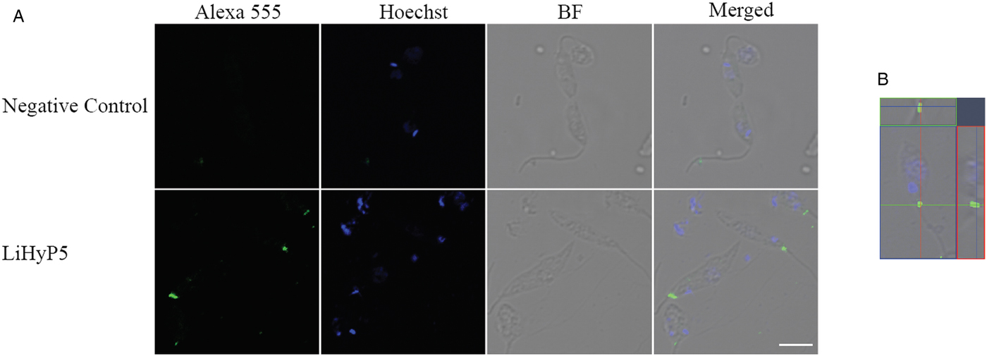
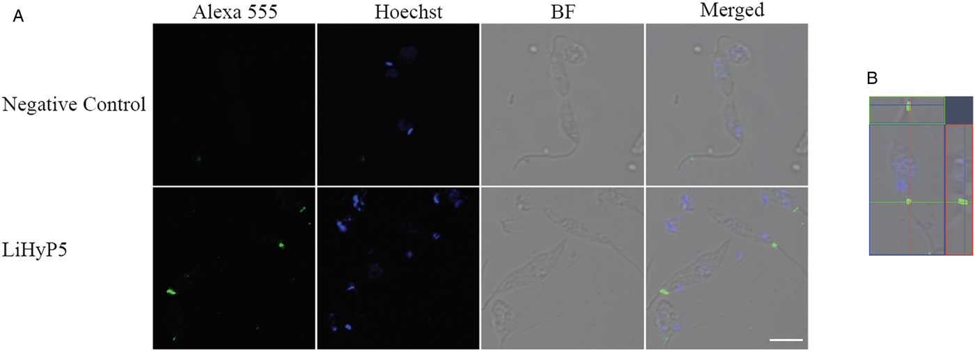
Immunofluorescence assay was performed as described by Faria et al. (Reference Faria, Andrade, Goes, Rodrigues and Gomes2016). Briefly, parasites were double-labelled with the anti-rLiHyS polyclonal IgG antibody (at a 1:50 dilution) and incubated with a secondary antibody conjugated to an Alexa Fluor® 555 Goat anti-mouse IgG antibody (1:1000 diluted, Life Technologies®, USA), together with Hoechst 33258 (1 µg mL−1, Life Technologies, USA), for 1 h at room temperature. Next, samples were washed three times in PBS 1× for 10 min, and then mounted with hydromount (Electron Microscopy Sciences). The negative control was included in all reactions by omitting primary antibodies. Images were collected using a Zeiss LSM 880 confocal microscopy (Carl Zeiss, Jena, Germany). The Zeiss Efficient Navigation (ZEN) software was used for orthogonal projections (XY, XZ, YZ), and image adjustments were performed according to the negative controls.
Infection of murine macrophages using pre-treated parasites
The inhibitory effect of anti-rLiHyS polyclonal antibody on the L. infantum invasion in murine macrophages was evaluated. For this, stationary-phase promastigotes of parasites (5 × 10⁶ cells) were pre-incubated with different concentrations (5·0, 10·0 and 50·0 µg mL−1) of anti-rLiHyS IgG antibody or a non-immune IgG for 4 h at 24 °C. After incubation, parasites were washed three times with RPMI 1640 medium, then quantified and incubated for 24 h with macrophages (in a ratio of 10 parasites per one macrophage). As a control, the same amount of non-treated parasites was used in the infection. Then, the cells were washed, set and stained to determine the percentage of infected macrophages by counting 200 cells in triplicate. In addition, the number of amastigotes per infected cell was evaluated. Data shown are representative of two independent experiments, which presented similar results.
Statistical analysis
The results were entered into Microsoft Excel (version 10·0) spreadsheets and analysed by using GraphPad Prism™ (version 6·0 for Windows). Statistical analyses were performed by one-way analysis of variance, followed by Bonferroni's post-test for comparisons between the groups. Differences were considered significant with P < 0·05. In addition, receiver operating characteristic (ROC) curves were constructed to obtain the sensitivity, specificity and area under the curve values for the diagnostic antigens, as well as the lower limit of positivity (cut-off). Vaccination results showed in this study are representative of two independent experiments, which presented similar results.
Results
Antigenicity and immunogenicity of the rLiHyS protein
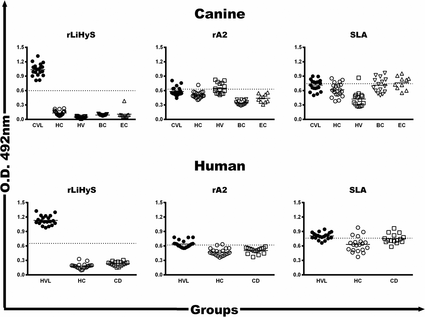
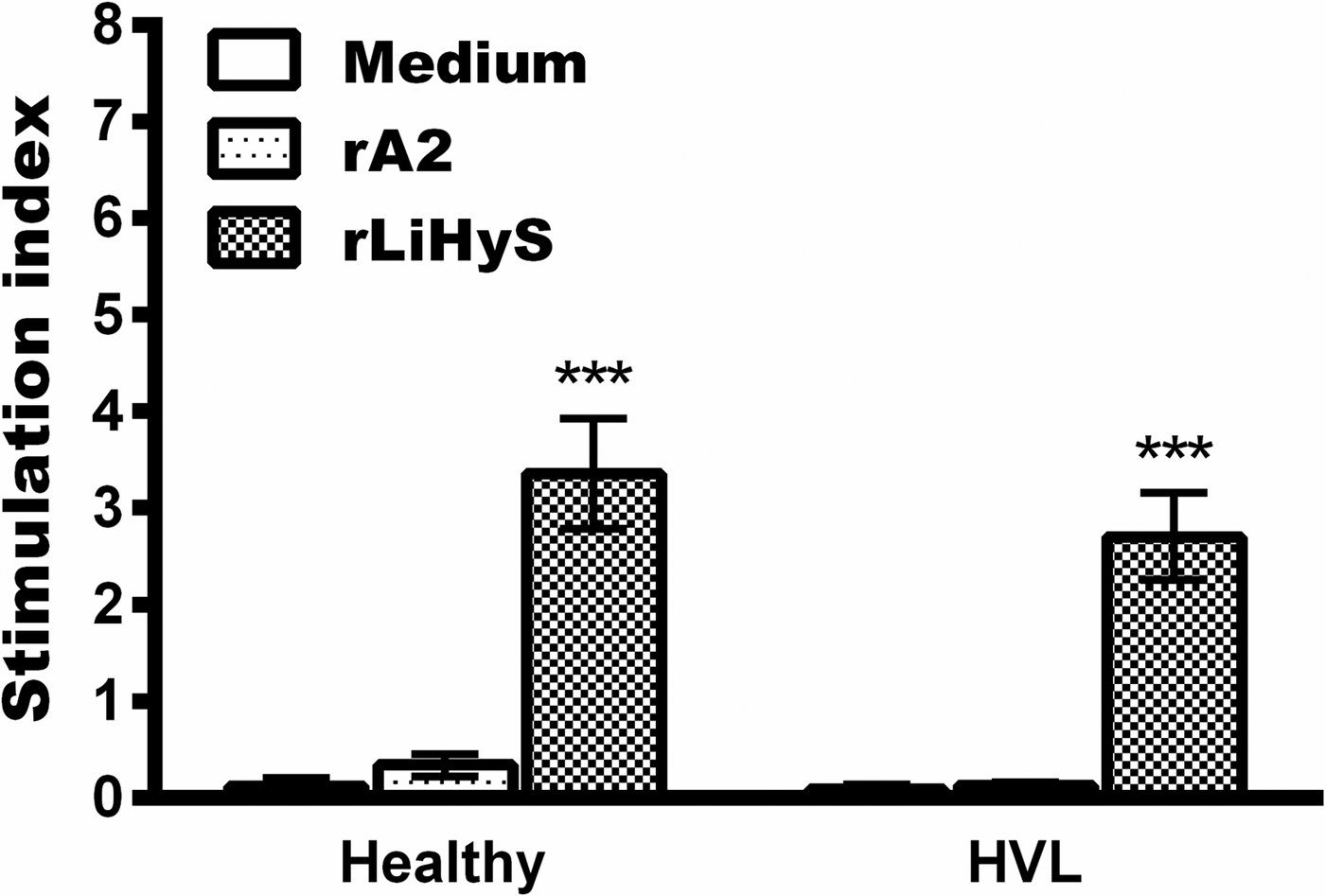
First, the antigenicity of rLiHyS was evaluated. For this, immunoblottings were performed, and the anti-protein reactivity was observed using both asymptomatic and symptomatic VL dog sera, as well as VL patient sera. However, sera were not collected from healthy dogs or humans living in an endemic area of disease (Supplementary Fig. S1). In addition, ELISA assays were developed using a human and canine serological panel. Cut-off values for negative and positive sample discrimination were constructed using ROC curves. In the results, both L. infantum-infected dogs and humans (not other groups’ sera) were highly reactive against rLiHyS, while the worst results of sensitivity and specificity were found when using rA2 or L. infantum SLA as antigens in the plates (Fig. 1). Then, the immunogenicity of rLiHyS was evaluated by means of a lymphoproliferation index in purified PBMCs from healthy subjects and VL patients (Fig. 2). In the results, this cell population showed high proliferative values when it was incubated with rLiHyS in both individuals, as compared to the results obtained when the in vitro cultures were unstimulated or stimulated with the rA2 antigen (P < 0·0001).
Reactivity of the diagnostic antigens by ELISA using a serological panel. The individual optical density (O.D.) of each serum sample against rLiHyS, rA2, or L. infantum SLA is shown here. Sera samples of VL dogs (n = 20), from healthy animals living in an endemic area of disease (n = 20), from Leish-Tec®-vaccinated animals (n = 15), or those experimentally infected with Ehrlichia canis (n = 15) or Babesia canis (n = 10) were used. Sera from VL patients (n = 20), from healthy subjects living in an endemic area of disease (n = 20), and from Chagas disease patients (n = 15) were employed in the assays. The cut-off values (dotted lines) were calculated by using ROC curves. CVL, canine visceral leishmaniasis; HC, healthy controls; HV, healthy vaccinated animals; BC, Babesia canis; EC, Ehrlichia canis; HVL, human visceral leishmaniasis; CD, Chagas disease.

Lymphoproliferative index in human PBMCs using different in vitro stimuli. Blood samples of healthy subjects (n = 10) and VL patients (n = 8) were collected; PBMCs were purified and either unstimulated (medium) or separately stimulated with rLiHyS or rA2 (10 µg mL−1, each). The incubation was carried out in a 5% CO2 atmosphere for 5 days at 37 °C, at which time the lymphoproliferation index was evaluated by flow cytometry. Results are shown as mean ± standard deviation using the different in vitro stimuli. ***Indicate statistically significant difference in relation to the other stimuli (P < 0·0001).

The rLiHyS/saponin vaccine is immunogenic in BALB/c mice
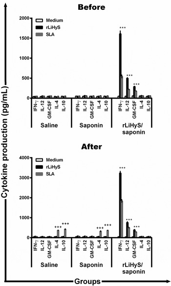
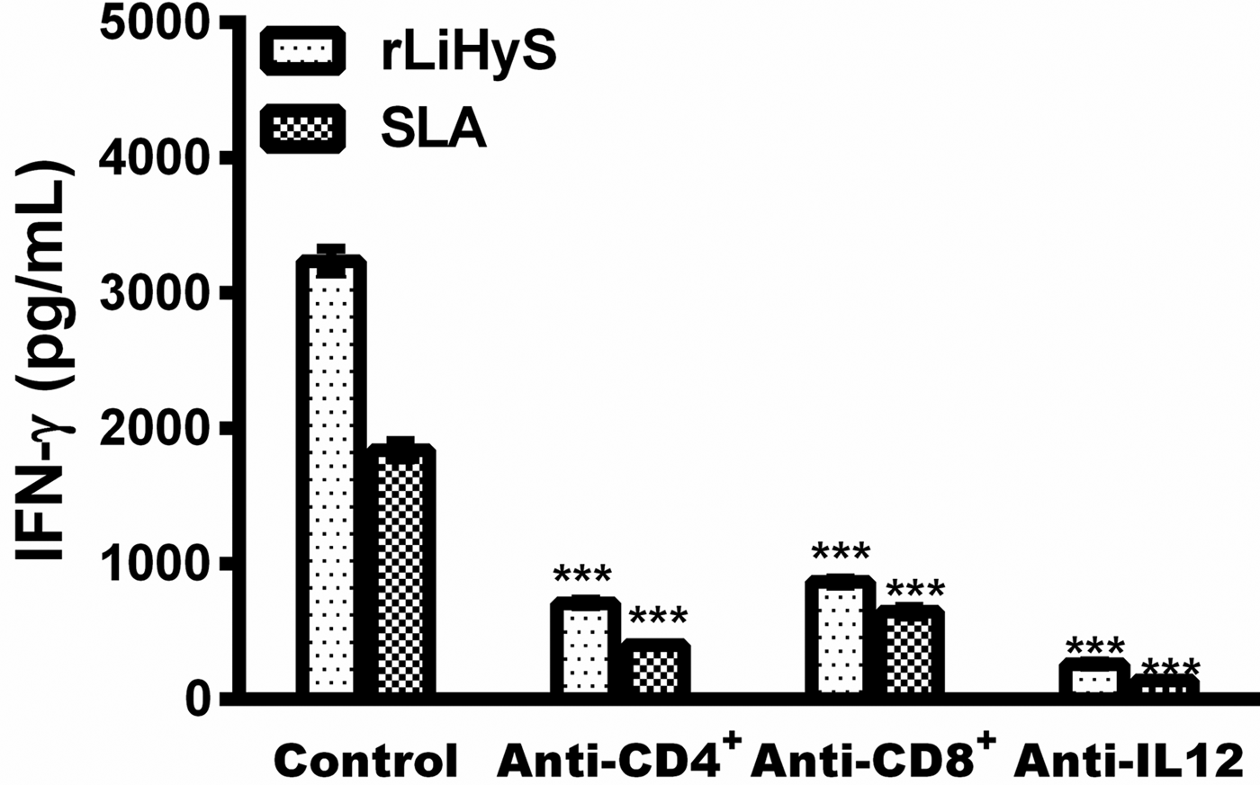
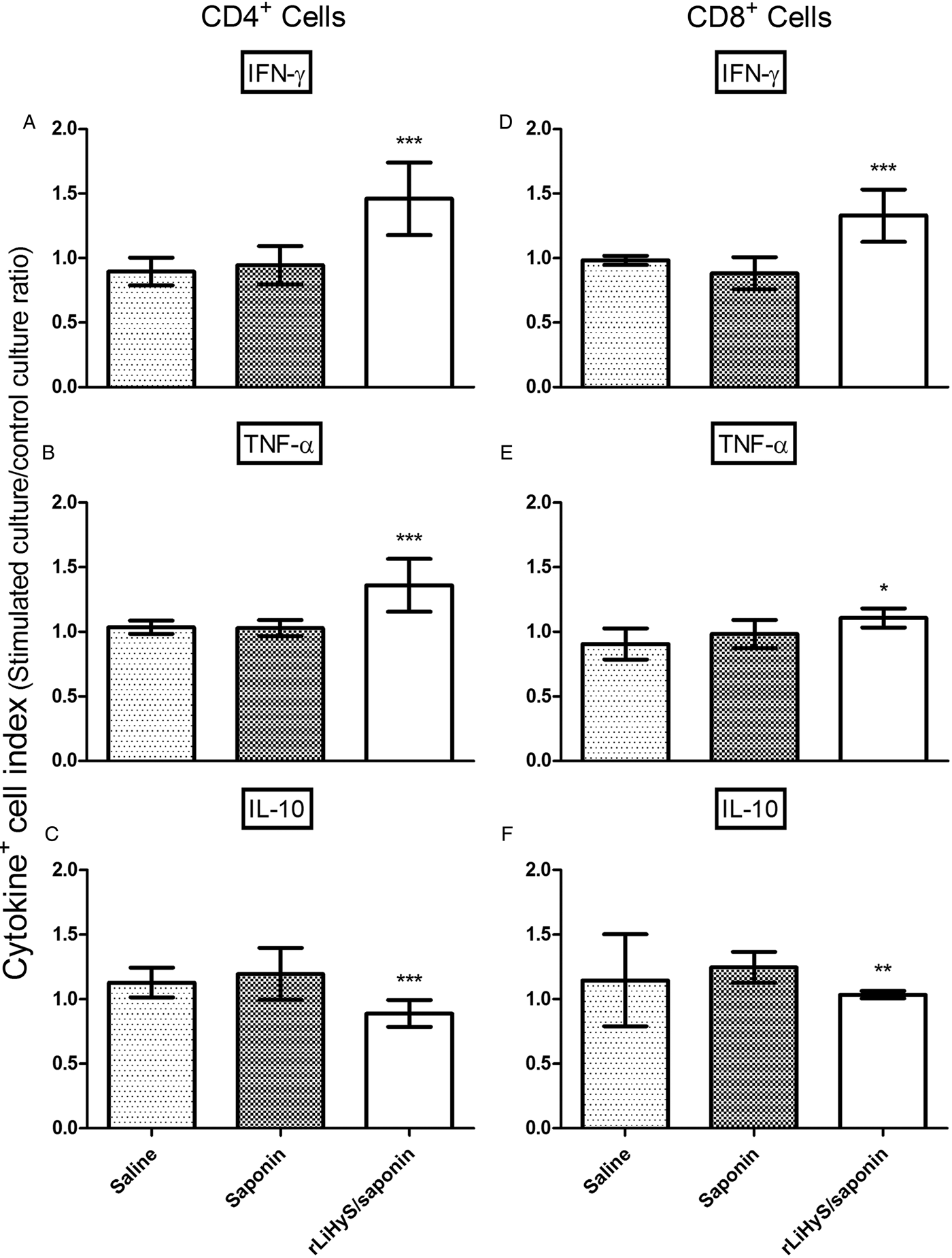
In this case, rLiHyS was used to immunize BALB/c mice in an attempt to evaluate its protective efficacy against VL. To achieve this, animals were vaccinated three times with the protein, associated or not with saponin, and 30 days after the last vaccine dose. The cytokine production was then evaluated based on the protein- and parasite-specific IFN-γ, IL-4, IL-10, IL-12 and GM-CSF production. In the results, spleen cells from rLiHyS/saponin-vaccinated mice produced significantly higher levels of IFN-γ, IL-12 and GM-CSF than did those secreted by cells from saline and saponin group mice (Fig. 3; P < 0·0001). No IL-4 and IL-10 production was detected in any experimental group. The animals were then challenged with L. infantum, and 60 days after infection, their splenocytes were in vitro stimulated with both antigens, and the cytokine production was again evaluated. The immune profile was maintained in the rLiHyS/saponin group, since higher levels of protein and parasite-specific IFN-γ, IL-12 and GM-CSF were found, which were associated with a low production of IL-4 and IL-10 (Fig. 3; P < 0·0001). By contrast, spleen cells of the mice from the saline and saponin groups produced significantly higher levels of anti-parasite IL-4 and IL-10 (P < 0·0001). Mice that were immunized with rLiHyS alone showed a mixed Th1:Th2 profile, which was observed before and after infection. As an additional immunological parameter, the participation of CD4+ and CD8+ T cells in the IFN-γ production was evaluated in the rLiHyS/saponin group. In the results, the production of this cytokine was significantly diminished when either an anti-IL-CD4 or an anti-CD8 antibody was added to the in vitro cultures (Fig. 4; P < 0·0001). The intracytoplasmic cytokine profile, which was evaluated by a flow cytometry assay (Fig. 5), also showed that rLiHyS/saponin-vaccinated mice presented higher levels of antileishmanial IFN-γ +- and TNF-α +-producing CD4+ and CD8+ T cells (P < 0·05), which were corroborated with the low presence of IL-10+-producing T cells (P < 0·05), as compared to the results found in the control groups, denoting the polarized Th1 profile found in the rLiHyS/saponin group mice.
Cell response induced in the rLiHyS/saponin-vaccinated BALB/c mice, before and after infection. Splenocytes were obtained from the spleen of mice, 30 days after the last immunization (Before) and 60 days after infection (After). Cells were unstimulated (medium) or stimulated with rLiHyS or SLA (10 and 25 µg mL−1, respectively) for 48 h at 37 °C in 5% CO2. IFN-γ, IL-12, GM-CSF, IL-4 and IL-10 levels were measured in culture supernatants by ELISA capture. Bars represent the mean ± standard deviation of the groups. ***Indicate statistically significant difference in relation to the saline and saponin groups (P < 0·0001). +++Indicate statistically significant difference in relation to the rLiHyS/saponin group (P < 0·0001).

Participation of the CD4+ and CD8+ T cells in the IFN-γ production in the protected animals. The anti-IL-12, anti-CD4 and anti-CD8 monoclonal antibodies were added to the spleen cell cultures of the infected and vaccinated animals, and the IFN-γ production was evaluated in the cell supernatants after incubation for 48 h at 37 °C in 5% CO2. The rLiHyS and SLA were used as stimuli (10 and 25 µg mL−1, respectively). Cytokine levels were measured by an ELISA capture in the absence (positive control) or presence of the monoclonal antibodies. Bars represent the mean ± standard deviation of the groups. ***Indicate statistically significant difference in relation to the control group (P < 0·0001).

Intracytoplasmic cytokine indexes in splenocytes after in vitro stimulation with the soluble Leishmania antigenic extract. Cytokine indexes were calculated as the ration of cytokine+ cells observed in SLA-stimulated cultures divided by the control culture (SLA/CC ratio). Mice received saline (saline: white rectangle), or were immunized with saponin (saponin: light grey rectangle), or rLiHyS/saponin (rLiHyS/saponin: black rectangle), and later challenged with Leishmania infantum promastigotes. After 60 days, their splenocytes were collected and in vitro stimulated with SLA. Results were reported as cytokine indexes (stimulated culture/control culture ratio) in A, B and C for CD4+ T cells and in D, E and F for CD8+ T cells, and were expressed as mean ± standard deviation of the groups. *Indicates statistically significant difference in relation to the saline group (P < 0·05). **Indicate statistically significant difference in relation to the saponin group (P < 0·05). ***Indicate statistically significant difference in relation to the saline and saponin groups (P < 0·05).

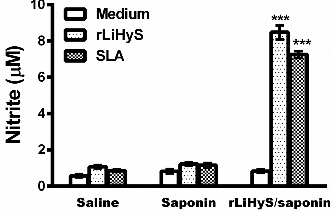
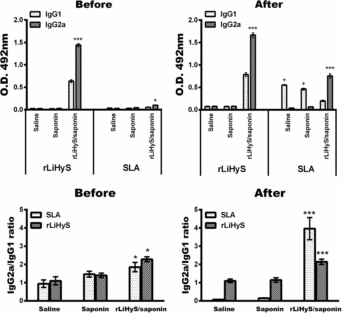
As an indicator of NO production, the nitrite presence was assayed in the in vitro cultures after challenge (Fig. 6). In the results, the nitrite secretion was significantly higher in rLiHyS/saponin-vaccinated mice, when either rLiHyS or SLA was used as a stimulus (P < 0·0001), as compared to the results found in the saline or saponin groups, in which a minimum nitrite presence was verified. Regarding humoral response, the protein- and parasite-specific IgG1 and IgG2a isotype productions were evaluated before and after infection (Fig. 7), and results showed that, in both conditions, rLiHyS/saponin-vaccinated mice produced higher IgG2a levels, when compared to the IgG1 levels (P < 0·05 and P < 0·0001, respectively). Moreover, the ratio between the IgG2a/IgG1 isotypes was calculated, and results showed a polarized Th1 immune response (P < 0·05 and P < 0·0001, respectively) in the protected animals. On the other hand, isotype production in the saline and saponin groups before infection was similar when both antigens were tested, although the anti-parasite IgG1 isotype levels were higher after challenge, when compared to the IgG2a levels in these groups (P < 0·0001).
Nitrite production. Spleen cells obtained from the saline, saponin, or rLiHyS/saponin-vaccinated mice, 60 days after challenge infection, were unstimulated (medium) or stimulated with rLiHyS or SLA (10 and 25 µg mL−1, respectively), for 48 h at 37 °C in 5% CO2. Next, nitrite production was analysed by the Griess method. Bars represent the mean ± standard deviation of the groups. ***Indicate statistically significant difference in relation to the saline and saponin groups (P < 0·0001).

Production of protein- and parasite-specific IgG2a and IgG1 isotype antibodies before and after infection. BALB/c mice received saline or were immunized with saponin or rLiHyS/saponin. Thirty days after the last immunization, sera samples were collected (Before). In addition, remaining animals were challenged with Leishmania infantum promastigotes and followed for 60 days, at which time sera samples were collected (After). The anti-protein and anti-parasite IgG2a and IgG1 isotypes antibody levels were calculated, as were the ratios between IgG2a and IgG1 values. Bars represent the mean ± standard deviation of the groups. *Indicates statistically significant difference in relation to the saline and saponin groups (P < 0·05). ***Indicate statistically significant difference in relation to the saline and saponin groups (P < 0·0001). +Indicates statistically significant difference in relation to the rLiHyS/saponin group (P < 0·01).

The rLiHyS/saponin vaccine induces partial protection in BALB/c mice against L. infantum
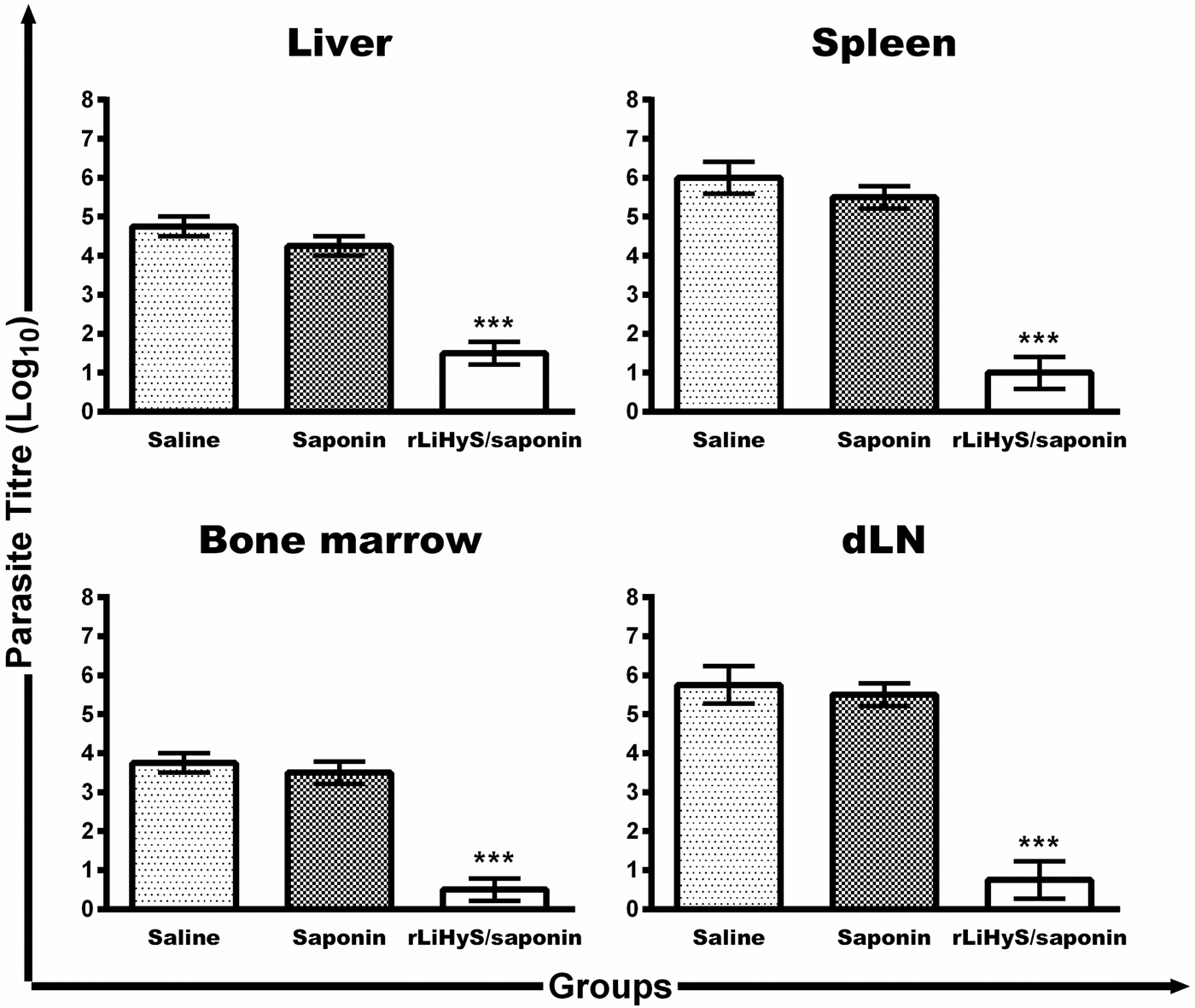
The protective efficacy induced by immunization with rLiHyS/saponin in BALB/c mice was investigated against an L. infantum challenge through the evaluation of parasite burden, 60 days after infection. In the results, using the limiting dilution technique (Fig. 8), rLiHyS/saponin-vaccinated mice showed significant reductions in the parasite load in the liver (3·3- and 2·8-log reductions, respectively), spleen (5·0- and 4·5-log reductions, respectively), BM (3·3- and 3·0-log reductions, respectively) and dLNs (5·0- and 4·8-log reductions, respectively), when compared to the saline and saponin groups (P < 0·0001). Evaluating the results obtained by immunization with rLiHyS administered with or without an adjuvant, rLiHyS/saponin-vaccinated and challenged mice showed significant reductions in the parasite load in the liver, spleen, BM and dLNs (2·8-, 4·2-, 2·7- and 4·5-log reductions, respectively), as compared to the results found in the rLiHyS group (P < 0·0005). Results obtained using the limiting dilution technique were corroborated by qPCR assay when the spleen of the animals was evaluated, since the immunization with rLiHyS/saponin reduced the parasite load in this organ in the order of 66·0% and 65·0%, when compared to the values found in the saline and saponin groups, respectively (P < 0·0001).
Parasite burden evaluated in the infected and vaccinated animals. BALB/c mice inoculated with saline or immunized with saponin or rLiHyS/saponin were infected with Leishmania infantum promastigotes. Sixty days after, the parasite load in the liver, spleen, bone marrow (BM) and paws’ draining lymph nodes (dLNs) was evaluated by a limiting-dilution technique. Bars represent the mean ± standard deviation of the groups. ***Indicate statistically significant difference in relation to the saline and saponin groups (P < 0·0001).

Cell localization of LiHyS in the L. infantum parasite
To investigate the cell location of LiHyS in L. infantum, an immunofluorescence experiment was performed using stationary-phase promastigotes and an anti-rLiHyS IgG antibody. In the results, confocal images showed that this protein is possibly located in the parasite's flagellar pocket (Fig. 9). In this context, it could be speculated that it would become part of the Leishmania flagellum machinery, and its location could be additionally investigated by using other flagellum apparatus proteins in new studies.
Cell location of LiHyS protein. Confocal fluorescence images were obtained using a negative control (upper panel), and the location of LiHyS in Leishmania infantum promastigotes is shown (green colour), stained with Hoechst (blue colour, lower panel). BF represents the bright field. The merged image shows the location of LiHyS (A). The three-dimensional reconstruction (X–Z sections) is shown at the top, and Y–Z sections are shown at the right for each image (B). Images are representative of two independent experiments. Scale bar: 5 µm.
LiHyS inhibits the Leishmania infectivity in murine macrophages
Aiming to show an immunotherapeutic role of LiHyS on the Leishmania invasion in mammalian cells, an experiment was performed using murine macrophages and parasites that were pre-treated with the anti-rLiHyS antibody. In the results, anti-rLiHyS IgG pre-treated promastigotes infect 46·6, 54·8 and 63·2% of the macrophages, and the number of recovered amastigotes was of 1·0 ± 0·3, 1·5 ± 0·4 and 2·0 ± 0·2, respectively, when IgG concentrations of 50·0, 10·0 and 5·0 µg mL−1 were used. On the other hand, when using a non-immune IgG antibody, the percentage of infection was of 84·5, 88·7 and 90·6%, and the number of recovered amastigotes was of 5·6 ± 0·4, 6·2 ± 0·3 and 6·9 ± 0·6, respectively, when the same IgG concentrations were used. Parasites that were not pre-incubated with antibodies were able to infect 94·5% of the macrophages, and 7·8 amastigotes per phagocytic cell were recovered.
Discussion
Leishmania antigenic proteins expressed in the L. infantum promastigote and amastigote stages were recently identified by an immunoproteomic approach using antibodies in VL dog sera, including some characterized as hypothetical (Coelho et al. Reference Coelho, Oliveira, Valadares, Chávez-Fumagalli, Duarte, Lage, Soto, Santoro, Tavares, Fernandes and Coelho2012). The fact that antibodies in infected animal sera, but not those from healthy animals, had recognized these molecules indicates that they are expressed by the parasites during the active disease and can be considered relevant to the infected host's immune response (Fernandes et al. Reference Fernandes, Coelho, Machado-Coelho, Grimaldi and Gazzinelli2012). In this light, in the present work, the LiHyS gene, which was identified in L. infantum promastigotes in the cited immunoproteomic study, was cloned, and its recombinant version was evaluated as a diagnostic or vaccine candidate against VL.
Studies have showed that Leishmania virulence factors can be found in the parasite promastigote stage (Yao et al. Reference Yao, Li, Donelson and Wilson2010; Dupé et al. Reference Dupé, Dumas and Papadopoulou2015). In this line, Leishmania macromolecules, such as tubulins (Werbovetz et al. Reference Werbovetz, Brendle and Sackett1999), protein disulphide isomerase (Achour et al. Reference Achour, Chenik, Louzir and Dellagi2002), sphingolipids (Heung et al. Reference Heung, Luberto and Del Poeta2006), lipophosphoglycans (Forestier et al. Reference Forestier, Gao and Boons2015), among others, have been characterized as virulence factors. Taking this fact into account, the protective efficacy achieved by immunization with promastigote antigens often results in a more effective immune response, which will eliminate these forms soon after parasite infection by the infected sandflies. In this context, several studies have been developed using promastigote proteins as vaccine candidates against leishmaniasis (Bhowmick et al. Reference Bhowmick, Ravindran and Ali2007; Chamakh-Ayari et al. Reference Chamakh-Ayari, Bras-Gonçalves, Bahi-Jaber, Petitdidier, Markikou-Ouni, Aoun, Moreno, Carrillo, Salotra, Kaushal, Negi, Arevalo, Falconi-Agapito, Privat, Cruz, Pagniez, Papierok, Rhouma, Torres, Lemesre, Chenik and Meddeb-Garnaoui2014; Thakur et al. Reference Thakur, Kaur and Kaur2015; Petitdidier et al. Reference Petitdidier, Pagniez, Papierok, Vincendeau, Lemesre and Bras-Gonçalves2016).
In the present work, LiHyS was identified in L. infantum promastigotes and by an immunofluorescence assay, which proved to possibly be located in the parasite's flagellar pocket, in turn allowing one to speculate about its involvement in Leishmania motility and, consequently, as a parasite virulence factor. In fact, when an anti-rLiHyS IgG antibody was pre-incubated with Leishmania promastigotes, and these pre-treated parasites were used to infect murine macrophages, significant reductions in the infection percentage were obtained, and a lower number of amastigotes was recovered by the infected phagocytic cells, when compared with the values found when parasites were untreated or pre-treated with a non-immune IgG antibody. As a consequence, a role of rLiHyS in the L. infantum infectivity could be postulated, although additional studies are necessary to be performed to elucidate this protein's biological action.
It has been reported that, to achieve a protective immunity against VL, a Th1 cell response, based on the production of cytokines such as IFN-γ, IL-12, TNF-α, GM-CSF, among others, is required (Zadeh-Vakili et al. Reference Zadeh-Vakili, Taheri, Taslimi, Doustdari, Salmanian and Rafati2004; Mizbani et al. Reference Mizbani, Taheri, Zahedifard, Taslimi, Azizi, Azadmanesh, Papadopoulou and Rafati2009; Saljoughian et al. Reference Saljoughian, Taheri, Zahedifard, Taslimi, Doustdari, Bolhassani, Doroud, Azizi, Heidari, Vasei, Namvar, Papadopoulou and Rafati2013; Martins et al. Reference Saljoughian, Taheri, Zahedifard, Taslimi, Doustdari, Bolhassani, Doroud, Azizi, Heidari, Vasei, Namvar, Papadopoulou and Rafati2016; Reed et al. Reference Reed, Coler, Mondal, Kamhawi and Valenzuela2016). On the other hand, susceptibility to disease is associated with the reduction of this immune profile, in parallel to the development of Th2 immunity, based on the production of cytokines, such as IL-4 and IL-10 (Goto et al. Reference Goto, Bhatia, Raman, Liang, Mohamath, Picone, Vidal, Vedvick, Howard and Reed2011; Agallou et al. Reference Agallou, Smirlis, Soteriadou and Karagouni2012; Gupta et al. Reference Gupta, Kumar, Kushawaha, Tripathi, Joshi, Sahasrabuddhe, Mitra, Sundar, Siddiqi and Dube2014). In our study, the association between rLiHyS and saponin proved to be immunogenic in vaccinated mice, since higher levels of IFN-γ, IL-12 and GM-CSF were found before and after infection. These findings are related to a decrease in the parasite load in the infected and rLiHyS/saponin-vaccinated animals, represented by significant reductions in parasitaemia in the liver, spleen, BM and dLNs, when compared to results obtained in the other groups. Although rLiHyS alone was not protective against the challenge, most of the tested immunogens, when used in different mammalian hosts, normally require the association of immune adjuvants to improve their protective efficacy (Bayih et al. Reference Bayih, Daifalla and Gedamu2014; Eskandari et al. Reference Eskandari, Talesh, Parooie, Jaafari, Khamesipour, Saberi, Abbasi and Badiee2014; Thakur et al. Reference Thakur, Kaur and Kaur2015; Petitdidier et al. Reference Petitdidier, Pagniez, Papierok, Vincendeau, Lemesre and Bras-Gonçalves2016). In this context, our antigen also fulfils the requirements to be considered as a vaccine candidate against VL.
An ideal vaccine will have the potential to protect mammalian hosts against Leishmania infection, as well as to reduce transmission between reservoir hosts, such as dogs and humans, in areas where parasite transmission is considered zoonotic, in addition to allowing for a more effective control of the disease (Saldarriaga et al. Reference Saldarriaga, Travi, Park, Perez and Melby2006; Stockdale and Newton, Reference Stockdale and Newton2013; Sundar and Singh, Reference Sundar and Singh2014). In the human disease, a depressed cell-mediated immune response, characterized by the failure of patients’ PBMCs to respond and proliferate when stimulated by Leishmania antigens, is commonly found, as is the production of cytokines, such as IFN-γ and IL-12 (Peruhype-Magalhães et al. Reference Peruhype-Magalhães, Martins-Filho, Prata, Silva, Rabello, Teixeira-Carvalho, Figueiredo, Guimarães-Carvalho, Ferrari and Correa-Oliveira2005). On the other hand, PBMCs from cured and treated patients usually produce pro-inflammatory cytokines when in vitro stimulated by parasite antigens (Singh et al. Reference Singh, Stober, Singh, Blackwell and Sundar2012).
The present study's data suggest that rLiHyS could be considered a potential target for an intervention in humans, due to the fact that a high lymphoproliferation index was found when PBMCs purified from healthy subjects or VL patients were in vitro stimulated with the recombinant protein. Although the experiments developed in this study do not allow one to infer that rLiHyS is immunogenic for human PBMCs, even though mitogen activity in cells derived from both individual classes was identified, additional studies are warranted in an attempt to clarify these findings, in turn allowing one to infer the possibility of an immunogenic action of this recombinant protein in human VL, thus opening the door to this antigen's use as a future immunogen to fight against this disease in this mammalian host.
Serological diagnosis for VL has used the rK39 antigen in clinical practice (Matlashewski et al. Reference Matlashewski, Das, Pandey, Singh, Das, Ghosh, Pandey and Das2013; Ghosh et al. Reference Ghosh, Hasnain, Ghosh, Hossain, Baker, Boelaert, Rijal and Mondal2015). However, studies have shown that this protein was not highly efficient, exhibiting cross-reactivity with leishmaniasis-related pathologies, as well as with non-infected subjects living in endemic areas of disease, in turn leading to false-positive results and hampering the specificity of the serological tests (Gadisa et al. Reference Gadisa, Custodio, Cañavate, Sordo, Abebe, Nieto, Chicharro, Aseffa, Yamuah, Engers, Moreno and Cruz2012; Aronson, Reference Aronson2017). In addition, since this commercial kit was developed to detect protein-specific antibodies for the L. donovani complex, false-negative results have been found when L. infantum-infected subject sera are tested (Mohammadiha et al. Reference Mohammadiha, Mohebali, Haghighi, Mahdian, Abadi, Zarei, Yeganeh, Kazemi, Taghipour and Akhoundi2013; Costa et al. Reference Costa, Salles, Santos, Ramos, Lima, Lima, Portela, Chávez-Fumagalli, Duarte, Menezes-Souza, Machado-de-Ávila, Silveira, Magalhães-Soares, Goulart and Coelho2017). In our study, VL patient sera recognized rLiHyS with high specificity and sensitivity values, highlighting the possibility of using our antigen to diagnose disease. However, the low number of patient sera, as well as sera from leishmaniasis-related diseases developed in patients, such as malaria, tuberculosis, among others, could be considered limitations of our work, since new samples could alter the sensitivity and specificity values found to rLiHyS. As a consequence, additional experiments using a larger and more varied serological panel should be performed in an attempt to corroborate the diagnostic efficacy found in this study regarding this recombinant protein.
In conclusion, the present study's data indicated that LiHyS, a Leishmania conserved protein, besides its antigenicity in canine and human VL, was also immunogenic and able to confer partial protection against L. infantum infection in BALB/c mice. In addition, a lymphoproliferation response was found when rLiHyS was used to stimulate PBMCs from healthy subjects and VL patients, thus highlighting the possibility to evaluate it as an immunogen against human disease.
Supplementary material
The supplementary material for this article can be found at https://doi.org/10.1017/S0031182017001731.
Financial support
This work was supported by grants from Pró-Reitoria de Pesquisa da Universidade Federal de Minas Gerais (Edital 02/2017), FAPEMIG (CBB-APQ-00819-12 and CBB-APQ-01778-2014) and CNPq (APQ-482976/2012-8, APQ-488237/2013-0 and APQ-467640/2014-9). MAC-F is a grant recipient of FAPEMIG/CAPES. EAFC is a grant recipient of CNPq. The authors would like thank the CAPI (Image Acquisition and Processing Center), from ICB, UFMG; where microscopic data showed in this work were obtained.