Paediatric pulmonary hypertension, once thought to be a rare condition, has been seem to have increased prevalence in the last 10 years.Reference Abman, Hansmann and Archer 1 This increase has been driven in part by a larger number of children surviving with what were previously life-threatening conditions such as CHD and chronic lung disease of prematurity.Reference Mourani, Sontag and Younoszai 2 , Reference Kozlik-Feldmann, Hansmann, Bonnet, Schranz, Apitz and Michel-Behnke 3 The incidence of pulmonary hypertension among children with chronic lung disease of prematurity alone is as high as 40%. The secondary diagnosis of pulmonary hypertension has also been noted in association with other disorders as well.Reference Zuckerman and Rosenzweig 4 , Reference Hutson, Baerg, Deming, St Peter, Hopper and Goff 5 Despite recognition of the linkage between cancer and pulmonary hypertension in adults and reporting of the pulmonary side effects from cancer and treatment in children, little information exists on the association of pulmonary hypertension and cancer in children.Reference Versluys and Bresters 6 , Reference Ranchoux, Günther and Quarck 7 Here we report on a cohort of children whose cancer treatment was complicated by pulmonary hypertension, emphasising the effectiveness of pulmonary hypertension therapy and recoverability.
Methods
We retrospectively evaluated the clinical records of patients with evidence of elevated pulmonary pressures and previously identified paediatric tumors. Patients with evidence of significant left ventricular systolic dysfunction or large pulmonary thromboembolisms were not analysed in this report. A total of four patients were found to have evidence of pulmonary hypertension after a diagnosis of paediatric malignancy. We reviewed clinical history, laboratory studies, echocardiograms, cardiac catheterisations, and CT scans. We tabulated dosing of chemotherapeutic drugs and radiation to the chest. This retrospective review was approved by the Human Research Protection Office at Washington University.
Results
Patient 1 was a 2-year-old former 24-week estimated gestational age female with history of mild bronchopulmonary dysplasia but was currently at home without the need for respiratory support. She presented to the emergency room with respiratory distress and seizures and was found to have a nasopharyngeal mass. After excisional biopsy and additional imaging, she was diagnosed with nasopharyngeal embryonal rhabdomyosarcoma with metastasis to multiple skeletal sites. Her echocardiogram before chemotherapy showed normal biventricular function with no signs of elevated pulmonary arterial pressures. She underwent chemotherapy that included induction with vincristine, irinotecan, and steroids, with subsequent two rounds of cyclophosphamide, doxorubicin, etoposide, ifosfamide, and vincristine. After 4 months and her fifth round of chemotherapy, she was admitted to the ICU with fever and neutropenia and signs of cardiogenic shock and cardiomegaly on a chest radiograph. An echocardiogram at this time revealed a dilated right ventricle with decreased function, mild tricuspid regurgitation though insufficient to estimate right ventricular pressure, and bowing of the interventricular septum into the left ventricle, consistent with suprasystemic right ventricular pressures. Her B-type natriuretic peptide was 5054 pg/ml (normal 0–39 pg/ml). On initiation of inhaled nitric oxide, milrinone, and epinephrine, clinical and echocardiographic indicators improved within 24 hours. Upon weaning of inhaled nitric oxide and inotropes, she again showed clinical and echocardiographic signs of elevated pulmonary pressures. With re-initiation of inhaled nitric oxide and starting a methylprednisone burst, her echocardiogram again normalised. She was then started on sildenafil and tolerated weaning of nitric oxide. She was subsequently found to desaturate during the night and she was discharged on nasal cannula oxygen and sildenafil and no further evidence of pulmonary hypertension. She underwent additional chemotherapy with vincristine and irinotecan as well as nasopharyngeal directed radiation with no worsening of echocardiograms. Though she was able to wean off the supplemental oxygen, she had relapse of her cancer and passed away before attempted wean of her sildenafil.
Patient 2 was a 3-month-old male who presented with an enlarging head circumference found to have a pineal blastoma. An initial attempt at surgical resection was aborted, owing to the highly vascular nature of the tumour and subsequently developed seizures with a small subdural haematoma. Initial echocardiogram showed normal ventricular function with no signs of elevated right ventricular pressures. He was given inpatient chemotherapy consisting of methotrexate, vincristine, etoposide, cyclophosphamide, and cisplatin. During his hospitalisation, which was complicated by hepatic dysfunction with hepatic veno-occlusive disease and systemic hypertension treated with amlodipine, he developed increased work of breathing and a B-type natriuretic peptide of 1900 pg/ml and was admitted to the ICU. A chest CT scan performed showed “ground glass opacities.” At this time, an echocardiogram showed signs of systemic right ventricular pressures including a dilated right ventricle with decreased function, a flattened interventricular septum, and right to left shunting across the patent foramen ovale confirmed by an agitated saline contrast study. A study performed 1 month previously showed no signs of elevated right ventricular pressure. He was started on inhaled nitric oxide through a high-flow nasal cannula and intravenous milrinone. He appeared to improve on inhaled nitric oxide over the next few days but developed methemoglobinemia. Upon weaning off inhaled nitric oxide, he developed worsening right ventricular dysfunction, flattened interventricular septum, and an elevated tricuspid regurgitation velocity suggesting systemic right ventricular pressures. At this point, he was then started on sildenafil and a methylprednisone burst with improvement. He was weaned off steroids and sildenafil over the subsequent month and never developed further clinical or echocardiographic signs of pulmonary hypertension despite additional chemotherapy of cisplatin and vincristine. He had progression of his oncologic illness with subsequent death at 13 months of age.
Patient 3 was a 4-year-old male who presented with systemic hypertension and found to have a large mass arising from his left adrenal gland and encasing his left renal artery. He was diagnosed with grade IV metastatic neuroblastoma with local extension and metastasis to his spleen and bones. He was initiated on cyclophosphamide and topotecan and then six rounds of chemotherapy with cisplatin, etoposide, doxorubicin, and vincristine. His multiple echocardiograms to this point showed no evidence of right ventricular dysfunction or elevated pressures. He then underwent an autologous stem cell transplant with subsequent 1 month of radiation therapy directed to the left infra-diaphragmatic region (3600 centigray) with only a small portion of the left lung receiving significant exposure. One week after completion of radiation treatment, he presented to oncology clinic with tachycardia, tachypnoea, and increased work of breathing and was admitted to the ICU. He had a chest CT that showed pulmonary oedema, bilateral pleural effusions, and cardiomegaly without a pulmonary embolism or interstitial pneumonitis. Though only three months after a normal echocardiogram, this study showed a large pericardial effusion, right atrial and right ventricular dilatation with decreased function, bowing of the ventricular septum causing “pancaking” of the left ventricle, giving evidence of supra-systemic pulmonary hypertension. There was also evidence for mild left ventricular diastolic dysfunction on tissue Doppler imaging. As he was in cardiogenic shock, he was started on inhaled nitric oxide, epinephrine, milrinone, and aggressive diuresis. His blood count revealed no signs of microangiopathic haemolytic anaemia and was inconsistent with thrombotic microangiopathy. He was also started on intravenous methylprednisone for pulmonary hypertension. He had gradual improvement in his examination and echocardiogram allowing inotropes and inhaled nitric oxide to be discontinued. He subsequently was weaned off steroids and remained on only a small amount of diuretics. After the clinical stabilisation, he was taken for a cardiac catheterisation which showed mild pulmonary hypertension with a mean pulmonary artery pressure of 26 mmHg, indexed pulmonary vascular resistance of 3.2 Woods units×m2, and pulmonary capillary wedge pressures of 8 mmHg. As his pulmonary pressures were nearly normal during the catheterisation, no acute vasodilator testing was performed. He continued to do well during the hospitalisation with no additional signs of cardiovascular compromise and discharged for outpatient treatment.
He was admitted to the ICU during treatment with dinutuximab and interleukin-2 administration because of concern for fluid shifts associated with this medication and his recent history of cardiovascular dysfunction. He had a normal baseline echocardiogram and a Swan-Ganz catheter was placed. After 24 hours of medication, he had fever, tachycardia, tachypnoea, and increased mean pulmonary artery pressure up to 54 mmHg, pulmonary wedge pressures of 9 mmHg, and indexed pulmonary vascular resistance of 14.0 Woods units×m2. A repeat echocardiogram showed no pericardial effusion, but increased tricuspid regurgitation with an elevated TR jet, and normal right ventricular function. He had no improvement with nasal cannula oxygen but saw immediate improvement on initiation of inhaled nitric oxide to mean pulmonary artery pressure of 40 mmHg, pulmonary wedge pressure of 8 mmHg, and indexed pulmonary vascular resistance of 5.5 Woods units×m2. These further normalised over the next 12 hours. He was started on sildenafil after the completion of the chemotherapeutics to minimise effects of pulmonary hypertension during subsequent chemotherapy. Subsequent rounds of treatment with dinutuximab and interleukin-2 again worsened his pulmonary hypertension, which was again responsive to inhaled nitric oxide and resolved upon completion of the chemotherapy cycle. In between rounds of therapy, he remained stable without clinical or echocardiographic signs of pulmonary hypertension.
Patient 4 was a 14-month-old female with haematuria and a right flank mass eventually diagnosed with stage IV high-risk neuroblastoma. Before chemotherapy, she had an echocardiogram that showed normal biventricular function and no signs of elevated right ventricular pressure. She received induction with carboplatin and etoposide and subsequently received cyclophosphamide, topotecan, cisplatin, etoposide, doxorubicin, and vincristine. Owing to persistence of disease, she then underwent an autologous stem cell transplant, local control with surgery, and photon beam radiation to her pelvis. After 9 months of adjuvant chemotherapy with dinutuximab and isotretinoin, she was admitted to the ICU with new desaturation, hypotension, and respiratory distress. Her physical examination revealed hepatomegaly and laboratory studies showed an elevated B-type natriuretic peptide of 3378 pg/ml. Her blood counts were inconsistent with thrombotic microangiopathy. Despite a normal echocardiogram 3 weeks before admission, a repeat study showed increased right atrial size though normal velocity to tricuspid regurgitation jet. CT scan of her chest showed no pulmonary embolus and scattered ground glass opacities. She was intubated, started on nitric oxide and epinephrine, with improved perfusion and oxygenation. With diuresis and initiation of methylprednisone for possible interstitial pneumonitis, she had improved ventilation, oxygenation, and systemic perfusion. During attempts to wean off steroids and nitric oxide, she had worsening clinical, laboratory, and echocardiographic signs of pulmonary hypertension prompting evaluation by cardiac catheterisation. At that time she was found to have mean pulmonary artery pressure of 30 mmHg, left ventricular end diastolic pressure of 9 mmHg, and indexed pulmonary vascular resistance of 5.6 Woods units×m2, which improved with inhaled nitric oxide testing to mean pulmonary artery pressure of 21 mmHg, no change in left ventricular end diastolic pressure, and indexed pulmonary vascular resistance of 3.1 Woods units×m2. With addition of amlodipine and continued sildenafil, she was weaned off inhaled nitric oxide. She underwent subsequent rounds of radiation and chemotherapy with interleukin-2 and dinutuximab without worsening of pulmonary hypertension. She has had no further issues and is currently weaning off sildenafil.
Discussion
This report is the first to outline the severity of pulmonary hypertension symptoms in children undergoing treatment for cancer. All four of these patients presented with cardiovascular compromise requiring intensive care support and prompt initiation of pulmonary hypertension-directed therapy. Despite the small cohort of children reported, important features regarding their malignancies, therapeutic regimens, as well as treatment course may prove applicable to any child undergoing chemotherapy.
Why these children developed pulmonary hypertension is unclear. Interestingly, all four had solid tumors with three of them of neuroectodermal origin. However, the fact that all four children had normal echocardiograms free of pulmonary hypertension findings, before initiation of treatment, makes the chemotherapeutic drugs more likely the cause of their pulmonary hypertension rather than underlying disease. Previous studies have implicated several chemotherapeutic agents in association with pulmonary hypertension, including alkylating agents, antimetabolites, kinase inhibitors, and cytotoxic antibiotics agents.Reference Ranchoux, Günther and Quarck 7 , Reference Ballout, Manshad and Okwuosa 8 Alkylating agents, in particular cyclophosphamide, have been most often implicated in previous reports and were used in greater than half of the chemotherapy-induced cases of pulmonary hypertension in a report from the French Pulmonary Hypertension Network.Reference Ranchoux, Günther and Quarck 7 Cyclophosphamide and other alkylating agents were part of the treatment protocols for each of the patients reported in this cohort. However, the vast majority of children treated with alkylating drugs do not develop pulmonary hypertension making the association less clear.
Notably, these patients had a normal echocardiogram without signs of elevated right ventricular pressures for a median of 2 months before the initial diagnosis. Despite the severe initial presentations requiring intensive care admissions, the severe pulmonary hypertension symptoms were remarkably responsive to a combination of halting current chemotherapeutic regimens and initiation of inhaled nitric oxide, sildenafil, and prednisone. Kinase inhibitors, in particular dasatinib, have described side effects of pulmonary complications including rapid onset of pulmonary hypertension with rapid resolution on discontinuation of medications in adults.Reference Ballout, Manshad and Okwuosa 8 , Reference Montani, Bergot and Günther 9 However, none of the children in this cohort were exposed to dasatinib or other kinase inhibitors. Two of the patients had medical history that may be associated with the development of pulmonary hypertension. Although she was clinically without signs of pulmonary dysfunction before her cancer diagnosis, Patient 1 did have bronchopulmonary dysplasia, a known risk factor for the development of pulmonary hypertension.Reference Al-Ghanem, Shah, Thomas, Banfield, El Helou, Fusch and Mukerji 10 This may indicate that chemotherapeutic agents were an additional trigger for this patient. Also, of note, Patient 2 was also found to have hepatic veno-occlusive disease shortly before pulmonary hypertension symptoms and diagnosis along with radiographic evidence with findings consistent with pulmonary veno-occlusive disease. Though it seems possible that pulmonary veno-occlusive disease caused the pulmonary hypertension, the marked symptomatic improvement with pulmonary vasodilators is inconsistent with a post-capillary pulmonary hypertension disease aetiology. Definitive diagnosis by pathology was not pursued given overall pulmonary hypertension improvement. Paediatric oncologic patients have been associated with thrombotic microangiopathy after haematopoietic stem cell transplant with pulmonary hypertension, though in these patients the disease process was unresponsive to pulmonary vasodilators and heralded rapid clinical deterioration.Reference Jodele, Hirsch, Laskin, Davies, Witte and Chima 11
Glucocorticoids were used as part of the pulmonary hypertension treatment protocols and were linked to the rapid improvement. The association between inflammation and pulmonary hypertension has been established in human autopsy studies, animal models of pulmonary hypertension, and referenced by numerous studies.Reference Price, Wort and Perros 12 In fact, pulmonary hypertension in animal models caused by an alkylating agent, monocrotaline, has both identified inflammation as a pathway for development of pulmonary hypertension and demonstrated therapeutic benefit through treatment with glucocorticoids.Reference Wang, Wang, Chen, Li, Hao, Jin and Han 13 , Reference Price, Montani and Tcherakian 14 Though not commonly used, the modulatory effect and clinical improvement in pulmonary hypertension has previously been reported.Reference Ogawa, Nakamura and Mizoguchi 15 , Reference Aggarwal and Grady 16 After considering the effects on oncologic therapeutic regimens, a short course of glucocorticoids may be an important auxiliary option for these children.
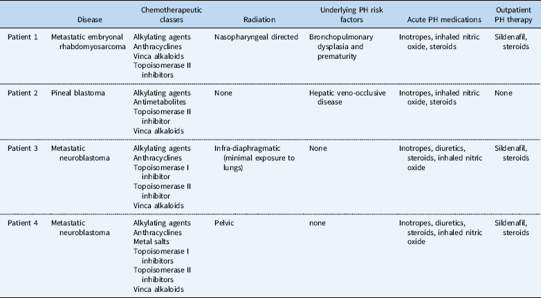
This single-centre retrospective description of cancer and pulmonary hypertension is limited by a small patient cohort and is unable to isolate an incidence of the disease. However, these cases highlight an important association previously unreported in the paediatric literature, despite significant attention to pulmonary complications.Reference Versluys and Bresters 6 A singular pathophysiological relationship will unlikely be identified given the heterogeneous patient medical history, tumour types, and treatment courses of patients who develop this complication. However, this report indicates that children treated for solid tumours who develop respiratory distress, evidence of right heart failure, or cardiogenic shock must be evaluated for pulmonary hypertension. After ruling out pulmonary embolism, echocardiography is able to provide rapid, non-invasive screening for pulmonary hypertension to help guide further clinical decisions. Prompt diagnosis of pulmonary hypertension and treatment with pulmonary vasodilators and steroids may allow rapid resolution of symptoms allowing chemotherapy and radiation regimens to continue (Table 1).
Important patient features including underlying risk factors and pulmonary hypertension treatment regimens.

Acknowledgements
None.
Financial Support
This research received no specific grant from any funding agency, commercial, or not-for-profit sectors.
Conflicts of Interest
None.
Ethical Standards
The authors assert that all procedures contributing to this work comply with the ethical standards of the relevant national guidelines on human experimentation and with the Helsinki Declaration of 1975, as revised in 2008, and has been approved by the Washington University School of Medicine institutional committees.