




No CrossRef data available.
Article contents
Adults acquire filarial infection more rapidly than children: a study in Indonesian transmigrants
Published online by Cambridge University Press: 12 July 2001
Abstract
To dissociate the influence of host age from length of exposure on acquisition of filarial infection, we examined the development of microfilaraemia and anti-filarial IgG4 in all ages of a naive population that became suddenly exposed to Brugia malayi as a result of transmigration. Responses in 247 transmigrants, who had settled for periods of several months up to 6 years in their new homesteads, were compared with those of 133 life-long residents. As shown in earlier studies, anti-filarial IgG4 increased with age in the indigenous population, whose age is equivalent to length of exposure. However, by examining transmigrants, it became clear that development of specific IgG4 was influenced by age, since levels of this antibody were consistently higher in transmigrant adults than in transmigrant children, despite an equal length of exposure to filarial infection. Examining microfilaraemia, it was confirmed that infection establishes more rapidly in adults than in children.
Information
- Type
- Research Article
- Information
- Copyright
- © 2001 Cambridge University Press
INTRODUCTION
In areas endemic for lymph dwelling nematodes of Wuchereria or Brugia species, filarial infections are persistent and result in a spectrum of clinical manifestations that depend on several factors. In utero exposure to filarial parasites (Weil et al. 1983; Lammie et al. 1991; Steel et al. 1994; King et al. 1998), genetic make-up of the host (Ottesen et al. 1981; Chan et al. 1984; Yazdanbakhsh et al. 1995) and, in addition to these pre-determined factors, age or increasing exposure can influence acquisition of filarial infection and development of pathology (Day, Gregory & Maizels, 1991a; Day et al. 1991b). In a recent study we have demonstrated that reactivity to filarial antigens begins early in life (18 months) as measured by anti-filarial IgG4 and IgE and is differentially influenced by gender and transmission intensity (Terhell et al. 2000). Other studies have shown that the intensity of infection levels off with age or even in some cases declines in older age groups, which may indicate acquired immunity (Maizels et al. 1993). Longitudinal measurement of circulating antigens in children versus adults has shown that whereas children acquire new infections, adults with low or high worm burden resist new incoming larvae (Day et al. 1991b). One way to interpret these data is that with increasing exposure to filarial infection, immunity develops to larval stages. However, as the immune system matures and evolves with increasing age (Wade, Green-Johnson & Szewczuk, 1988; Horan & Ashcroft, 1997), the observed changes in filarial infection over years of exposure (from childhood to adulthood) may be caused by an age-dependent mechanism that is not necessarily exposure driven. With the increasing interest in the role that innate immune mechanisms play to control infection (Fearon & Locksley, 1996), understanding of how host age per se may influence acquisition of infection becomes relevant to defining components which contribute to immunity.
Most studies on the dynamics of filarial infection have been carried out in populations that are life-long residents of endemic areas. In such situations, history of exposure to parasites cannot be dissociated from age-related variation in immunity to filarial infection. This is only possible when a naive population of all ages becomes exposed to filarial infection abruptly. In Indonesia, the governmental transmigration programme relocates groups of people from overcrowded islands such as Java and Bali to under-populated and less-developed areas on more remote islands, which are often endemic for a multiplicity of infections. This transmigration policy provides a unique situation that allows the study of development of infection and immunity in individuals with varying age but with an equal length of exposure to infections.
The objective of the present study was to investigate the acquisition of infection with increasing length of exposure in new settlers, by measuring not only microfilariae in night blood but also by examining anti-filarial IgG4, which is an immunological marker of active filarial infection and has increased sensitivity compared to classical parasitological assays (Ottesen et al. 1985; Kurniawan et al. 1993; Haarbrink et al. 1995). We have studied 6 transmigration units in South-Sulawesi, Indonesia, which were established between 1990 and 1996 and 2 neighbouring villages of native Sulawesians in a region endemic for B. malayi. Both children and adults were included in the study in an attempt to dissociate the influence of age from exposure in development of filarial infection.
MATERIALS AND METHODS
Description of the study area
The survey was conducted in Budong-budong, a district of Mamuju Regency in South-Sulawesi, Indonesia, which is endemic for periodic nocturnal B. malayi (Slee, 1930; Partono et al. 1977). The transmigrants had travelled to their new homesteads in groups coming from the same village or region in Bali or Lesser Sunda islands as part of the government-sponsored relocation programme. Each year a new settlement was founded close to the former one (between 10 and 20 kilometres) which accommodated groups of transmigrants from 2–3 different regions together with migrants from Polmas, an over-populated area in South Sulawesi, to promote integration of different tribes. A total of 6 transmigrant units, settled between several months and 6 years prior to the survey, were included in the study together with 2 villages of indigenous Sulawesians, which were situated closely to the transmigration areas. Transmigrants from areas where filariasis is endemic (South Sulawesi and Lesser Sunda islands (Joesoef & Cross, 1978) were excluded from the analysis.
In co-operation with the medical doctors and health workers of the local District Health Centre and the head of each transmigration unit or village, all residents were informed about the study and invited to participate. Informed consent was obtained from all study participants or parents of underage children before parasitological studies and blood withdrawal in accordance with the guidelines of the Indonesian Department of Health and Human Services.
A total number of 247 transmigrants and 133 life-long residents (LLR) were enrolled in the study. Transmigrants were grouped together according to year of arrival in the new area. Table 1 shows the number of cases, number of children (aged[les ]15 years) to adults (aged 16 years and older), sex distribution and microfilaria prevalence of transmigrants (according to length of residence) and indigenous Sulawesians. The number of children that participated in the survey varied per village and more males than females registered for the study. However, the average age of transmigrant children (range 6–15, mean = 10.2) was comparable to the indigenous children (range 4–15, mean = 10.4), and the same was true for adults in the transmigrant (range 16–70, mean = 32.6) and indigenous study population (range 16–60, mean = 31.2). For logistic reasons blood was collected during daytime in 2 villages, therefore microfilaria counts are lacking in a subset of study participants. Due to constraint of material microfilaria prevalence in the 3 year residents could be determined in 20 out of 67 study participants only. After parasitological examination and blood withdrawal all individuals with circulating microfilariae were treated with a 12-day course of diethylcarbamazine (DEC).
Table 1. Description of the study population (Residents from 6 transmigration units were grouped according to length of residence in the new settlement. Life-long residents (LLR) from 2 neighbouring villages were included in the study as a reference group.)

Blood collection
Venous blood samples of 10 ml were collected between 20.00h and 24.00 h. Ethylenediaminetetraacetic acid (EDTA) was added at a final concentration of 0.05M. The tubes were centrifuged and plasma was stored at −20 °C for several months until shipment to the Netherlands, where it was stored at −70 °C until use.
Parasitological examination
After centrifugation and removal of plasma, 10ml of distilled water was added to the remaining blood pellet and was shaken firmly. The next day this suspension was filtered through a Millipore® membrane with a pore size of 5 μm. The filters were air dried, fixed with methanol, stained with Giemsa's stain and examined by light microscopy for the presence of microfilariae.
Parasite antigen
Adult B. malayi worms were purchased from TRS labs, Athens, Georgia. Female worms were freeze dried, ground to powder, dissolved in phosphate-buffered saline (PBS), homogenized and slowly stirred overnight at 4 °C. The protein concentration was determined by 2,2′-biquinoline-4,4′-dicarboxylic acid disodium salt hydrate (BCA) method before storage at −20 °C.
Enzyme-linked immunosorbent assay for detection of IgG4
Anti-filarial IgG4 levels were determined in an enzyme-linked immunosorbent assay (ELISA), which has been described elsewhere (Terhell et al. 1996, 2000). OD values of patient plasma were converted into arbitrary units (A.U.) by drawing a standard curve of a control plasma from a donor positive for B. malayi in Central Sulawesi, Indonesia. A cut-off value for BmA specific IgG4 antibodies was determined by taking the mean IgG4 reactivity (A.U.) plus 3 times standard deviations of 20 healthy Dutch donors at the Blood Bank in Leiden.
Statistical analysis
Statistical analysis was performed in SPSS for Windows version 8.0. For specific IgG4 levels a log10 transformation was used to obtain normally distributed data; in this paper specific IgG4 is the transformed log10 (specific IgG4) level. Levels of specific IgG4 were compared by t-test. Chi-square test was used for comparison of proportions; if indicated, Fisher's exact test was used instead. The relationship between anti-filarial IgG4 and length of residence was investigated by calculating Spearman's rank correlation.
The influence of independent variables age, gender, and length of residence on the dependent variable specific IgG4 level (in A.U.) was evaluated by performing multiple linear regression analysis. Various transformations of age were modelled (e.g. linear, natural logarithm, quadratic, square root) before selection of the natural logarithm of age (ln(age)) in the final models, which provided the best fit.
RESULTS
Anti-filarial IgG4 as a function of length of exposure in adults and children
The prevalence of anti-filarial IgG4 in transmigrants and LLR, classified as children ([les ]15 years) and adults (16 years and older) as a function of length of exposure is shown in Fig. 1A. The levels of this antibody produced by IgG4+ individuals are depicted in Fig. 1B. In transmigrants, there was a clear correlation between levels of anti-filarial IgG4 and length of settlement in the area (ρ = 0.37, P<0.001, n = 247). In this group, the prevalence of specific IgG4 (Fig. 1A) increased with duration of exposure from 0% in new arrivals ([les ]1 month) and 8.3% in recent settlers (2–4 months) up to 63% in 5–6 year residents. Moreover, a clear increase in antibody levels produced by IgG4+ individuals was observed (Fig. 1B). Considering age, LLR adults showed higher levels of anti-filarial IgG4 than children, although this was not statistically significant (P = 0.26). Several studies have indicated a higher production of anti-filarial IgG4 in adults than in children, and this pattern has often been attributed to the increasing exposure experienced from childhood to adulthood, leading to an accumulation of filarial infection in adults. However, in transmigrants, specific IgG4 prevalence was also higher in adults than in children despite equal length of exposure to filariasis (P = 0.05 for 3–4 year residents and P = 0.02 for 5–6 year residents), except for recent settlers (2–4 months, P = 0.32). In addition, IgG4+ adults were able to produce higher levels of anti-parasite IgG4 than IgG4+ children were. These results indicate that given an equal length of exposure, higher levels of anti-filarial IgG4 were found in adults than in children.

Fig. 1. Prevalence of anti-filarial IgG4 (A) and specific IgG4 levels produced by IgG4+ subjects (B) in transmigrants and life-long residents, classified as children ([les ]15 years; [squf ]) and adults (16 years and older; ▩) as a function of length of exposure. In (B) the number of IgG4+ individuals is given above each bar.
After 5–6 years of exposure, mean IgG4 levels in all transmigrant children (mean IgG4 = 4.56) and adults (mean IgG4 = 5.25) had reached levels that were comparable to native residents (mean IgG4children = 4.85; mean IgG4adults = 5.10; P = 0.13 and P = 0.52 respectively). However, anti-filarial IgG4 prevalence in the transmigrant children (47%) remained lower than in LLR children (73%, P<0.01). A lower prevalence of anti-filarial IgG4 in transmigrant adults was seen compared with LLR (83%) but this difference was not statistically significant (P = 0.32). In addition, after 5–6 years of exposure, IgG4+ transmigrants produced higher levels of anti-filarial IgG4 than LLR (P = 0.02) (Fig. 1B).
Anti-filarial IgG4 as a function of age
The development of anti-filarial IgG4 in different age groups was analysed as a function of length of exposure (Fig. 2). A gradual increase in levels of anti-filarial IgG4 was observed with increasing age in the transmigrant population, with varying lengths of residence in the area, as well as in LLR. Despite the higher IgG4 prevalence in children, who were born in the area, compared to children of transmigrants that had settled 5–6 years prior to this study, the shape of the age-curve of mean IgG4 levels in these transmigrants was comparable to the indigenous population. In both groups, anti-parasite IgG4 was maximal in subjects aged 21–40 years, and decreased slightly after the age 40 years.

Fig. 2. Anti-filarial IgG4 levels in the transmigrant and indigenous population in different age groups as a function of length of residence in the area. (The straight line represents the cut-off value for specific IgG4 reactivity.)
Multiple linear regression analysis of anti-filarial IgG4
To test statistically for the patterns observed so far, multiple linear regression models were constructed to dissect the influence of length of exposure and host age on the development of anti-filarial IgG4. In the transmigrant population, the linear regression model for specific IgG4 (F = 17.5, d.f. = (3,244), P<0.001) confirmed that development of anti-filarial IgG4 was not only dependent on duration of exposure (in years) (b = 0.20, P[les ]0.001) but also on age (bln(age) = 0.21, P = 0.03). Furthermore, there was a strong influence of gender on levels of specific IgG4, with males showing higher anti-filarial IgG4 (bmales = 0.27, P = 0.03 with females as a reference group). When IgG4+ subjects were considered separately (n = 92, F = 7.3, d.f. = (2,89), P<0.001), levels of specific IgG4 were less strongly associated with age (bln(age) = 0.24, P = 0.13) but increased with length of residence in the area (b = 0.22, P = 0.001), and no differences between sexes were observed.
In LLR, regression analysis on specific IgG4 levels (F = 6.5, d.f. = (2,130), P = 0.002) confirmed the increase of specific IgG4 with age (bln(age) = 0.35, P = 0.006), and levels of specific IgG4 were higher in males than in females (bmales = 0.33, P = 0.04 with females as a reference group). Anti-filarial IgG4 levels produced by IgG4+ individuals (n = 102, F = 5.5, d.f. = (1,100), P = 0.02) were only associated with age (bln(age) = 0.26, P = 0.02).
Relationship between microfilaria prevalence and age
Microfilariae could first be detected after 6 years of residence in previously unexposed individuals. Fig. 3 shows the prevalence of microfilaraemia in different age groups of transmigrants, after 6 years of settlement, and in LLR. Although children, who were born in the area, developed microfilaraemia at earlier age than transmigrants, the overall prevalence was comparable in both populations (12.8% versus 12.0%, P = 0.89). Interestingly, the increase in microfilaria prevalence with age, which was observed in the indigenous population, was also evident in the transmigrant population. Within the transmigrant population, prevalence in adults aged 16 years and older (21%, 6/28) was significantly higher than in children ([les ]15 years; 0%, 0/22, P = 0.02), similar to the differences between children (8%, 7/86 and adults (21%, 10/47) observed in LLR (P = 0.03). These results indicate that adults acquire infection more rapidly than children.

Fig. 3. Microfilaria prevalence (%) in transmigrants, settled in the area for 6 years and in life-long residents in different age groups. The number of subjects in age groups is given above each bar.
DISCUSSION
The present study was undertaken to investigate the development of microfilaraemia and anti-filarial IgG4, an immunological marker of active filarial infection, in Indonesian transmigrants to determine the role that age per se plays in acquisition of B. malayi infection. The dissection of the influence of age from that of length of exposure is possible in transmigrant groups because both adults and children have been exposed to filarial infection for identical periods of time since their arrival in the endemic region. The results showed that specific IgG4 in transmigrants increased with increasing length of residence in the area. Migrants living for 5–6 years in the area had higher levels of specific IgG4 than those residing in the area for 4 years or less. After 6 years of settlement, both anti-filarial IgG4 and microfilaria prevalence in the transmigrant population had reached levels that were comparable to life-long residents. The data show that in this particular area with microfilaria prevalence of 13%, 5–6 years of exposure is needed for the adult worm burden to reach an equilibrium in the population at a level that allows microfilariae to be detected.
We observed that despite equal length of residence in the B. malayi endemic area of all age groups, anti-filarial IgG4 in trasmigrants was consistently lower in children than in adults. In addition, microfilaria burden, detectable in the 6-year-long residents was significantly higher in adults than in children, which is in agreement with previous studies performed in transmigrants on Sulawesi (Tesch, 1937; Partono et al. 1972), and confirms that filarial infection establishes more rapidly in adults. Interestingly, the shapes of the age-curves of anti-filarial IgG4 and microfilaria prevalence in transmigrants were similar to the indigenous population. These findings indicate that the development of anti-filarial IgG4 and microfilaraemia is not only determined by length of exposure but depends strongly on intrinsically age-related factors.
Although development of specific IgG4 has not been investigated in a naive population before, 2 previous reports have studied antibody profiles in transmigrants. Piessens, Wadee & Kurniawan (1987) found an increase of IgG directed against soluble B. malayi microfilarial antigen between 3 and 14 months after settlement, which is in agreement with the present findings. In another study performed in Indonesia (Kurniawan et al. 1990) anti-filarial IgG antibodies failed to correlate to length of exposure (3–6 years), but the number of subjects examined was relatively small.
Several possibilities may account for the observed difference in acquisition of infection between children and adults. First, exposure to infective larvae might be different between the two groups, as a result of either different behavioural patterns or attractiveness for mosquitoes (Takken, 1991). In this area, the main vectors for filarial transmission are Anopheles barbirostris and Mansonia uniformis (Slee, 1930; Partono et al. 1977), which are both indoor-resters and bite mainly at night. Adults may present a greater body-surface, and thereby be subject to greater exposure than children. Secondly, the results could be explained by physiological differences between adults and children, allowing adults to host a greater worm burden than children, resulting in higher microfilaria prevalence and anti-filarial IgG4 levels in older subjects, despite equal exposure. Third, differences in innate immune response between adults and children could underlie the more rapid acquisition of infection by the adult population. The cells and tissues of the immune system are affected, both quantitatively and qualitatively, by maturation and ageing (Wade et al. 1988; Horan & Ashcroft, 1997). This is reflected in a greater number of natural killer cells as well as T- and B-lymphocytes in children than in middle-aged people (Sansoni et al. 1993). However, the ratios of CD4/CD8 T-cells have been reported to decline from young age onwards (Aldhous et al. 1994; Osugi et al. 1995). This relative decline of CD4 may be of importance as these cells are essential in animal models for protection against tissue dwelling helminths (Urban, Katona & Finkelman, 1991).
It is interesting to note that studies performed in recent foci for schistosomiasis in Africa revealed that age–intensity profiles of schistosome infection were similar to those seen in communities with life-long exposure to the parasite (Stelma et al. 1993; Ouma et al. 1998). Therefore it was suggested that age-related phenomena and not prolonged exposure (>10 years) would account for the observed age-stratified immunity. In lymphatic filariasis we found a similar specific IgG4-age curve for life-long residents and transmigrants; an initial increase which levels off after the age of 20 years. If the observed levelling off of the specific IgG4 curve (rather than a continual increase), is related to the acquisition of immunity in older age groups, then the similar patterns of IgG4 in transmigrants and native residents, in analogy with the schistosomiasis studies, would support the observations that under certain conditions it is possible that prolonged and cumulative experience of exposure is not a pre-requisite for acquired immunity. Direct evidence for acquired immunity in humans has come from studies measuring changes in infection levels over time, which showed an increase of parasite burden in children but not in adults (Day et al. 1991b). This immunity is thought to be mediated by anti-L3 surface antibodies that are present only in older individuals (Day et al. 1991a). More recently, Michael & Bundy (1998) have developed a mathematical model using data from 19 different published studies which could effectively explain the variations in observed age-prevalence patterns by the occurrence of an exposure-driven acquisition of herd immunity. To reconcile our observations that in a naive population infection establishes more rapidly in adults than in children, with the latter studies showing a higher resistance to incoming L3 in adults, the following needs to be considered. If acquired immunity is dependent on quantitative experience of L3 or adult worms (which could be measured by anti-filarial IgG4 since L3 and adult worms contain cross-reactive epitopes), then an initially faster acquisition of infection in adults may lead to a more rapid build up of immunity to incoming L3. Therefore, although adults may acquire infection more rapidly due to intrinsic age-related factors they may, after an initial period of exposure, build up immunity more rapidly. To prove this, it would be essential to study transmigrants longitudinally in the same manner as was done by Day et al. (1991b). It is interesting to note that in studies in transmigrants on Irian Jaya adults developed effective immunity to Plasmodium falciparum more quickly than infants (Baird et al. 1993).
It was noted that after 5–6 years of exposure IgG4+ transmigrants were able to produce even higher levels of anti-filarial IgG4 than LLR. This could indicate that immunity in transmigrant adults might need a longer time-span to reach its plateau in the population. However, it could also reflect an immunologically naive response, which is uninfluenced by in utero exposure to filarial antigens (Weil et al. 1983; Lammie et al. 1991; Steel et al. 1994; King et al. 1998).
Many studies in filariasis endemic areas have indicated a higher prevalence of lymphatic filarial infection in males than in females by detection of microfilariae or specific IgG4 (Hitch, Lammie & Eberhard, 1989; Brabin, 1990; Chanteau et al. 1995; Haarbrink et al. 1995; Michael, Bundy & Grenfell, 1996), and this difference might be hormonally mediated. In the present study prevalence of anti-filarial IgG4 was higher in males than in females, and this was true for both transmigrants and native Sulawesians. This finding indicates that development of infection in previously unexposed individuals is not only regulated by age-related factors, but also by gender, in a similar manner as for the indigenous population.
In conclusion, we have shown that development of anti-filarial IgG4 correlates with duration of exposure in previously unexposed individuals, resulting in development of infection levels comparable to the indigenous population within 5–6 years after settlement in this filarial endemic area. However, infection is established more rapidly in the adult population than in children, which might indicate that either differences in exposure exist between children and adults, or alternatively that age-dependent factors such as physiological or immunological differences regulate this differential gain of filarial infection.
The authors wish to thank Communicable Disease Centre (CDC) Ujung Pandang, Sulawesi, for providing the infrastructure for fieldwork. The support of Dr M. Asri, Mr Edy Wadillah and staff members was of great value. We thank A. Bahri, M.D., M. Risal, M.D., Mr S. Syukur and nurses of the Local District Health Centres (PUSKESMAS) of Babana and Topoyo, district Budong-budong, Mamuju Regency, Sulawesi, for their technical assistance and kind co-operation. The participation of inhabitants of the study villages was greatly appreciated. Grant support was provided by the Netherlands Foundation for the Advancement of Tropical Research (WOTRO, grant no. W93-242) and the Scientific Directorate of the Commission of the European Community under the Science and Technology for Development Programme INCO/DC (ERBIC8CT970245).
References
ALDHOUS, M. C., RAAB, G. M., DOHERTY, K. V., MOK, J. Y., BIRD, A. G. & FROEBEL, K. S. (1994). Age-related ranges of memory, activation, and cytotoxic markers on CD4 and CD8 cells in children. Journal of Clinical Immunology 14, 289–298.CrossRefGoogle Scholar
BAIRD, J. K., PURNOMO, BASRI, H., BANGS, M. J., ANDERSEN, E. M., JONES, T. R., , MASBAR, S., , HARJOSUWARNO, S., , SUBIANTO, B. & ARBANI, P. R. (1993). Age-specific prevalence of Plasmodium falciparum among six populations with limited histories of exposure to endemic malaria. American Journal of Tropical Medicine and Hygiene 49, 707–719.CrossRefGoogle Scholar
BRABIN, L. (1990). Sex differentials in susceptibility to lymphatic filariasis and implications for maternal child immunity. Epidemiology and Infection 105, 335–353.CrossRefGoogle Scholar
CHAN, S. H., DISSANAYAKE, S., MAK, J. W., ISMAIL, M. M., WEE, G. B., SRINIVASAN, N., SOO, B. H & ZAMAN, V. (1984). HLA and filariasis in Sri Lankans and Indians. Southeast Asian Journal of Tropical Medicine and Public Health 15, 281–286.Google Scholar
CHANTEAU, S., GLAZIOU, P., PLICHART, C., LUQUIAUD, P., MOULIA-PELAT, J. P., N'GUYEN, L. & CARTEL, J. L. (1995). Wuchereria bancrofti filariasis in French Polynesia: age-specific patterns of microfilaremia, circulating antigen, and specific IgG and IgG4 responses according to transmission level. International Journal for Parasitology 25, 81–85.CrossRefGoogle Scholar
DAY, K. P., GREGORY, W. F & MAIZELS, R. M. (1991a). Age-specific acquisition of immunity to infective larvae in a bancroftian filariasis endemic area of Papua New Guinea. Parasite Immunology 13, 277–290.Google Scholar
DAY, K. P., GRENFELL, B., SPARK, R., KAZURA, J. W. & ALPERS, M. P. (1991b). Age specific patterns of change in the dynamics of Wuchereria bancrofti infection in Papua New Guinea. American Journal of Tropical Medicine and Hygiene 44, 518–527.Google Scholar
FEARON, D. T & LOCKSLEY, R. M. (1996). The instructive role of innate immunity in the acquired immune response. Science 272, 50–53.CrossRefGoogle Scholar
HAARBRINK, M., TERHELL, A., ABADI, K., VAN BEERS, S., ASRI, M. & YAZDANBAKHSH, M. (1995). IgG4 antibody assay in the detection of filariasis [letter]. Lancet 346, 853–854.CrossRefGoogle Scholar
HITCH, W. L., LAMMIE, P. J. & EBERHARD, M. L. (1989). Heightened anti-filarial immune responsiveness in a Haitian pediatric population. American Journal of Tropical Medicine and Hygiene 41, 657–663.CrossRefGoogle Scholar
HORAN, M. A & ASHCROFT, G. S. (1997). Ageing, defence mechanisms and the immune system. Age Ageing 26 (Suppl. 4), 15–19.CrossRefGoogle Scholar
JOESOEF, A. & CROSS, J. H. (1978). Distribution and prevalence of cases of microfilaraemia in Indonesia. Southeast Asian Journal of Tropical Medicine and Public Health 9, 480–488.Google Scholar
KING, C. L., MALHOTRA, I., MUNGAI, P., WAMACHI, A., KIOKO, J., OUMA, J. H. & KAZURA, J. W. (1998). B cell sensitization to helminthic infection develops in utero in humans. Journal of Immunology 160, 3578–3584.Google Scholar
KURNIAWAN, A., YAZDANBAKHSH, M., VAN REE, R., AALBERSE, R., SELKIRK, M. E., PARTONO, F. & MAIZELS, R. M. (1993). Differential expression of IgE and IgG4 specific antibody responses in asymptomatic and chronic human filariasis. Journal of Immunology 150, 3941–3950.Google Scholar
KURNIAWAN, L., BASUNDARI, E., FUHRMAN, J. A., TURNER, H., PURTOMA, H. & PIESSENS, W. F. (1990). Differential recognition of microfilarial antigens by sera from immigrants into an area endemic for brugian filariasis. Parasite Immunology 12, 213–228.CrossRefGoogle Scholar
LAMMIE, P. J., HITCH, W. L., WALKER ALLEN, E. M., HIGHTOWER, W. & EBERHARD, M. L. (1991). Maternal filarial infection as risk factor for infection in children. Lancet 337, 1005–1006.CrossRefGoogle Scholar
MAIZELS, R. M., BUNDY, D. A., SELKIRK, M. E., SMITH, D. F. & ANDERSON, R. M. (1993). Immunological modulation and evasion by helminth parasites in human populations. Nature, London 365, 797–805.CrossRefGoogle Scholar
MICHAEL, E. & BUNDY, D. A. (1998). Herd immunity to filarial infection is a function of vector biting rate. Proceedings of the Royal Society London, Series B 265, 855–860.CrossRefGoogle Scholar
MICHAEL, E., BUNDY, D. A. & GRENFELL, B. T. (1996). Re-assessing the global prevalence and distribution of lymphatic filariasis. Parasitology 112, 409–428.CrossRefGoogle Scholar
OSUGI, Y., HARA, J., KURAHASHI, H., SAKATA, N., INOUE, M., YUMURA-YAGI, K., KAWA-HA, K., OKADA, S. & TAWA, A. (1995). Age-related changes in surface antigens on peripheral lymphocytes of healthy children. Clinical and Experimental Immunology 100, 543–548.CrossRefGoogle Scholar
OTTESEN, E. A., MENDELL, N. R., MACQUEEN, J. M., WELLER, P. F., AMOS, D. B. & WARD, F. E. (1981). Familial predisposition to filarial infection-not linked to HLA-A or -B locus specificities. Acta Tropica 38, 205–216.Google Scholar
OTTESEN, E. A., SKVARIL, F., TRIPATHY, S. P., POINDEXTER, R. W. & HUSSAIN, R. (1985). Prominence of IgG4 in the IgG antibody response to human filariasis. Journal of Immunology 134, 2707–2712.Google Scholar
OUMA, J. H., FULFORD, A. J., KARIUKI, H. C., KIMANI, G., STURROCK, R. F., MUCHEMI, G., BUTTERWORTH, A. E. & DUNNE, D. W. (1998). The development of schistosomiasis mansoni in an immunologically naive immigrant population in Masongaleni, Kenya. Parasitology 117, 123–132.CrossRefGoogle Scholar
PARTONO, F., HUDOJO, OEMIJATI, S., NOOR, N., BORAHINA, CROSS, J. H., CLARKE, M. D., IRVING, G. S. & DUNCAN, C. F. (1972). Malayan filariasis in Margolembo, South Sulawesi, Indonesia. Southeast Asian Journal of Tropical Medicine and Public Health 3, 537–547.Google Scholar
PARTONO, F., OEMIJATI, S., HUDOJO, JOESOEF, A., SAJIDIMAN, H., PUTRALI, J., CLARKE, M. D., CARNEY, W. P. & CROSS, J. H. (1977). Malayan filariasis in Central Sulawesi (Celebes), Indonesia. Southeast Asian Journal of Tropical Medicine and Public Health 8, 452–458.Google Scholar
PIESSENS, W. F., WADEE, A. A. & KURNIAWAN, L. (1987). Regulation of immune responses in lymphatic filariasis. Ciba Foundation Symposium 127, 164–179.Google Scholar
SANSONI, P., COSSARIZZA, A., BRIANTI, V., FAGNONI, F., SNELLI, G., MONTI, D., MARCATO, A., PASSERI, G., ORTOLANI, C. & FORTI, E. (1993). Lymphocyte subsets and natural killer cell activity in healthy old people and centenarians. Blood 82, 2767–2773.Google Scholar
SLEE, W. VAN. (1930). Onderzoek naar het voorkomen van filaria te Mamoedjoe. Geneeskundig Tijdschrift voor Nederlands-Indië 70, 444–450.Google Scholar
STEEL, C., GUINEA, A., MCCARTHY, J. S. & OTTESEN, E. A. (1994). Long-term effect of prenatal exposure to maternal microfilaraemia on immune responsiveness to filarial parasite antigens. Lancet 343, 890–893.CrossRefGoogle Scholar
STELMA, F. F., TALLA, I., POLMAN, K., NIANG, M., STURROCK, R. F., DEELDER, A. M. & GRYSEELS, B. (1993). Epidemiology of Schistosoma mansoni infection in a recently exposed community in northern Senegal. American Journal of Tropical Medicine and Hygiene 49, 701–706.CrossRefGoogle Scholar
TAKKEN, W. (1991). The role of olfaction in host-seeking of mosquitos: a review. Insect Science and Applications 12, 287–295.Google Scholar
TERHELL, A. J., PRICE, R., KOOT, J. W. M., ABADI, K. & YAZDANBAKHSH, M. (2000). The development of specific IgG4 and IgE in a paediatric population is influenced by filarial endemicity and gender. Parasitology 121, 535–543.CrossRefGoogle Scholar
TERHELL, A. J., HAARBRINK, M., ABADI, K., BRONNEBERG, D. C., TIELEMAN, M. C., ASRI, M. & YAZDANBAKHSH, M. (1996). A filter paper technique for the detection of anti-filarial IgG4 in lymphatic filariasis. Transactions of the Royal Society of Tropical Medicine and Hygiene 90, 196–198.CrossRefGoogle Scholar
TESCH, J. W. (1937). Filariasis and elephantiasis in an imported Javanese population in Sulawesi (Celebes). Geneeskundig Tijdschrift voor Nederlands-Indië 77, 1434–1461.Google Scholar
URBAN, J. F. JR., KATONA, I. M. & FINKELMAN, F. D. (1991). Heligmosomoides polygyrus: CD4+ but not CD8+ T cells regulate the IgE response and protective immunity in mice. Experimental Parasitology 73, 500–511.CrossRefGoogle Scholar
WADE, A. W., GREEN-JOHNSON, J. & SZEWCZUK (1988). Functional changes in systemic and mucosal lymphocyte repertoires with age: an update review. Aging: Immunology and Infectious Diseases 1, 65–97.Google Scholar
WEIL, G. J., HUSSAIN, R., KUMARASWAMI, V., TRIPATHY, S. P., PHILLIPS, K. S. & OTTESEN, E. A. (1983). Prenatal allergic sensitization to helminth antigens in offspring of parasite-infected mothers. Journal of Clinical Investigations 71, 1124–1129.CrossRefGoogle Scholar
YAZDANBAKHSH, M., SARTONO, E., KRUIZE, Y. C., KURNIAWAN, A., PARTONO, F., MAIZELS, R. M., SCHREUDER, G. M., SCHIPPER, R. & DE VRIES, R. R. (1995). HLA and elephantiasis in lymphatic filariasis. Humoral Immunology 44, 58–61.CrossRefGoogle Scholar

Table 1. Description of the study population
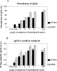
Fig. 1. Prevalence of anti-filarial IgG4 (A) and specific IgG4 levels produced by IgG4+ subjects (B) in transmigrants and life-long residents, classified as children ([les ]15 years; [squf ]) and adults (16 years and older; ▩) as a function of length of exposure. In (B) the number of IgG4+ individuals is given above each bar.
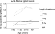
Fig. 2. Anti-filarial IgG4 levels in the transmigrant and indigenous population in different age groups as a function of length of residence in the area. (The straight line represents the cut-off value for specific IgG4 reactivity.)
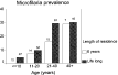
Fig. 3. Microfilaria prevalence (%) in transmigrants, settled in the area for 6 years and in life-long residents in different age groups. The number of subjects in age groups is given above each bar.
 You have
Access
- 7
- Cited by