Introduction
Adverse stimuli presented to mothers during gestation can permanently alter the structure and function of fetal tissues, a process known as fetal programming. The programming is based on the fact that alterations in the intrauterine environment can redirect developmental pathways during critical periods when tissues are still in proliferation and differentiation stages. Reference Chavatte-Palmer, Tarrade and Rousseau-Ralliard1-Reference Guilloteau, Zabielsky, Hammon and Metges3 These changes are exemplified by maternal malnutrition, smoking, alcohol consumption, drug abuse, emotional stress, Reference Cheong, Wlodek, Moritz and Cuffe4 and endocrine disorders. Reference Seckl5 As a result, exposure of the fetus or newborn to adverse environmental during gestation and/or throughout the lactation period, respectively, may trigger a number of diseases in adulthood. Reference Tarrade, Panchenko, Junien and Gabory6,Reference Zandi-Nejade, Luyckx and Brenner7
The thyroid secretes two major hormones, thyroxin (T4) and triiodothyronine (T3). Although T4 is produced in greater amounts, this hormone will be later converted by different tissues into another active hormone, T3. Since these hormones are able to increase the body metabolic rate, their deprivation promotes a significant fall in basal metabolism. Reference Berne and Levy8,Reference Guyton and Hall9 A deficiency of thyroid hormones is called hypothyroidism, a very common and condition which is most prevalent in women, the elderly, and in some ethnic groups. Among their symptoms are fatigue, bradycardia, weight gain, muscle weakness, and hypertension. Reference Patel, Mishra, Pawar, Ranvir, Sundar and Dabhi10,Reference Erdogan, Canataroglu, Ganidagli and Kulaksızoglu11 Consequences of the untreated hypothyroidism during pregnancy may be a miscarriage, preterm birth, preeclampsia, complications at birth, neonatal hypothyroidism, changes in fetal brain development, and growth, besides congenital hypothyroidism. Reference Ajmani, Aggarwal, Bhatia, Sharma, Sarabhai and Paul12,Reference Stoian, Pantea and Margan13 In this context, thyroid hormones have an important participation in early developmental processes, controlling cell growth, maturation, and the metabolism of virtually all tissues. Reference Forhead14
Patients with hypothyroidism have alterations associated with gastrointestinal (GI) function, presenting symptoms such as constipation and flatulence. Reference Daher, Yazbeck, Jaoude and Abboud15 Studies conducted in animals with hypothyroidism showed a reduction in the stomach, small intestine, and the motor activity in the colon, Reference Yalylali, Kirac and Yilmaz16 an alteration which can lead to constipation, atony, and intestinal obstruction. Reference Maser, Toset and Roman17,Reference Middleton18 Albino male rats with hypothyroidism, for instance, have been shown to be more susceptible to stress-induced gastritis and ulcers. Reference Abdel-Sater19 Since little is known about the long-term impact of maternal hypothyroidism on children’s health or offspring from Wistar rats, and considering the effects of thyroid hormones on GI function, we test the hypothesis that maternal hypothyroidism during gestation and/or lactation could produce a long-lasting imprint on the offspring’s GI tract.
Material and methods
Animals
The experiments were performed using male (300 g) and female (180 g) of Wistar rats. The animals were housed in a room at 21°C with 12 h light/dark cycles and were provided with free access to tap water and standard chow. All the protocols used were reviewed and approved by the Ethics Committee on Animal Use of the Federal University of Uberlândia (CEUA/UFU; process n° 069/17). Male animals (n = 10) were kept individually in their boxes at the end of the afternoon, and females were placed in the boxes with males in the proportion of two to three females for each male. Early morning of the following day, a vaginal smear was performed to detect any possible pregnancy, due to the presence of spermatozoa in the smear. Once pregnant, the females were placed in individual boxes, and after birth, the male pups were divided into the following groups:
Group C (Control). 30-day-old male offspring (n = 77) from dams (n = 18) that had free access to water normally during gestation until weaning, about 21 days after gestation.
Group E1 (Experimental Hypothyroid Gestation/Breastfeeding). 30-day-old male offspring (n = 63) from dams (n = 13) that had free access to 0.02% methimazole (MMI) in drinking water from the eighth day of gestation to the period of weaning, about 21 days after gestation.
Group E2 (Experimental Hypothyroid Gestation). 30-day-old male offspring (n = 63) from dams (n = 12) that had free access to 0.02% MMI in drinking water from the eighth to the last day of gestation.
All male pups from each dam were used in each study group, but only one to two male pups from each dam were randomly chosen and used in each type of analysis proposed to ensure maternal and offspring variability.
Drugs and reagents
For maternal hypothyroidism induction, a solution of 0.02% MMI (MMI diluted in drinking water) was used, from LEMMA Supply Solutions Ltda (São Paulo, SP, Brazil). The castor oil used was from Sigma-Aldrich Chemical Co. (San Louis, MO, USA), the activated carbon from Proquimios Comércio e Indústria Ltda (Rio de Janeiro, RJ, Brazil), and the sodium thiopental from Cristália Produtos Químicos e Farmacêuticos Ltda (Itapira, SP, Brazil). The other reagents were supplied by the Federal University of Uberlândia.
Analyses performed on dams
Water and food consumption and maternal body weight
The liquid and food intakes of the pregnant rats were carried out from the eighth day of gestation, for a period of 6 days. In addition, the bodyweight (BW) gain during gestation (from the 1st to the 12th day of pregnancy) was evaluated (control n = 7; E1 n = 8; E2 n = 7).
Hormone concentrations
Samples of 1 ml of tail blood were collected from dams (control n = 7; E1 n = 8; E2 n = 7) of three studied groups, 5 days postpartum, for the measurement of free T3 and T4 thyroid hormones to confirm the euthyroid state of dams from the C and E2 groups and the hypothyroid state of dams from the E1 group. The method used to measure the hormones was electrochemioluminescence. The kits used were Elecsys FT3 IIII and Elecsys FT4 III from Roche Diagnostics (Basel, Switzerland). In addition, for free T3, values below the white limit are indicated as <0.4 pmol/l and values above the measurement range as >50 pmol/l. For free T4, values below the detection limit are indicated as <0.5 pmol/l and values above the measurement range as >100 pmol/l.
After the weaning period, the dams were euthanized by overdose of anesthetics. The anesthetic used was sodium thiopental at a dose of 160 mg/kg of BW intraperitoneally.
Analyses performed on the pups
BW and number of pups
At 30 days old, the pups of all the groups were weighed and counted to determine the total litter and the proportion between the males and females.
Hormone concentrations
Samples of 1 ml of tail blood were collected from 30-day-old male pups of three studied groups (control n = 11; E1 n = 9; E2 n = 10), for the determination of free T3 and T4 thyroid hormones by the electrochemioluminescence method, and after collection, the animals were euthanized by cervical dislocation.
Organ analysis
In the 30-day-old male pups of the three groups studied, gastric analyses (control n = 14; E1 n = 10; E2 n = 8) were performed, such as total stomach area, BW, and the relationship between stomach weight (SW) and BW. In relation to the small intestine (control n = 37; E1 n = 32; E2 n = 36), we performed the analysis of the total length, organ weight, and the relationship between the intestinal weight and BW.
Evaluation of ulcer susceptibility
The model of gastric lesions induced by hydroalcoholic solution was used to analyze the susceptibility to ulcers in 30-day-old male pups (control n = 14; E1 n = 10; E2 n = 8) from Wistar rats. The animals were fasted for 12 h prior to intragastric administration, and 0.8 ml/100 g of hydroalcoholic solution using a gavage needle was injected (0.3 M HCl/60% ethanol). After 1 h, the animals were euthanized by cervical dislocation after anesthesia with thiopental (0.2 ml/100 g i.p.). Then, a laparotomy was performed to remove the stomachs, which were opened through the greater curvature, emptied, weighed, washed in saline, and mounted between two glass plates. The material was scanned on the HP Scanjet 2400 Scanner, and images of the stomachs were analyzed in the Image J software (https://imagej.nih.gov/ij/), the results being expressed using the ulcer index (UI). To calculate the UI, the gastric lesions were classified and received scores according to their severity, as follows: area of the hemorrhagic lesions or ulceration proper (3); area of intense hyperemia (2); and mild/moderate hyperemia area (1). The UI was determined as previously described: Reference Prado, Silva, De Oliveira-Silva, Hiraki, Nunes and Bispo-da-Silva20 UI = 3X area of the hemorrhagic lesion (mm2) + 2X area of intense hyperemia (mm2) + 1X area of mild/moderate hyperemia (mm2).
Evaluation of GI motility using the activated carbon transit model
The 30-day-old male pups (control n = 15; E1 n = 16; E2 n = 16) were fasted for 12 h and then received 0.8 ml/100 g of activated carbon suspension (5% in 0.5% carboxymethylcellulose solution, m/v) orally. Following 30 min of carbon administration, the animals were euthanized by cervical dislocation after thiopental anesthesia (0.2 ml/100 g i.p.). Then the small intestine was removed to determine the distance traveled by the activated carbon (expressed in % of intestine length). Reference Harrison, Erlwanger, Elbrond, Andersen and Unmack21,Reference Uddin, Shilpi, Alam, Alamgir, Rahman and Sarker22 After checking small intestine motility, the organ was emptied and weighed.
Evaluation of intestinal fluid (enteropooling) produced by castor oil
The 30-day-old male pups were fasted for 12 h and received (gavage) castor oil (control n = 13; E1 n = 9; E2 n = 9) and saline (0.8 ml/animal) (control n = 12; E1 n = 11; E2 n = 10) orally. Reference Doherty and Hancock23,Reference Robert, Nezamis, Lancaster, Hanchar and Klepper24 After 30 min, the animals were anesthetized with thiopental (0.2 ml/100 g i.p.) and euthanized by cervical dislocation. A laparotomy was performed followed by tying and insulation of the small intestine, which was weighed. The intestinal contents were removed and the intestine weighed again. The difference between the full intestinal (FI) and empty (EI) weights was used as an indicator of the relative intestinal contents, where enteropooling = (FI−EI)/animal weight (mg/g × 1000).
Study of distal colon motility
A 2-mm-diameter glass sphere was introduced into the distal colon (distance 2.5 cm) of the 30-day-old male pups (control n = 14; E1 n = 10; E2 n = 14) with the aid of a plastic probe. The time for the expulsion of the sphere was determined for each animal and served as a parameter to infer changes in distal colon motility. Reference Koslo, Burks and Porreca25
Statistical analysis
The statistical analysis was performed using GraphPad Prism Version 5.00 software (Trial; San Diego, CA, USA). The level of significance was set at p < 0.05. The tests used for each parameter studied are cited below and in the legend of each table/figure in the item Results.
-
(A) Maternal parameters
-
– BW, food and water intake: one-way analysis of variance (ANOVA) with Tukey’s post-test.
-
– Free T3 and T4: Kruskal–Wallis with Dunn’s post-test.
-
-
(B) Offspring parameters
-
– BW, number of pups and free T4, small intestine weight: BW ratio (SIW:BW ratio), UI, enteropooling and small intestine and distal colon motility: Kruskal–Wallis with Dunn’s post-test.
-
– Free T3, stomach area, SW, SW:BW ratio, SIW, and length: one-way ANOVA with Tukey’s post-test.
-
Results
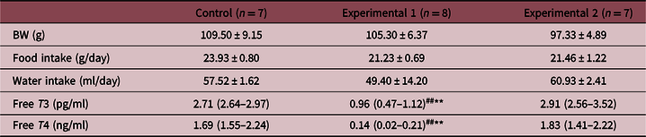
There were no differences between the groups for maternal weight gain, water, and food intake during gestation. The evaluation of maternal thyroid hormones, five days after delivery, showed that free T3 and T4 plasma levels were lower in the E1 group when compared to the C and E2 groups, confirming the postpartum hypothyroid state only of the dams from the E1 group (Table 1).
Maternal data: BW gain, food and water intake, and thyroid hormone levels (free T3 and T4) from control, experimental 1 and experimental 2 groups

Values are expressed as mean ± SEM for BW, food and water intake (one-way ANOVA with Tukey’s post-test) or median with percentiles 25 and 75 for free T3 and T4 (Kruskal–Wallis with Dunn’s post-test). The level of significance was set at p < 0.05. ##p < 0.01 versus control; **p < 0.01 versus experimental 2.
The total number of pups and the proportion between the males and females per litter were evaluated, and there was no difference between the groups. The 30-day-old pups from the E1 and E2 groups presented a significant reduction in BW when compared to the controls. However, weight reduction was higher in pups from E1 than in E2. The evaluation of the plasma levels of thyroid hormones from 30-day-old pups showed that there was no significant difference between the groups for free T3. However, free T4 levels were significantly lower in the E1 group when compared to the C and E2 groups (Table 2).
Offspring data: BW, total number of pups, and thyroid hormone levels (free T3 and T4) from control, experimental 1 and experimental 2 groups

Values are expressed as median with percentiles 25 and 75 for BW, number of pups, and free T4 (Kruskal–Wallis with Dunn’s post-test) or mean ± SEM for free T3 (one-way ANOVA with Tukey’s post-test). The level of significance was set at p < 0.05. ##p < 0.01 versus control; ###p < 0.001 versus Control; *p < 0.05 versus experimental 2.
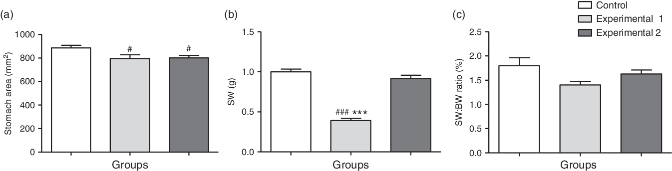
With regard to the gastric development of the offspring, it was observed that the stomach area of the 30-day-old pups was smaller in the E1 and E2 groups when compared to the C group (Fig. 1a), but the SW was lower only in the E1 group than in the other groups (Fig. 1b) and the SW:BW ratio was not different between the groups studied (Fig. 1c).
Stomach area and weight and SW:BW ratio of offspring from control, experimental 1, and experimental 2 groups.

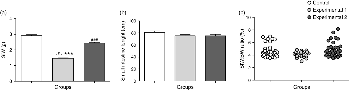
Regarding the development of the small intestine, it was observed that pups from the E1 and E2 groups presented with a lower intestinal weight when compared to group C. However, the intestinal weight in E1 was lower than in E2 (Fig. 2a). The intestinal length (Fig. 2b), as well as the intestinal weight:BW ratio (Fig. 2c), was not different between the groups.
Small intestine area and weight and SIW:BW ratio of offspring from control, experimental 1, and experimental 2 groups.

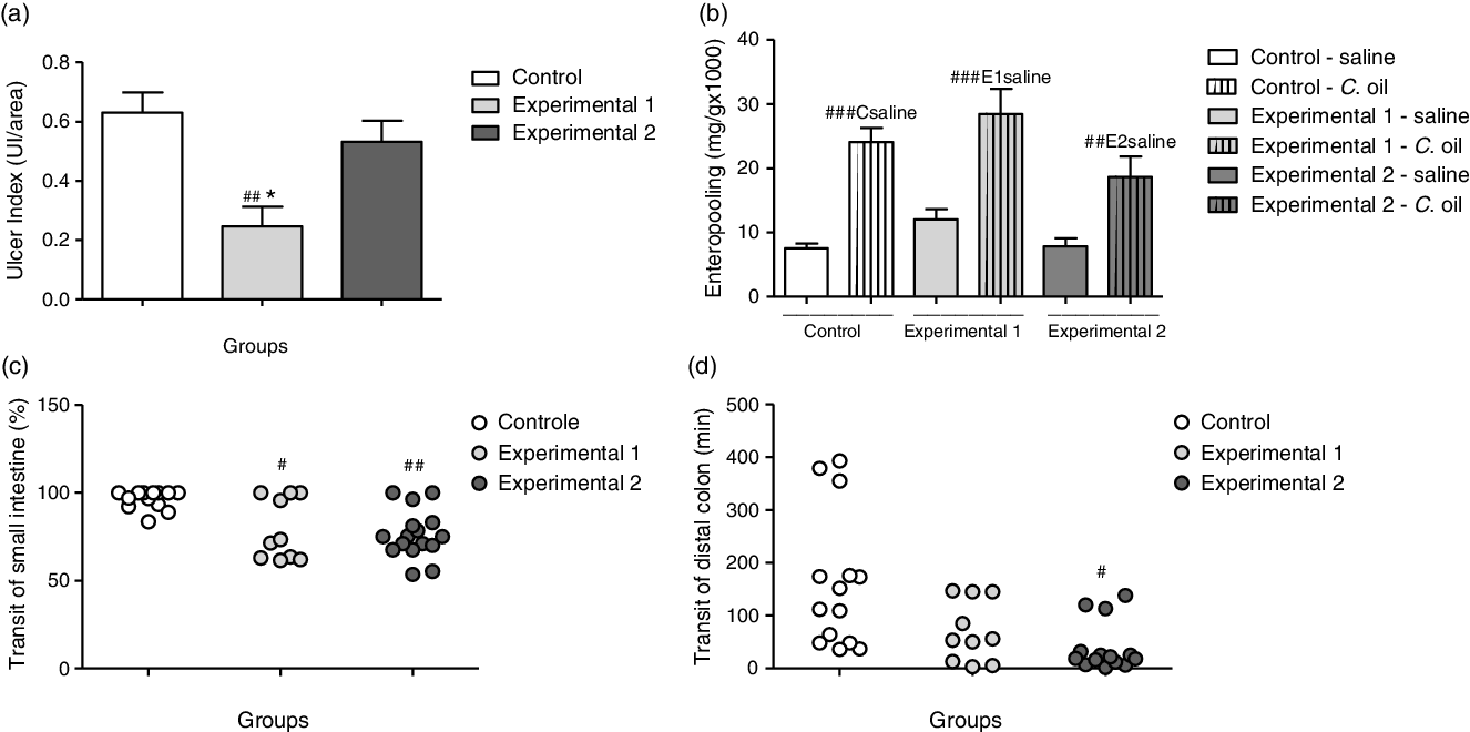
In response to the administration of the hydroalcoholic solution, the susceptibility to an ulcer was lower in group E1 when compared to groups C and E2 (Fig. 3a). In all the groups studied, intestinal fluid accumulation was greater in animals receiving castor oil than in those receiving the saline solution. There were no differences between the experimental groups and the control group for this parameter (Fig. 3b). Regarding intestinal motility, the transit of the small intestine was smaller in the experimental groups in relation to the control (Fig. 3c), and on the other hand, the distal colonic transit was increased only in E2 compared to the control (Fig. 3d).
UI, transit of small intestine and distal colon and enteropooling of offspring from control, experimental 1, and experimental 2 groups.

Discussion
In the present study, we aimed to evaluate any possible GI changes in the young offspring of mothers that ingested MMI during pregnancy and lactation, or only during the gestational period. The MMI is a thioamide drug that inhibits a key enzyme involved in thyroid hormones synthesis, the thyroid peroxidases. Reference Nambiar, Palanisamy and Okerberg26 Indeed, effective maternal hypothyroidism was induced by MMI administration, as suggested by reduced plasma levels of T3 and T4 observed in the dams of the E1 group. MMI administration was discontinued as soon as the E2 pups were born and 5 days postpartum, E2 dams were euthyroid, as indicated by T3 and T4 plasmatic levels normalization.
It is well established that MMI treatment induces weight loss in rats. Reference Cakic-Milosevic, Korać and Davidović27,Reference Cabello and Wrutniak28 Since the literature has long considered the pivotal role of T3 in the control of body growth, not only through direct and specific effects, but also as a mediator that potentiates growth hormone and insulin-like growth factor 1 activity, Reference Cabello and Wrutniak28 the slowed weight gain associated to hypothyroidism in rodents was simply attributed to T3 deficiency. However, this is not the only possible explanation. Indeed, it has shown that MMI-induced weight gain reduction can occur with no change in food consumed by the animals, observations that raised the question about what has happened with energy ingested but not built into the body mass. Reference Cakic-Milosevic, Korać and Davidović27 A proposed mechanism is that the energy was released owing to an increase in the brown adipose tissue mass and activity observed in rats treated with MMI. Reference Cakic-Milosevic, Korać and Davidović27 Similar to that described by Kobayashi et al. Reference Kobayashi, Kubota, Hojo and Miyagawa29 concerning the BW of rats exposed to propiltiuracil (PTU, another drug used to induce experimental hypothyroidism) during gestation and lactation, we observed no alteration in BW of dams treated with MMI, although they were hypothyroid and their food consumption was unchanged. Therefore, some maternal physiological adaption altered the effect of MMI concerning the BW loss. One possible explanation is that pregnancy may change the reported increases in brown adipose mass and activity associated to the experimental hypothyroidism. Corroborating this hypothesis, it has shown that brown adipose tissue thermogenesis is inhibited during late pregnancy and lactation in rats. Reference Abelenda and Puerta30,Reference Frontera, Pujol and Rodríguez-Cuenca31 An adaption that might explain the unchanged weight gain in dams from E1 and E2 groups reported in the present study. Alternatively or in addition, it is possible that more time is required for changes in weight gain to become evident after hypothyroidism induction. Indeed, it has shown that food intake decreased only 14 days post PTU treatment in both pregnant and male rats, Reference Karbalaei, Ghasemi, Hedayati, Godini and Zahediasl32,Reference Hwang, Jung and Kang33 while weight loss was observed only 3 weeks after hypothyroidism induction.
Thyroid hormones affect growth, Reference Soliman, De Sanctis, Bedair and Potluková34 and their effects during critical periods of cell proliferation and fetal development are evident in the reduced pups’ BW from dams of the E1 and E2 groups. A variety of mechanisms can contribute to the growth retardation associated with hypothyroidism, such as abnormalities on growth factor secretion, decreases in insulin-like growth factor I synthesis, and also a direct (but insufficient) action of low T3 levels on growth plate and skeletal growth. Reference Fraichard, Chassande and Plateroti35 Despite these mechanisms might be responsible for the general growth failure in pups from both the E1 and E2 groups, they cannot explain the reduced BW of pups from the E1 group compared to those from the E2 group. Indeed, pups from dams that received MMI during gestation and lactation (E1) and those from dams that were treated with MMI only throughout the gestational period (E2) were not hypothyroid, as suggested by their normal T3 plasma levels (the active hormone). The literature has reported that pregnant rats with hypothyroidism present reduced prolactin plasma levels, decreased milk production and ejection, and also changes in milk composition. Reference Hapon, Simoncini, Via and Jahn36,Reference Tamásy, Du, Vallerga, Meisami and Timiras37 Therefore, since the reduced BW observed in pups from the E1 group compared to those of pups from the C and E2 groups appears to be unrelated to the actions of the thyroid hormones, a malnutrition-like state in pups from the E1 group associated to alterations in the feeding processes (owing to the abnormalities described above) appears to be a reasonable explanation. In this regard, it is important to mention that postnatal growth is related to the size of the litter in rats Reference Wurtman and Miller38 ; however, maternal hypothyroidism did not affect the number of pups per litter nor the proportion between the male and female birth disproving, in both the E1 and E2 groups, that changes in feeding processes may be related to pups competition for milk. Concerning free T4 plasma levels (acting mainly as a prohormone), they were decreased only in pups that had fed in dams with hypothyroidism, probably reflecting the action of MMI in the sons’ thyroid due to its excretion in the milk. Reference Azizi39,Reference Taylor and Vaidya40 Altogether, T3 and T4 plasma levels observed in pups from the E1 group suggest a compensatory mechanism related to increases in T4 to T3 conversion and/or to decreases in T3 metabolism in this group, allowing the maintenance of the euthyroidism state.
As described above, thyroid hormones are important for normal development and hypothyroidism greatly impairs GI physiology in human and experimental animals; thus, we hypothesized that maternal hypothyroidism (during gestation and lactation) might be responsible for alterations in the offspring’s GI development and function. Despite pups from the dams treated with MMI presented with decreases in the stomach and SIW, in the E1 and E1/E2 groups, respectively, no alterations in the organ:BW ratio (relative weight) were observed. Moreover, maternal hypothyroidism decreased the stomach area in both the E1 and E2 groups without a change in the small intestine length. These data suggest that the fetus exposure to the intrauterine environment provided by maternal hypothyroidism caused a general decrease in body development and that the alterations in the absolute weight of the small intestine and in the stomach area reflect this general change. However, the postnatal development of the stomach concerning the organ mass seems to have occurred normally in offspring fed by euthyroid dams but was impaired in puppies from dams with hypothyroidism. Since the offspring’s BW from hypothyroid dams (i.e., E1) was smaller than that from euthyroid ones (i.e., C and E2 groups), the observed alteration in the SW of the E1 group probably reflects the postnatal impairment on the pups’ growth associated with the feeding processes, as argued above. In this respect, the only truly morphological alterations associated with maternal hypothyroidism observed in the present study appear to be the stomach mass thickness, that was seen to be smaller in the E1 and biggest in the E2 group, and the ratio between the small intestine mass and small intestine length that was smaller in both the E1 and E2 groups. A lighter small intestine that has a normal length strongly suggests a reduced amount of contractile elements throughout its length. This would possibly explain, at least partially, the decreased intestinal transit observed in the present study in both the experimental groups. Impairment in the excitation–contraction coupling mechanism in the smooth muscle of small intestine unrelated to the number of contractile elements should also be considered and needs further investigation. Interestingly, the motor activity of distal colon was increased in pups that had fed in dams with hypothyroidism only during the gestational period. This alteration could represent an adaptive response to deal with the decrease in the small intestine activity discussed above; moreover, to be fed in dams with hypothyroidism appears to delay the expression of this response, since the distal colon activity of the E1 group was not different from that of control. The mechanism responsible for this functional alteration still to be determined.
An ethanol-induced ulcer is a multifaceted process that involves, among other mechanisms, polymorphonuclear leukocytes migration into the inflammation zones, as suggested by increases in myeloperoxidase activity (MPO; a marker enzyme of leukocytes) in those areas; these cells appear to be responsible for the production of reactive oxygen species that damage the stomach mucosa, an action related to MPO activity. Reference Hamauzu, Forest, Hiramatsu and Sugimoto41 Moreover, pretreatment with MMI has been shown to protect the intestinal mucosa from injuries produced by trinitrobenzene sulfonic acid administration, an effect associated with decreases in inflammation and MPO activity. Reference Oren, Maaravi and Karmeli42 Finally, neutrophil chemotaxis was impaired in children with protein-energy malnutrition. Reference Ibrahim, Zambruni, Melby and Melby43 In our experimental conditions, the offspring from the E1 group appeared to have malnutrition, judging by their reduced body mass, and they also had ingested milk with MMI in an effective dose, as suggested by their low T4 plasma levels. Therefore, these two facts, along with the apparent small stomach mass thickness, can explain the decreased susceptibility to ethanol-induced ulcers in pups from the E1 group.
It is known that ricinoleic acid, released after hydrolysis of castor oil by lipases, is a laxative that decreases fluid absorption, increases electrolyte secretion, and produces changes in intestinal motility. Reference Capasso, Mascolo, Izzo and Gaginella44,Reference Tunaru, Althoff, Nüsing, Diener and Offermanns45 Indeed, castor oil administration increased intestinal fluid accumulation (i.e., produced enteropooling) in offspring, an effect unaltered by maternal hypothyroidism during gestation or gestation and lactation periods. These observations suggest that intestinal fluid absorption and/or the electrolyte secretion process are sensitive to ricinoleic acid, which is preserved in pups from hypothyroid dams.
Finally, pregnancy is associated with several physiological adaptations such as increased maternal thyroid gland activity due to increased thyroxine-binding globulins, altered maternal thyroid hormones peripheral metabolism, and increased chorionic gonadotropin levels. Thus, a new state of thyroid balance is achieved and maintained until the end of pregnancy, but this state may be compromised in women who develop hypothyroidism in pregnancy or were already hypothyroid when they became pregnant. Reference Glinoer46 The consequences of untreated maternal hypothyroidism during pregnancy range from preeclampsia to complications such as perinatal death, premature birth, among others. Reference Ajmani, Aggarwal, Bhatia, Sharma, Sarabhai and Paul12,Reference Stoian, Pantea and Margan13,Reference Blazer, Moreh-Waterman, Miller-Lotan, Tamir and Hochberg47 In our study, pups from hypothyroid mothers (E1 and E2) had lower weight at 30 days old compared to controls, as it reinforces the importance of maternal thyroid hormones for the proper development of offspring. In this context, from human studies, fetal sex also appears to interfere with intrauterine development, so that male fetuses are most exposed to macrosomia, whereas female fetuses more predisposed to intrauterine growth restriction. Reference Aiken and Ozanne48,Reference Melamed, Yogev and Glezerman49 Regarding maternal thyroid function, a human study showed that increased thyroid activity during pregnancy was more associated with low birth weight in male newborns, whereas subclinical maternal hypothyroidism was more related with higher weight in newborn from the same sex. Reference Vrijkotte, Hrudey and Twickler50 It is possible that maternal hypothyroidism may result in different GI changes or different intensities between male and female pups from Wistar rats, but this was not the objective of our study.
Conclusion
Maternal hypothyroidism impaired the total body development, as well as gastric and intestinal development, and interfered with the susceptibility to the ulcer of the offspring. Furthermore, reduced small intestine motility; this alteration appears to be associated to an adaptive increase in the distal colon motility. Thus, the reduced GI motility in euthyroid adults may be a consequence of intrauterine exposure to low thyroid hormone levels and of this exposure during breastfeeding in hypothyroid dams. The present study adds to our general understanding of GI disorders pointing to pup’s small intestine and stomach as pivotal sites associated to maternal hypothyroidism.
Acknowledgments
We are grateful to the UFU, who allowed the development of this work, and to REBIR-UFU (Rodent Biotteries Network from UFU) for supply and maintenance of the experimental animals.
Financial Support
This research did not receive any specific grant from any funding agency in the public, commercial or not-for-profit sector.
Conflicts of Interest
None.
Ethical Standards
The authors assert that all procedures contributing to this work comply with the ethical standards of relevant national guides on the care and use of laboratory animals and has been approved by the institutional committee of UFU (069/17).