Introduction
Epistaxis is the most common emergency presentation for otolaryngologists in the UK, experienced by 60 per cent of the general population and accounting for 25 000 acute presentations to National Health Service (NHS) hospitals each year.Reference Kelly, Committee, Ellis, Hall, Hardman and Mehta1 Of those experiencing epistaxis, 6 per cent will seek medical attention, and the majority of these can be managed conservatively with methods such as nasal packing or diathermy.Reference Tunkel, Anne, Payne, Ishman, Rosenfeld and Abramson2 Cases of epistaxis that cannot be managed conservatively will require more invasive management with surgical intervention methods. Embolisation techniques used for cases attributed to the sphenopalatine artery are not deemed suitable for the ethmoidal arteries because of both their size and their origin from the ophthalmic artery.Reference Tunkel, Anne, Payne, Ishman, Rosenfeld and Abramson2 The most common surgical management of ethmoidal epistaxis is ligation, which can be approached either intra-nasally or intra-orbitally. Opportunities for trainee exposure to anterior ethmoidal artery ligation are limited, with a recent national audit demonstrating that 3 of 1152 cases of epistaxis were managed with this technique.Reference Kelly, Committee, Ellis, Hall, Hardman and Mehta1
Traditionally, surgical training has followed a master–apprentice model, in which the individual will learn and acquire the skills required to perform procedures from their seniors in the operating theatre. In recent years, the time available for junior surgeons to learn in the operating theatre has been reduced, and with a constant emphasis on increasing efficiency in the operating theatre, there is less time for consultants to teach inexperienced surgeons. In order to train surgeons to the same standard without the same degree of exposure in the operating theatre, the future is likely to hold an increasing demand for surgical simulation. Through the use of simulation, surgeons have the opportunity to develop their skills without the risk of mistakes jeopardising patient safety.
The use of surgical simulation can broadly be categorised into: use of anatomical models; use of animal tissue or cadavers; virtual reality and computerised simulation; and mock scenarios with simulated patients. Anatomical model simulation in ENT training is already well established and has been used to teach bronchoscopy, tracheostomy and temporal bone dissection.Reference Abou-Elhamd, Al-Sultan and Rashad3,Reference Rose, Kimbell, Webster, Harrysson, Formeister and Buchman4 For managing epistaxis, the use of model simulation is limited to conservative management methods such as nasal packing.Reference Pettineo, Vozenilek, Kharasch, Wang and Aitchison5 Sphenopalatine artery and anterior ethmoidal artery ligation is most often taught either directly from clinical cases in the operating theatre or by using functional endoscopic sinus surgery cadaveric training courses, which are expensive and limited in availability. In this study, we describe an alternative for anterior ethmoidal artery ligation training that is cheaper and does not depend on the availability of clinical cases or cadavers.
Materials and methods
Building a prosthesis
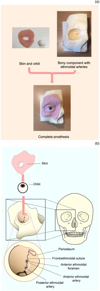
The base of the model consisted of the bony component formed of plaster. The frontoethmoidal suture was deliberately constructed to be prominent because it is an important anatomical landmark for locating the ethmoidal arteries with reference to the ‘24-12-6 rule’.Reference Naidoo and Wormald6 The soft tissue components (orbit, fat, periosteum, skin) were prepared from a soft rubber (Ecoflex), commonly used for anatomical tissue models, and the vasculature was prepared from bell wire (Fig. 1). The cost of the materials used to build one prosthesis is estimated to be close to £10 (this excludes the costs of labour and assembly).
(a) Components and (b) stages of prosthesis assembly.

When designing the prosthesis, it was important to consider the anatomy and physiology that would be feasible to replicate within our budget and also relevant to the procedure. The only physiological response considered for replication was bleeding from the tissue or vasculature; however, this would have added a significant layer of complexity and increased the costs. As it was not a necessity, bleeding was not incorporated.
Assessment of prosthesis
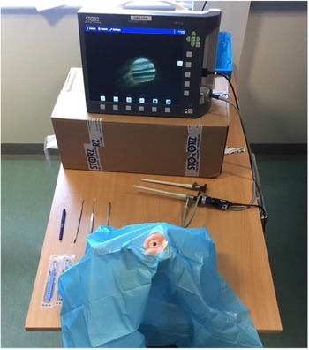
Current surgical otolaryngology training in the UK most commonly comprises formal assessments exclusively at either end of training, beginning with Membership of the Royal College of Surgeons and finishing with Fellowship of the Royal College of Surgeons, to complete higher surgical training. In the North-West, further annual formative assessments to evaluate progression throughout surgical training have been implemented in the form of objective, structured, clinical examination.Reference Stobbs, Khwaja, Khwaja and Kumar7 In order to assess the benefit of this prosthesis to a surgical otolaryngology trainee, 1 of the 20 stations in 2018 consisted of this procedure. Surgeons undergoing the formal assessment were at varying stages of training, from specialty trainee 3 to specialty trainee 6. At the station, individuals would be asked to complete an endoscopic anterior ethmoidal artery ligation of the right eye during the 8 minutes provided (Fig. 2).
Objective structured clinical examination (OSCE) station for simulation of procedure.

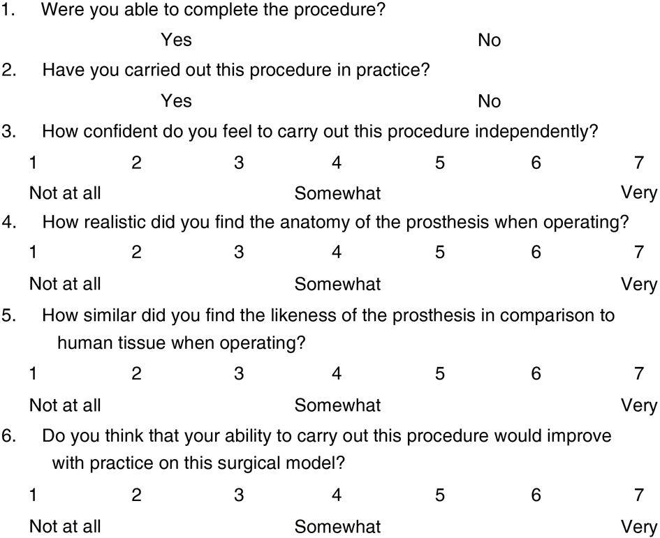
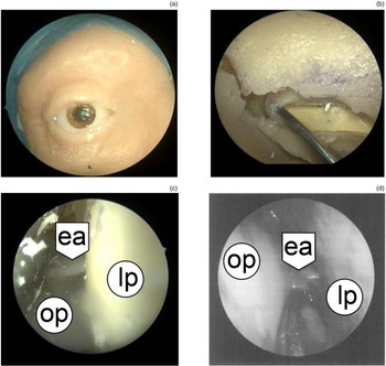
In order to assess the prosthesis, the participants who had operated on the models were asked to fill out a face validity questionnaire to provide feedback on the prosthesis following the examination (Fig. 3). The questionnaire consisted of two yes or no questions to ask about the surgeon's ability to complete the procedure during the objective, structured, clinical examination and their experience of the procedure in practice. These were followed by five Likert scale questions to assess surgical knowledge and to provide feedback on the prosthesis’ fidelity and benefit to surgical training. During the assessment, the endoscopic view of the procedure was taped and recorded for comparison against the procedure in vivo (Fig. 4).
Questionnaire to assess face validity of prosthesis.

Endoscopic stages of anterior ethmoidal artery ligation using a simulation model. (a) View of prosthesis prior to incision. (b) access to sub-periosteal plane following incision. (c) comparison of endoscopic view at point of identification of anterior ethmoidal artery (ea) between orbital periosteum (op) and lamia papyracea (lp) in (c) prosthesis and (d) patient.

Results
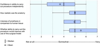
The four questions in the format of a Likert scale were analysed, followed by calculations of statistical averages, documented as a box plot (Fig. 5).
Questionnaire results from Likert scale questions to assess surgical trainee ability and response to the prosthesis following formative objective, structured, clinical examination assessment. Median averages and inter-quartile range are shown by dark vertical lines and blue boxes, respectively. Mean averages are indicated by O. No results exceeded 1.5 times that of the interquartile ranges, so no outliers were plotted.

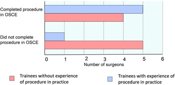
During the assessment, 9 of the 15 (60 per cent) participants were able to complete the procedure in the 8 minutes given. Trainees scored their confidence to carry out the procedure independently, with a mean of 3.67, but this was based on a broad range of results with a large discrepancy between those with and those without experience of the procedure in practice, with means of 4.83 and 2.89, respectively (see Fig. 6). Five of the six (83 per cent) participants with experience of the procedure in the operating theatre completed the procedure in the objective, structures, clinical examination station in comparison with only 4 of the 9 (44 per cent) trainees without experience completing the procedure (Fig. 6). Both the accuracy of the anatomy replication and the likeness to human tissue when operating were scored highly, with means of 5 and 4.93, respectively (Fig. 5). During the assessment, the endoscopic view was recorded, which can be seen in comparison with a clinical procedure in the operating theatre at the point of anterior ethmoidal artery identification before ligation in Fig. 5.
Comparison between surgical trainees with and without experience of procedure in practice in their ability to complete the procedure in 8 minutes at an objective structured clinical examination (OSCE) station.

The trainees were asked whether practice on the model would improve their ability to carry out the procedure. This aspect of the questionnaire received the highest ranking, with a mean of 5.53 (Fig. 5). Written feedback was also offered in the questionnaire, which provided two comments: firstly in reference to the lack of bleeding and secondly regarding the lack of adhesion between periosteum and bone.Reference Browning and Gilchrist8
Discussion
This study demonstrated the development and application of a high-fidelity, low-cost orbital prosthesis suitable for the simulation of anterior ethmoidal artery ligation. Otolaryngology trainees have limited exposure to this procedure and predominantly low levels of confidence that correlated with a lack of first-hand experience.
Procedural confidence
The effect that prior experience of the procedure in practice had on the surgeon's confidence in their ability to carry out the procedure independently was substantial; those with prior experience scored their confidence as almost twice that of those without (means of 4.83 and 2.89, respectively). This level of confidence was reflected in the surgeon's ability to complete the procedure: 5 (83 per cent) of the 6 participants with experience completed the procedure in the objective, structured, clinical examination station in comparison with only 4 (44 per cent) of the 9 surgeons without. These findings support the necessity of first-hand exposure to this procedure for surgeons to develop proficiency. The benefit to be gained from this model was also scored highly (5.53), which would suggest that this prosthesis would be a suitable teaching tool.
Model fidelity
The anatomy and likeness of the prosthesis to human tissue were both ranked highly, with means of 5 and 4.93, respectively (Fig. 5), indicating a high level of fidelity. The written feedback that the models received from the survey was regarding the anatomy: a lack of bleeding and a lack of adhesion between bone and periosteum. The unrealistic complexity of adding bleeding to the models has already been addressed in the methodology. Lack of adhesion between periosteum and bone could likely have been improved with the use of a simple adhesive.
Limitations
With any Likert scale face validity study, both response and central-tendency bias may give rise to misleading results. As the mean responses were consistently positive and did not deviate more than 1.53 from the central 4, both may have contributed to the results of this study.
• This study demonstrated the development and application of a high-fidelity, low-cost orbital prosthesis suitable for the simulation of endoscopic anterior ethmoidal artery ligation
• Current otolaryngology trainee exposure to anterior ethmoidal artery ligation is minimal and associated with low levels of confidence
• The use of an artificial anatomical model is both cheaper and more convenient than current training methods as it does not depend upon either the availability of clinical cases or cadavers
• To the authors’ knowledge, this is the first documentation of simulated surgical epistaxis management using an artificial anatomical model
When considering the relevant anatomy for replication, it is important to consider natural variation that may be seen between cases in clinical practice; for this procedure, a relevant example may be the presence of a middle ethmoidal artery, seen in up to 30 per cent of patients.Reference Wang, Youseef, Al Qahtani, Gun, Prevedello and Otto9 Such small anatomical variations are difficult to account for when designing a prosthesis, but without them a surgeon may develop false levels of confidence. This is one advantage of cadaveric models that may be impossible to parallel when using artificial simulation models.
A further consideration regarding the results is the validity of a surgical trainee's opinion. As discussed earlier, the level of fidelity for the models was scored highly, but as only 6 of the 15 trainees had experience of this procedure in practice, the opinions of more senior surgeons might have been of greater value.
Conclusion
The results of this study support the hypothesis that a simple anatomical prosthesis for the simulation of endoscopic anterior ethmoidal artery ligation can be created, with potential value to otolaryngology surgical training. To the authors' knowledge, this is the first documentation of simulated surgical epistaxis management through the use of an artificial anatomical model. The surgical trainees’ knowledge of this procedure was found to be low, indicating a potential need for modification to the current teaching of ligation of the ethmoidal arteries.
Further work
Since the use of this anatomical model, further work has been performed using the same design for a surgical simulation model for intra-orbital abscess drainage, which has been used on otolaryngology training courses in the North-West.
Acknowledgements
Thanks to Sadie Khwaja (ENT Consultant) for her guidance and overall supervision of this project, and to Patricia Healy for her work to design and build the prostheses.
Competing interests
None declared.