Introduction
Lyme disease, or Lyme borreliosis, is the most common tick-borne human infection in the northern hemisphere (carried by the tick Ixodes ricinus), occurring mainly in North America, Asia and Central Europe. There are 20 000 new cases every year (9.1 cases per 100 000 population) in the USA,1 whilst in Europe 65 500–85 000 cases are reported every year.Reference Rizzoli, Hauffe, Carpi, Vourćh, Neteler and Rosà2, Reference Hubálek3 Prevalence of borrelia infection in ticks in Europe is approximately 13.7 per cent.Reference Rizzoli, Hauffe, Carpi, Vourćh, Neteler and Rosà2, Reference Rauter and Hartung4 The first Portuguese human case of Lyme borreliosis was identified in 1989.Reference David de Morais, Filipe and Núncio5 Data have shown an incidence of 0.04 per 100 000 inhabitants, with the highest number of laboratory confirmed cases being from the Lisbon district.Reference Lopes de Carvalho and Núncio6
This report aims to describe a case of Lyme disease where the only manifestation was unilateral sudden sensorineural hearing loss (SNHL). It is a rare presentation of a rare disease in Portugal.
Case report
A 40-year-old woman presented with a 48-hour history of right SNHL, tinnitus and mild peripheral vertigo, with no other symptomatology. Pure tone audiometry revealed average hearing thresholds of 52.5 dB in the symptomatic ear, which was in accordance with Fetterman and colleagues' criteria for sudden SNHL.Reference Fetterman, Saunders and Luxford7 The patient was commenced on systemic corticosteroid (prednisolone 1 mg/kg/day for 10 days, followed by a tapering dose for 5 days). In addition, she received 10 sessions of hyperbaric oxygenation (2.5 ATA, 100 per cent oxygen for 90 minutes each session). Post-treatment audiometry revealed hearing thresholds of 20 dB in the right ear, demonstrating a recovery of 32.5 dB.
Our department has created a protocol (based on discussion of peer-reviewed articles) that includes laboratory tests for Borrelia burgdorferi; this protocol was produced with the aim of developing guidelines for the diagnosis and treatment of sudden SNHL. However, in order to prevent a long delay between the onset of the hearing loss and treatment, patients start corticosteroid and hyperbaric oxygen treatment as soon as possible.
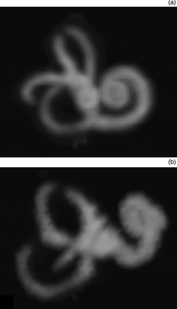
The three-dimensional (3D), T2-weighted magnetic resonance imaging (MRI) scans revealed focal loss of signal in the right semicircular canals, suggesting labyrinthitis (Figure 1).
Three-dimensional, T2-weighted magnetic resonance images of the right inner ear, showing: (a) focal loss of signal in the most superior aspect of the superior semicircular canal and in the entire posterior part of the lateral semicircular canal; and (b) focal loss of signal in the distal part of the posterior semicircular canal and a posterior view of the loss of signal in the superior semicircular canal.

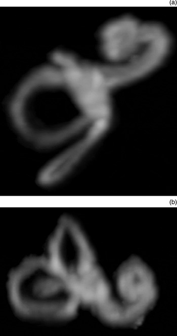
The B burgdorferi antibodies blood tests revealed positive findings for immunoglobulin M (IgM) and negative findings for immunoglobulin G (IgG) (based on the findings of an enzyme-linked immunosorbent assay test, which were confirmed by IgM and IgG Western blotting). As the patient had acute Lyme disease, she was treated with doxycycline 200 mg/day for 14 days. After antibiotic treatment, a full recovery of the hearing loss was observed and confirmed with audiological testing. Borrelia burgdorferi laboratory tests post-treatment were negative for IgM and positive for IgG. Five months after treatment, MRI showed resolution of all signs of labyrinthitis (Figure 2).
Three-dimensional, T2-weighted magnetic resonance images revealed lack of occlusion of the semicircular canal in the right ear with canal permeabilisation after treatment: (a) shows the superior and posterior semicircular canal, and (b) shows the lateral semicircular canal.

Discussion
Sudden hearing loss affects 5 to 20 individuals per 100 000 population,Reference Mosnier, Bouccara and Sterkers8, Reference Stachler, Chandrasekhar, Archer, Rosenfeld, Schwartz and Barrs9 with spontaneous recovery in 32 to 83 per cent.Reference Byl10, Reference Byl11 Treatment of sudden SNHL is difficult as it is hard to establish an aetiological factor in most cases.Reference Chau, Lin, Atashband, Irvine and Westerberg12, Reference Merchant, Adams and Nadol13
Lyme disease is a tick-borne (ixodes spp) infection caused by an immune response to the spirochaete B burgdorferi sensu lato. It may lead to a variety of symptoms affecting different organs such as the skin, joints, heart muscle or nervous system.Reference Rizzoli, Hauffe, Carpi, Vourćh, Neteler and Rosà2, Reference Hoen, Margos, Bent, Diuk-Wasser, Barbour and Kurtenbach14 There are at least 18 species of B burgdorferi sensu lato.Reference Stanek and Reiter15 In the USA, B burgdorferi sensu stricto is the only species that causes disease.Reference Hoen, Margos, Bent, Diuk-Wasser, Barbour and Kurtenbach14, 16, Reference Stanek, Wormser, Gray and Strle17 In Europe, however, several of these species are pathogenic (Borrelia afzelii, Borrelia garinii, B burgdorferi, Borrelia spielmanii and Borrelia bavariensis).Reference Rizzoli, Hauffe, Carpi, Vourćh, Neteler and Rosà2, Reference Rauter and Hartung4, 16–Reference Rudenko, Golovchenko, Grubhoffer and Oliver18 In Portugal, Borrelia lusitaniae is the most frequently identified species; however, it is not considered pathogenic.Reference David de Morais, Filipe and Núncio5
Lyme disease is the most prevalent arthropod-borne disease in temperate regions of the northern hemisphere. It is considered endemic in many parts of the UK, particularly in woodland and heathland areas, and over 1000 serologically confirmed infections are reported annually in the UK.Reference Byl11 This disease has been considered a notifiable disease in Portugal since 1999, but only a few cases are reported each year.Reference David de Morais, Filipe and Núncio5 The authors believe that not all cases are reported.
Lyme borreliosis risk is specifically linked to tick abundance and exposure. Therefore, a higher risk is correlated not only with residency in rural areas, but also with occupation (forestry work and farming) and certain leisure activities. It can occur at any age, but there are 2 age groups mainly affected, in light of greater exposure: children aged 5–9 years and adults aged 35–55 years.Reference Rizzoli, Hauffe, Carpi, Vourćh, Neteler and Rosà2, Reference Byl11, Reference Hoen, Margos, Bent, Diuk-Wasser, Barbour and Kurtenbach14, Reference Stanek, Wormser, Gray and Strle17, Reference Ertel, Nelson and Cartter19 The species (of the vector) also varies according to geographical area, with Ixodes scapularis being the most frequent in the USA and I ricinus the most frequent in Europe.Reference Stanek, Wormser, Gray and Strle17 The transmission depends on the activity of the vector, which is higher in summer (from May to September), and does not usually occur within the first 24 hours of a blood meal, so immediate removal of ticks is a highly recommended preventive measure.Reference Rizzoli, Hauffe, Carpi, Vourćh, Neteler and Rosà2, Reference Chau, Lin, Atashband, Irvine and Westerberg12, Reference Hoen, Margos, Bent, Diuk-Wasser, Barbour and Kurtenbach14
Borrelia burgdorferi infection can be asymptomatic. Symptomatic cases are potentially progressive and the clinical course has customarily been considered in terms of three stages: early, localised disease; early, disseminated disease; and late disease. The initial phase usually sees erythema migrans at the site of the tick bite, characterised by an erythematous, annular and expansive rash. The patient typically experiences ‘flu-like’ symptoms of malaise, headache, myalgia and arthralgia. However, erythema migrans can be absent in 20 to 50 per cent of cases,Reference Girschick, Morbach and Tappe20 with neuroborreliosis or arthritis being the first manifestation of Lyme disease.Reference Rizzoli, Hauffe, Carpi, Vourćh, Neteler and Rosà2, 16, Reference Stanek, Wormser, Gray and Strle17, Reference Girschick, Morbach and Tappe20
The early dissemination phase may occur weeks or months later; it usually presents with two or more cutaneous lesions, but it may occur as a manifestation of neuroborreliosis. In children, early dissemination, and especially neuroborreliosis, usually occurs earlier than in adults. This might be due to a different site of the tick bite – in children the upper trunk and the head are selected more often by the tick than in adults, potentially making the central nervous system more accessible to the spirochaete.Reference Huppertz, Girschick, Cimaz and Lehman21
Borreliae can disseminate haematogenously or directly to other organs and tissues, which heralds the late stage of the disease. It can cause general systemic symptoms and signs, affecting the nervous system (meningitis, facial and/or other cranial palsies, meningoradiculitis, or radiculopathy), skin (acrodermatitis chronica atrophicans), joints and heart (myopericarditis).Reference Rizzoli, Hauffe, Carpi, Vourćh, Neteler and Rosà2, Reference Hubálek3, Reference David de Morais, Filipe and Núncio5, Reference Hoen, Margos, Bent, Diuk-Wasser, Barbour and Kurtenbach14, 16, Reference Coumou, van der Poll, Speelman and Hovius22 It would appear that symptoms depend on the species involved. In Portugal, neuroborreliosis is the most common clinical manifestation of Lyme disease.Reference David de Morais, Filipe and Núncio5
The correlation between Lyme disease and sudden SNHL is not well known. However, sudden SNHL can be seen in neuroborreliosis. In a group of 165 patients with a diagnosis of sudden hearing loss, serology was positive for B burgdorferi in 12 per cent of cases; B burgdorferi was 6 times more prevalent in this patient group than in the general population residing in the same area.Reference Peltomaa, Pyykkö, Sappälä, Viitanen and Viljanen23
There is no international consensus regarding treatment of Lyme disease, nor of possible antibiotic resistance. Treatment is usually based on antibiotics, with dose, type, duration and administration route (intravenous or oral) dependent on the symptoms and stage of the disease.Reference Rizzoli, Hauffe, Carpi, Vourćh, Neteler and Rosà2, 16, Reference Stanek, Wormser, Gray and Strle17, Reference Girschick, Morbach and Tappe20, Reference Coumou, van der Poll, Speelman and Hovius22 Only a short course of antibiotics is required for most cases, with the exception of more severe and chronic cases. In the initial stages, doxycycline, amoxicillin, azithromycin or cefuroxime is administered for 14 days. Oral or intravenous ceftriaxone is recommended in cases of borreliosis in which the central nervous system or heart are affected, and in cases of borrelial lymphocytoma. When cranial nerve palsies are present, the suggested treatment is doxycycline 200 mg per day for 14 days. In late stage disease, the same antibiotics are recommended, but for a longer period (14 to 30 days). The treatment duration depends on the persistence of symptoms, although there seems to be no advantage in long-term medication.16, Reference Stanek, Wormser, Gray and Strle17, Reference Girschick, Morbach and Tappe20, Reference Coumou, van der Poll, Speelman and Hovius22 There is no reference in the literature, however, to treatment for Lyme disease in cases with labyrinthitis.
Most symptoms resolve with medication, but symptoms such as marked fatigue, fibromyalgia, arthralgia, impaired concentration or short-term memory may persist in some patients. Persistent symptoms that last longer than six months are classified as post-Lyme disease syndrome and antibiotics are not recommended.16, Reference Stanek, Wormser, Gray and Strle17, Reference Girschick, Morbach and Tappe20, Reference Logigian, Kaplan and Steere24 The factors that cause this syndrome have not been defined, but it is thought that it results from the chronic lesions which develop in the affected organs during various stages of the disease. The diagnosis of this syndrome is made using serological tests.Reference Stanek, Wormser, Gray and Strle17, Reference Girschick, Morbach and Tappe20
In the case presented, blood tests revealed recent Lyme infection (a positive enzyme-linked immunosorbent assay IgM result, which was confirmed via Western blotting) and MRI showed evidence of labyrinthitis.Reference Verbist25 In acute labyrinthitis, the perilymphatic space becomes filled with inflammatory cells. At this stage, changes are still reversible (before bone formation starts, leading to permanent obliteration of the inner ear). Once fibroblasts fill the labyrinth, signal loss will be seen on thin-sliced, 3D, T2-weighted images.Reference Verbist25 In the present case, the 3D, T2-weighted MRI scans showed focal loss of signal in the most superior aspect of the superior semicircular canal, in the distal part of the posterior semicircular canal and in the entire posterior part of the lateral semicircular canal, suggesting labyrinthitis. Therefore, the patient was treated with doxycycline for 14 days, as the sudden SNHL was considered a symptom of the initial stage of Lyme disease. After antibiotics, a total recovery of the hearing loss was observed, and MRI scans demonstrated resolution of the signs which initially suggested labyrinthitis.
• Lyme disease is an uncommon condition, usually presenting with erythema migrans
• Sudden onset sensorineural hearing loss (SNHL) is a rare manifestation of the disease, often associated with other otological symptoms
• Magnetic resonance imaging showed signs of labyrinthitis
• Serological confirmation of Borrelia burgdorferi allowed correct treatment to be commenced promptly and hearing loss recovered completely
• Lyme disease should always be considered in the diagnostic testing of SNHL
A clinical practice guideline was recently published regarding sudden hearing loss.Reference Stachler, Chandrasekhar, Archer, Rosenfeld, Schwartz and Barrs9 According to the panel, obtaining routine laboratory tests in patients with sudden SNHL is not recommended. However, Lyme disease should be considered in higher risk individuals and/or in areas where incidence is high.
Conclusion
Sudden SNHL is a rare symptom of Lyme disease. It can occur with or without a neuropathy. In this study, MRI suggested labyrinthitis and blood tests were positive for B burgdorferi.
Although borreliosis has a low incidence in Portugal, it should be considered as an aetiological factor in sudden SNHL. Adequate treatment may provide total recovery and prevent more severe forms of Lyme disease.