Introduction
Chronic suppurative otitis media (CSOM) is an infection of the middle ear that persists for more than two months, continuously or intermittently, with perforation of the tympanic membrane and secretion from the ear.Reference Gupta, Harshvardhan and Samdani1, Reference Mahdiani, Lasminingrum and Anugrah2 This tympanic membrane perforation can lead to hearing loss. A tympanoplasty is often performed to repair a perforated tympanic membrane and restore hearing in cases of chronic otitis media (COM) without cholesteatoma.Reference Hsu, Kuo and Huang3 Perforations can be repaired using a variety of surgical procedures and graft materials. The most used grafts are those made from conchal cartilage or the temporalis fascia.Reference Bhardwaj and Singh4
Traditionally, surgery on the middle ear has been conducted using microscopy. This method offers a somewhat limited visualization of the surgical field. Endoscopic middle ear surgery was pioneered in the 1990sReference Thomassin, Duchon-Doris, Emram, Rud, Conciatori and Vilcoq5 and has gradually become more widely used in clinical practice. Endoscopy offers a superior surgical field of the middle ear cavity, allowing the visualization of hidden areas of the middle ear cavity.Reference Lee, Kang, Lee, Kim and Lee6 Endoscopy also allows for a more minimally invasive surgical approach, as the transcanal approach can be achieved via a tympanometry flap, reducing additional incisions and dissections.Reference Choi, Noh, Park, Lee, Yook and Choi7
Several previous studies have compared microscopic and endoscopic surgery in the context of tympanoplasty to treat CSOM. In 2016, a systematic review and meta-analysis by Tseng et al.Reference Tseng, Lai, Wu, Yuan and Ding8 It was found that across four studies analyzed out of the 49 screened initially (with 266 patients), microscopic and endoscopic surgery yielded closure rates and post-operative audiological outcomes that were not significantly different. However, patients who had received endoscopic surgery had significantly lower canaloplasty rates and achieved superior cosmetic results. These latter findings were, however, derived from just two studies with 80 patients in total.
A 2019 meta-analysis by Han et al.Reference Han, Lee, Chung and Kim9 covering four studies also concluded that microscopic and endoscopic tympanoplasty surgery had similar graft success rates. Although not part of the formal meta-analysis, they also observed that most of the 19 studies analyzed reported similar post-operative audiological outcomes with both techniques.
A 2023 systematic review by Stefan et al.,Reference Stefan, Stefanescu, Vlad, Zainea and Hainarosie10 found that graft success rates were not significantly different between the two methods across the six studies analyzed out of the 292 screened initially (with 859 patients in total). Mean operating time was significantly lower with the endoscopic method across all studies analyzed. Audiological outcomes were somewhat mixed; one study analyzed by Stefan et al. observed a significant difference in audiological outcomes in favor of the endoscopic method at the one-month follow-up, but this difference had disappeared by the 12-month follow-up, while another study observed a significant difference in favor of the microscopic method. Self-reported post-operative pain ratings were also mixed, with favorable results for the endoscopic method reported in two studies.
Individual studies have also reported similar incidences of delayed facial palsy between microscopic and endoscopic surgery,Reference Li, Chan, Chen, Kuo, Wang and Chang11 as well as reduced intra-operative blood loss,Reference Li, Zhang, Xie, Ren and Wang12 reduced total hospitalization time,Reference Gulsen and Baltacı13 lower total hospital costsReference Kim, Lee, Choi, Oh, Kong and Lee14, Reference Kuo and Wu15 and superior quality of life outcomes for patients who had undergone endoscopic surgery.Reference Li, Ke, Yang, Zhang, Pan and Xin16
The primary objective of the present monocentric retrospective study was to compare the audiological outcomes after tympanoplasty using endoscopic versus microscopic surgery in patients with central perforation following CSOM. The secondary objectives were to compare the surgical duration, post-operative pain incidence and severity and the failure rate of grafting (residual perforation) with endoscopic versus microscopic tympanoplasty.
Materials and methods
Study design
A retrospective review was conducted on all patients with central tympanic perforation following CSOM who underwent tympanoplasty at King Abdullah Medical City (KAMC) between January 2020 and April 2022. Records were obtained from our clinical database.
Ethical standards
As a retrospective review of data obtained from routine clinical practice, this study received an exemption from ethics review by the institutional review board approval of the KAMC (Institutional Review Board [IRB] code 22-967). All patients provided informed consent for the medical treatments described in this study.
Inclusion and exclusion criteria
This study included all patients who presented at the ENT clinic of the KAMC between January 2020 and April 2022 with central perforation following a CSOM and who subsequently received tympanoplasty surgery using either the endoscopic or microscopic method. Tympanoplasty surgery must have been performed using a tragal cartilage, conchal cartilage or temporalis fascia graft (regardless of graft size). Additionally, it was required to have had a post-surgery follow-up for assessment of hearing and graft uptake. Patients were excluded if they had cholesteatoma, polyps or granulations or had had previous tympanoplasty surgery.
Data collection and management
The data were collected from patient records as anonymized electronic data forms. Patients were pseudonymized with study codes. Initials linked to the patient’s name and medical record number were kept in a separate identification log sheet in a safe, locked place. Two different persons performed data transcription. After verification of data transcription, the data were transferred to a statistical database.
Surgery
Microscopic tympanoplasty is done through a posterior auricular skin incision, harvesting temporalis fascia, which is used as a graft placed medial to the rim of the perforated tympanic membrane, while in endoscopic tympanoplasty tragal cartilage, its inner part used as graft and placed same as in microscopic approach medial to the rim of perforation of the tympanic membrane. In the microscopic approach, temporalis fascia was primarily used (but not always). In endoscopic use, tragal cartilage is usually but not always present. Different surgeons have no specific surgeon for a particular approach.
Audiometry
Audiometry is done operatively to determine the type and severity of hearing loss; follow-up audiometry is done three months after the surgical procedure to determine the hearing gain and benefit of the operation.
Residual perforation
Detected during follow-up in outpatient visits after surgical operation by microscopic examination and documented the location and size of residual perforation.
Post-operative pain management
By reviewing patients’ charts during their stay in the hospital until discharge, those asking and required.
• Post-operatively, we prescribe paracetamol 500 mg every 6 hours as needed (PRN) for pain.
• Some noted they still complained of pain despite receiving their dose of paracetamol; in such a case, we administered dual painkillers.
• Also, we asked the patients to scale their pain (How do you scale the pain post-operatively? Mild, moderate or severe?).
Results and analysis
Statistical Analysis
Data entry was performed using Microsoft Excel 2016 (Microsoft Corporation). Statistical analyses were performed using SPSS statistics version 28.0 (IBM). Statistical comparisons were made using the Mann–Whitney U test and t-tests for independent and paired samples. The results were assessed at confidence intervals within 95 per cent and a significance level of p less than 0.05.
Clinical features
The study cohort consisted of 58 patients. 29 patients (19 female) underwent endoscopic tympanoplasty via a transcanal approach, all of whom had a tragal cartilage graft. Twenty-nine patients (22 female) underwent microscopic tympanoplasty via two different types of approaches (transcanal [n = 8] or post-auricular [n = 21]) and three different types of grafts (tragal cartilage [n = 13], conchal cartilage graft [n = 1] or temporalis muscle fascia [n = 15]) (Table 1).
Clinical features of the endoscopic and microscopic groups

Audiological outcomes
At the pre-operative interval, the endoscopic group had a mean (±SD) air conduction threshold of 40.53 (±14.12) dB HL, with a mean air–bone gap of 22.34 (±7.43) dB hearing loss (HL). The microscopic group had a mean air conduction threshold of 55.04 (±22.1) dB HL with a mean air–bone gap of 26.73 (±9.12) dB HL.
At the six- to eight-week post-operative interval, the endoscopic group had a mean air conduction threshold of 31.33 (±15.58) dB HL with a mean air–bone gap of 11.7 (±9.81) dB HL. Pre- to post-operative mean air conduction hearing thresholds improved significantly by 9.21 dB HL (p = 0.004), and the mean air–bone gap significantly improved by 10.64 dB HL (p < 0.001).
The microscopic group had a mean air conduction threshold of 43.04 (±26.71) dB HL with a mean air–bone gap of 15.00 (±11.82) dB HL. Pre- to post-operative mean air conduction hearing thresholds improved significantly by 12.0 dB HL (p < 0.001), and the mean air–bone gap significantly improved by 10.74 dB HL (p < 0.001).
The differences in air conduction threshold improvement between the two groups were not significant (p = 0.472), nor were the differences in air–bone gap improvement (p = 0.973).
Surgery duration
The mean surgery time was 75.46 (±21.04) minutes for the endoscopic group and 126.66 (±34.27) minutes for the microscopic group. The difference between the mean surgery times for the two groups was statistically significant (p < 0.001) (Figures 1 and 2).
Distribution of durations for the microscopic tympanoplasty procedures.

Distribution of durations for the microscopic endoscopic procedures.

Residual perforation and post-operative pain
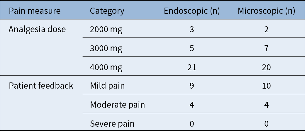
At the follow-up interval, residual perforation was present in 12 patients (41.4 per cent) within the endoscopic group and 17 patients (58.6 per cent) in the microscopic group. No significant differences were observed in the incidence of residual perforation between the two groups (p = 0.790). No significant differences were observed between the distributions of dosages of post-operative analgesia administered to each group (Table 2) (p = 0.681). No significant differences were observed between the self-reported pain ratings of each group each group (Table 2) (p = 0.902).
Distributions of post-operative analgesia dosages and of self-reported pain by surgery type

Post-hoc power analysis
The power analysis showed that a sample size of 29 cases per group was sufficient to detect the true difference in the average surgical duration between study groups (75.46 ± 21.04 minutes) vs. (126.66 ± 34.27 minutes) for endoscopic vs. microscopic tympanoplasty respectively, achieving 99.9 per cent power using a detected Cohen’s d effect size = 1.8.
Therefore, the large effect size (conventional effect size from Cohen, 1982) for the primary outcome of the study (surgical duration) between the two groups indicated that the total sample of 58 cases would be of sufficient, and high power to prove it.
Discussion
Here, we conducted a retrospective monocentric review of 58 cases of CSOM treated by tympanoplasty, divided evenly into those conducted via endoscopic and microscopic procedures. The primary objective was to compare the audiological outcomes after tympanoplasty using endoscopic versus microscopic surgery. The secondary objectives were to compare the surgical duration, post-operative pain incidence and severity and the failure rate of grafting (residual perforation) with endoscopic versus microscopic tympanoplasty.
No statistically significant differences were observed in audiological outcomes between the two groups. Both achieved, however, statistically significant improvements in air conduction thresholds of 9.21 dB HL and 12.0 dB HL for the endoscopic and microscopic groups, respectively, as well as significant improvements in the air–bone gap of 10.64 dB HL and 10.74 dB HL for the endoscopic and microscopic groups, respectively. These findings show that both endoscopic and microscopic surgery are similarly effective at achieving satisfactory post-operative audiological outcomes.
The incidence of graft failure, as measured by the presence of residual perforation at follow-up, was 41.4 per cent for the endoscopic group and 58.6 per cent for the microscopic group. This difference was not found to be statistically significant. These findings are in line with what has previously been reported.Reference Tseng, Lai, Wu, Yuan and Ding8–Reference Stefan, Stefanescu, Vlad, Zainea and Hainarosie10
The incidence of post-operative pain did not differ significantly between the two groups, as measured both by the dosage of analgesia administered or through self-reported pain ratings. The incidence of pain overall was relatively low, and no patients reported severe post-operative pain in either group. Previous studies have found mixed results about the incidence of post-operative pain,Reference Stefan, Stefanescu, Vlad, Zainea and Hainarosie10 with some finding significantly less pain for patients who underwent endoscopy, while others observed no difference. Our findings are in accord with the latter.
The surgical duration differed significantly between both groups, with mean durations of 75.46 minutes and 126.66 minutes for the endoscopic and microscopic groups, respectively. As equivalent outcomes are achieved with substantially shorter operative time, this finding suggests a key advantage for endoscopic surgery. Previous studies have observed that endoscopy leads to a lower overall medical cost,Reference Gupta, Harshvardhan and Samdani14, Reference Gupta, Harshvardhan and Samdani17 which may be attributable in part to reduced operating room time consumption.
Some disadvantages of endoscopic surgery have previously been noted. One is that the endoscope requires less-efficient one-handed surgery.Reference Choi, Noh, Park, Lee, Yook and Choi7 This necessitates a degree of adaptation and experience to become proficient. Another potential disadvantage is the thermal effect produced by the greater light intensity of the microscope. Intra-operative sensations of vertigo are a frequent complication of middle-ear endoscopy, and this has been attributed to the thermic heating of the lateral semicircular canal.Reference Bottrill, Perrault and Poe18 Practical recommendations have been made to minimize potential thermal risks from middle ear heat exposure during endoscopy.Reference Mitchell and Coulson19 Despite these potential disadvantages, the equivalence in audiological outcomes, complication rates and incidence of post-operative pain documented here and elsewhere demonstrates that these issues do not hamper overall success rates.
Endoscopic techniques have been successfully employed in other types of middle ear surgery, including cholesteatomaReference Li, Zhou, Wang, Wang and Zou20, Reference Nair, Aishwarya, Vasu, Karthikeyan and Shalini21 and stapes surgery.Reference Nikolaos, Aikaterini, Dimitrios, Sarantis, John and Eleana22–Reference Ho, Patel, Ballard, Rosenfeld and Chandrasekhar25 As with tympanoplasty, these endoscopic procedures have generally been found to produce equivalent surgical success rates and audiological outcomes compared to traditional microscopic surgery but have the advantage of reduced operative time and a less invasive surgical approach overall.Reference Tseng, Lai, Wu, Yuan and Ding8–Reference Stefan, Stefanescu, Vlad, Zainea and Hainarosie10
This study has some limitations which should be noted. Due to the retrospective nature, some procedural differences were present between the groups. These are related to the surgical approach and graft material used. As detailed in Table 1, all patients in the endoscopy group underwent the transcanal approach, while in the microscopic group, the transcanal approach was used in eight (27.5 per cent) cases and a post-auricular approach was used in 21 (72.5 per cent) of cases. For the graft material, all patients in the endoscopic group received grafts from tragal cartilage, while those in the microscopy group received tragal cartilage (n = 13; 44.8 per cent), conchal cartilage (n = 1, 3.4 per cent) or temporalis muscle fascia (n = 15; 51.7 per cent). These differences may, in principle, have influenced outcome measures, especially surgery duration, which was the only outcome that differed significantly between the two groups. Nevertheless, the reduced operative time with endoscopic surgery reported here is in line with that generally reported previously. A future randomized trial study is recommended to determine which approach is superior.
• Endoscopic tympanoplasty appears to be an effective alternative to the microscopic approach
• Both techniques result in equivalent post-operative audiological outcomes and with similar complications rates
• Endoscopic surgery had a shorter operative duration and may therefore be superior when operating room time consumption is taken into account
• Previous studies have found mixed results regarding the incidence of post-operative pain, with some finding significantly less pain for patients who underwent endoscopy, while others observed no difference. Our findings are in accord with the latter
Conclusion
Endoscopic tympanoplasty appears to be an effective alternative to the microscopic approach. Both techniques result in equivalent post-operative audiological outcomes and with similar complication rates. Endoscopic surgery has a shorter operative duration and may, therefore, be superior when operating room time consumption is taken into account.
Data availability
The datasets used and analyzed in the current study are available from the corresponding author upon reasonable request.
Acknowledgments
None.
Funding
No funding was received for conducting this study.
Competing interests
The authors have no competing interests to declare that are relevant to the content of this article.
Ethics approval
The study was conducted in accordance with the Declaration of Helsinki and approved by the local institutional review board of the KAMC (IRB code 22-967).
Consent
All patients provided their informed consent for the medical treatments described in this study.
Author contributions
Conceptualization: Khalid Badr and Sarah Radwan; Methodology: Mohammed Shaheen and Basil Alqarni; Formal analysis and investigation: Mutep Aljahdali and Saeed Alghamdi; Writing - original draft preparation: Asail Alghamdi and Fares Alghamdi; Writing - review and editing: Syed Raza; Supervision: Khalid Badr and Syed Raza.