Highlights
-
Actigraphy differentiates central hypersomnolence disorders by objectively assessing key sleep parameters.
-
Variability in actigraphy methods hinders result consistency, highlighting the need for standardized protocols to enhance reliability in hypersomnolence research.
-
Actigraphy aids hypersomnolence diagnosis in overestimated sleep but needs validation for broader clinical use.
Introduction
Daytime sleepiness, according to the International Classification of Sleep Disorders, Third Edition (ICSD-3), corresponds to difficulty staying awake or alert during the day, which can cause a person to fall asleep involuntarily and is more common in monotonous environments.1 Excessive daytime sleepiness (EDS) is not only a risk factor for cardiovascular and neurological disorders but also a public health problem with significant socio-economic consequences.Reference Pérez-Carbonell, Mignot, Leschziner and Dauvilliers2 ICSD-3 classifies the central disorders of hypersomnolence (CDH) into narcolepsy type 1 (NT1), narcolepsy type 2 (NT2), idiopathic hypersomnia (IH), Kleine–Levin syndrome (KLS) and insufficient sleep syndrome (ISS). Hypersomnia associated with medical or psychiatric disorders and medications or substances is also included in this group, and each of these disorders can be subdivided into different pathophysiological subtypes. CDH thus includes disorders in which EDS has a central origin, which implies the absence of sleep disturbances or appropriate treatment.1 Currently, European experts have recognized the limitations of ICSD-3 and are proposing a new CDH classification: “narcolepsy,” “IH” and “idiopathic excessive sleepiness.”Reference Lammers, Bassetti and Dolenc-Groselj3 As far as the objective assessment of EDS is concerned, tests such as actigraphy, polysomnography (PSG), the multiple sleep latency test (MSLT) and the maintenance of wakefulness test help to confirm the diagnosis and initiate the appropriate treatment.Reference Gandhi, Mansukhani, Silber and Kolla4 Actigraphy is a cost-effective, non-invasive and well-tolerated technique. Actigraphy devices can be used on the wrist of the non-dominant hand and allow conclusions to be drawn about the periods corresponding to sleep (absence of movement) and wakefulness (periods of activity) from the measurement of movement. The actigraph can be used by the patient at home for several days or weeks, and the sleep diary should also be completed. The actigraphy data are downloaded and analysed afterwards using special software, and information is collected.Reference Bassetti, McNicholas, Paunio and Peigneux5
The American Academy of Sleep Medicine (AASM) has published a clinical practice guideline that recommends actigraphy 7–14 days before PSG and MSLT in patients with CDH. The aim of actigraphy is to determine the total sleep time (TST) before MSLT and to rule out ISS and circadian rhythm sleep-wake disorders.Reference Smith, McCrae and Cheung6 Another indication for actigraphy is the measurement of sleep duration for the diagnosis of IH.1 Actigraphy has obvious advantages, such as recording activity/sleep over several days and in the patient’s home, and is not dependent on the patient’s memory. However, actigraphy also has its limitations, such as its limited suitability for assessing sleep onset latency (SOL) and the need for patients to complete sleep logs to improve the quality of the recording analysis.Reference Martin and Hakim7 Actigraphy is a simple instrument that should be used before MSLT. Therefore, its data are important to understand its role in the diagnosis of diseases causing drowsiness of central origin. The aim of this systematic review, which was largely carried out as part of a master’s thesis, was therefore to summarize the available actigraphic features of CDH.
Methods
Study design
A systematic review was conducted in accordance with the PRISMA (Preferred Reporting Items for Systematic Reviews and Meta-Analyses) guidelines.Reference Page, McKenzie and Bossuyt8 The eligibility criteria and search strategy were based on a PICOTS (Participants/Population, Interventions, Comparators/Controls, Outcomes, Timing and Study design) framework, as described in Table S1 in the supplementary material.
Search strategy
PubMed, EMBASE, Web of Science, Scopus, Cochrane and SciELO were the bibliographic databases searched for this review. The review was conducted on 2 March 2023 and last updated on 9 March 2023. The search strategy resulted from the combination of the controlled terms Medical Subject Headings (MeSH) and Emtree with uncontrolled terms. The following terms were combined to increase the sensitivity of the search: “Disorders of Excessive Somnolence”[Mesh] OR “Narcolepsy”[Mesh] OR “Idiopathic Hypersomnia”[Mesh] OR “Kleine-Levin Syndrome”[Mesh] OR “Sleep Deprivation”[Mesh] OR “Central Disorders of Hypersomnolence” OR “Narcolepsy type 1” OR “Narcolepsy type 2” OR “Idiopathic Hypersomnia” OR “Kleine-Levin Syndrome” OR “Hypersomnia due to a Medical Disorder” OR “Hypersomnia due to a Medication or Substance” OR “Hypersomnia associated with a Psychiatric Disorder” OR “Insufficient Sleep Syndrome” AND “Actigraphy”[Mesh] OR Actigraph*. Table S2 in the supplementary material contains a detailed description of the queries used to search the various databases. Duplicate publications were excluded using a reference manager.
Selection procedure
To reduce selection bias, the titles and abstracts were evaluated independently by SM and JIS using Rayyan, a web-based tool for systematic reviews,Reference Ouzzani, Hammady, Fedorowicz and Elmagarmid9 based on the following inclusion criteria:
-
Studies related to CDH diagnosis;
-
CDH should be classified according to ICSD-3. With regard to hypersomnia associated with medical or psychiatric disorders and drug- or substance-induced hypersomnia, the criteria for inclusion were the various pathophysiological subtypes listed in the ICSD-3. The subtypes included in this review are listed in Table S3 in the supplementary material;
-
Studies related to CDH diagnosis must mention the acquisition of actigraphy data;
-
Randomized controlled trials, cohort and case-control observational studies;
-
Adult participants;
-
Studies published after January 2015. ICSD-3 was published in 2014. For this reason, this time frame was chosen to avoid publications in which the CDH were classified according to a different version;
-
Studies written in English, Portuguese or Spanish;
-
If the same patient group participated in different studies, only the most recent publication was considered in order to avoid data duplication.
Studies that mentioned the following were excluded (Table S1 in the supplemental material):
-
CDH classification according to criteria other than ICSD-3;
-
Research without control or comparison groups;
-
Studies related to CDH therapies;
-
Actigraphy data not available;
-
Case reports, guidelines, commentaries and reviews;
-
Animal studies;
-
Paediatric participants;
-
Pregnancy;
-
Full text not available;
-
No information on the outcomes of interest.
Conflicting decisions on selection were resolved through consensus or by JCL. The full-text analysis of the remaining records was also performed by the same authors who met the previously defined criteria.
Data extraction
The data from the original studies were collected using a predefined Microsoft® Excel spreadsheet. Author, year of publication, country, study design, sample size, number of participants for each diagnosis and healthy controls (HC), gender, age, Epworth Sleepiness Scale (ESS) and sleep latency (SL) at MSLT were recorded. In relation to actigraphy, the following data were extracted (if available): manufacturer and model of the device, name and version of the software used to analyse the actigraphy data, epoch length, actigraphy duration, sleep diary completion, bedtime, rise time, time in bed (TIB), TST, sleep efficiency (SE), SOL, wake after sleep onset (WASO), sleep motor activity (SMA), number of awakenings (Awk), fragmentation index (FI), daytime motor activity (DMA), mean duration of longest sleep episodes per day (LNAP), interdaily stability (IS), intradaily variability (IV), relative amplitude (RA), discriminant score (DS) and new discriminant score (NDS). Data extraction was conducted by SM and reviewed by JIS. Disagreements were resolved by consensus or by JCL.
Assessment of study quality
Case-control and cohort studies were assessed using the Newcastle–Ottawa quality assessment scale.Reference Wells, Shea and O’Connell10 Each study was rated with stars in relation to various points, including selection, comparability and exposure or outcome for case-control or cohort studies. The scale ranges from 0 to 9, and the quality of the studies was categorized as low (0–3 stars), moderate (4–6 stars) and high (7–9 stars). Two authors (SM and JIS) independently scored each included study, and differing views on the quality of articles were resolved by consensus or JCL.
Data synthesis
Meta-analyses were conducted using the open-source software jamovi, version 1.6.23, with the METAFOR package,11–Reference Viechtbauer13 for all outcomes with at least three studies. The outcomes of the actigraphy were expressed as absolute mean differences and 95% confidence intervals (CI). A fixed-effects model was used to compare patients with NT1 with HC when heterogeneity was considered low (I 2 < 50%); otherwise, a DerSimonian–Laird random-effects model was used. Heterogeneity was assessed using the Higgins I 2 statistic and the Cochrane Q test. If significantly high heterogeneity was found in an analysis that included four or more studies, a sensitivity analysis was performed by removing one study at a time. Visual inspection of funnel plots was used to assess publication bias, and no statistical test was used as the number of publications was small. A p-value of less than 0.05 was considered statistically significant.
Results
Study selection
The database search identified 1737 publications, and after excluding duplicates, 1283 were analysed. In this step, titles and abstracts were screened, and 1248 studies were excluded. The remaining 35 studies were analysed for eligibility through a full-text review. Of these, 27 studies did not fulfil the inclusion criteria: 20 had no control group, 5 had insufficient data and 2 were case reports. Thus, eight studies were included in this systematic review.Reference Filardi, Pizza, Martoni, Vandi, Plazzi and Natale14–Reference Torstensen, Pickering, Kornum, Wanscher, Baandrup and Jennum21 The study selection flowchart is shown in Figure 1.
Flow diagram of study selection according to PRISMA guidelinesReference Page, McKenzie and Bossuyt 8 .

Systematic review
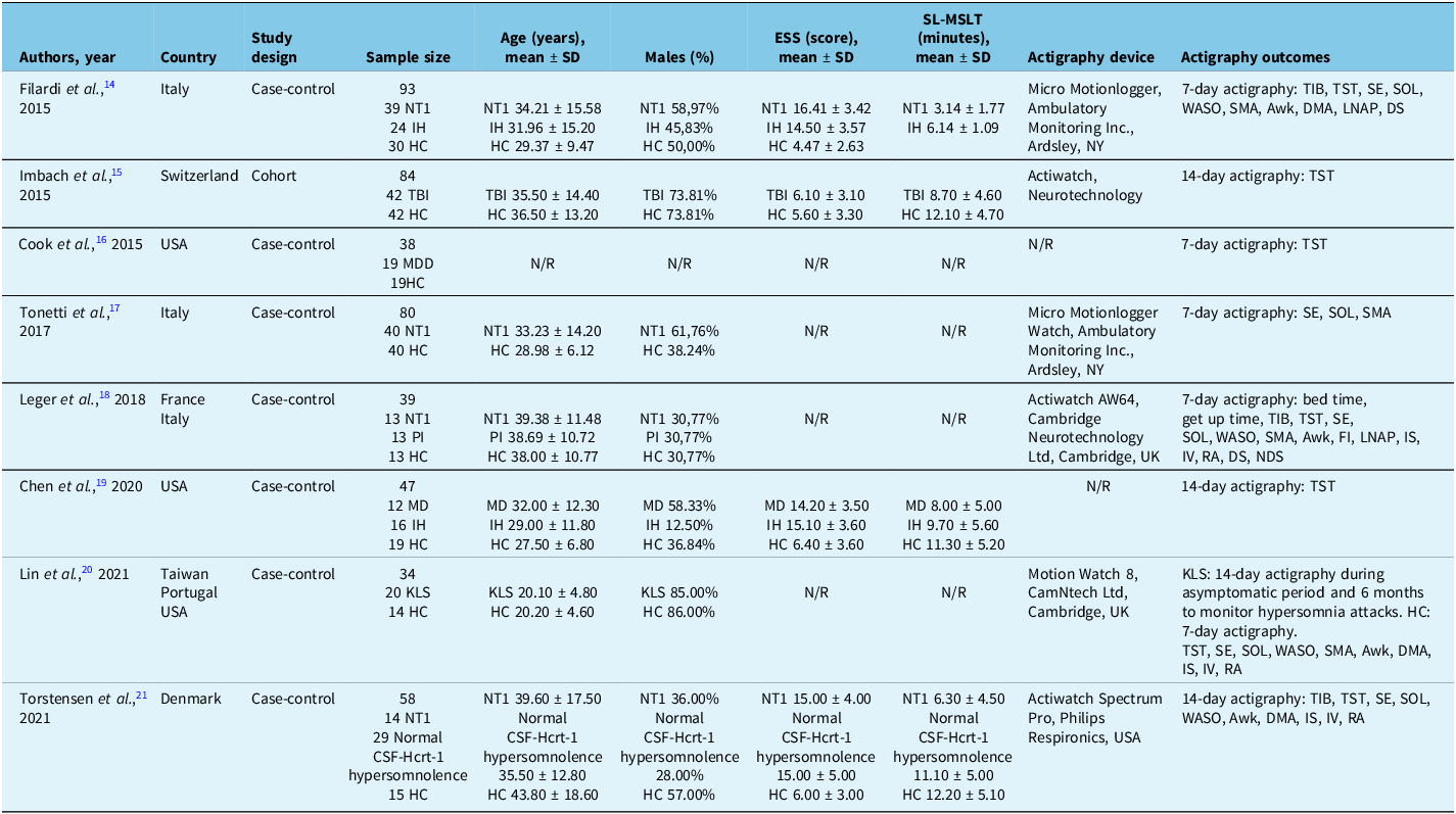
The selected studies were published between 2015 and 2021 and were conducted in different countries: Italy, Switzerland, the USA, France, Taiwan, Portugal and Denmark. In terms of study design, seven studies were case-control studies, and one was a cohort study. The main characteristics of all studies included in this review can be found in Table 1. The sample size ranged from 34 to 93 with a total sample size of 473 participants, consisting of 106 patients with NT1, 40 patients with IH, 29 patients with hypersomnolence and normal CSF hypocretin-1 (CSF-Hcrt-1), 20 patients with KLS, 42 patients with traumatic brain injury (TBI), 19 patients with major depressive disorder (MDD), 12 patients with myotonic dystrophy (MD), 13 patients with primary insomnia (PI) and 192 HC. Only one study did not specify the age and gender of the participants.Reference Cook, Goldstein and Plante16 The mean age was between 20 and 44 years, and the proportion of men ranged from 13% to 86%. Only four studies mentioned the ESS score and SL in MSLT.Reference Filardi, Pizza, Martoni, Vandi, Plazzi and Natale14 –Reference Imbach, Valko and Li15,Reference Chen, Trotti and Rye19,Reference Torstensen, Pickering, Kornum, Wanscher, Baandrup and Jennum21 Patients with NT1 had the highest mean ESS scores, and HC had the lowest scores (between 4 and 6). SL in MSLT was lower in patients with NT1.Reference Filardi, Pizza, Martoni, Vandi, Plazzi and Natale14,Reference Torstensen, Pickering, Kornum, Wanscher, Baandrup and Jennum21 In two studies, it was not mentioned whether the participants were taking medication that affects sleep.Reference Imbach, Valko and Li15,Reference Lin, Chin and Huang20 Four studies stated that all patients were medication-free or did not take any medication that affected their sleep.Reference Filardi, Pizza, Martoni, Vandi, Plazzi and Natale14,Reference Cook, Goldstein and Plante16–Reference Tonetti, Filardi, Plazzi and Natale17,Reference Torstensen, Pickering, Kornum, Wanscher, Baandrup and Jennum21 Two studies included patients who were taking medication (modafinil or sodium oxybate and flumazenil).Reference Leger, Gauriau and Tonetti18–Reference Chen, Trotti and Rye19
Characteristics of the studies included in the review

Awk = number of awake episodes; CSF-Hcrt-1 = CSF hypocretin-1; DMA = daytime motor activity; DS = discriminant score; ESS = Epworth Sleepiness Scale; FI = fragmentation index; HC = healthy control; IH = idiopathic hypersomnia; IS = interdaily stability; IV = intradaily variability, KLS = Kleine–Levin syndrome; LNAP = mean duration of longest sleep episodes during day; MD = myotonic dystrophy; MDD = major depressive disorder; MSLT = multiple sleep latency test; NDS = new discriminant score; N/R = not reported; NT1 = narcolepsy type 1; NY = New York; PI = primary insomnia; RA = relative amplitude; SD = standard deviation; SE = sleep efficiency; SL = sleep latency; SMA = sleep motor activity; SOL = sleep onset latency; TBI = traumatic brain injury; TIB = time in bed; TST = total sleep time; UK = United Kingdom; USA = Unites States of America; WASO = wake after sleep onset.
The Newcastle–Ottawa quality assessment scale for cohort and case-control studies was between five and eight stars. The quality ratings of the studies included in this review are shown in Tables S4 and S5 in the supplementary material.
Actigraphy
The actigraphs used in the studies analysed were Micro Motionlogger (Ambulatory Monitoring, Inc. NY),Reference Filardi, Pizza, Martoni, Vandi, Plazzi and Natale14,Reference Tonetti, Filardi, Plazzi and Natale17 Actiwatch (Neurotechnology),Reference Imbach, Valko and Li15 Actiwatch AW64 (Cambridge Neurotechnology Ltd., UK),Reference Leger, Gauriau and Tonetti18 Motion Watch 8 (CamNtech Ltd., UK)Reference Lin, Chin and Huang20 and Actiwatch Spectrum Pro (Philips Respironics, USA).Reference Torstensen, Pickering, Kornum, Wanscher, Baandrup and Jennum21 In two studies, the device used was not specified.Reference Cook, Goldstein and Plante16,Reference Chen, Trotti and Rye19 Regarding epoch length, three studies used 1-minute epochs,Reference Filardi, Pizza, Martoni, Vandi, Plazzi and Natale14,Reference Tonetti, Filardi, Plazzi and Natale17,Reference Lin, Chin and Huang20 while one study used 30-second epochs.Reference Torstensen, Pickering, Kornum, Wanscher, Baandrup and Jennum21 This issue was not mentioned in the other four studies.Reference Imbach, Valko and Li15–Reference Cook, Goldstein and Plante16,Reference Leger, Gauriau and Tonetti18–Reference Chen, Trotti and Rye19 In 4 studies, actigraphy recording lasted 7 daysReference Filardi, Pizza, Martoni, Vandi, Plazzi and Natale14,Reference Cook, Goldstein and Plante16–Reference Leger, Gauriau and Tonetti18 and 14 days in 3 studies.Reference Imbach, Valko and Li15,Reference Chen, Trotti and Rye19,Reference Torstensen, Pickering, Kornum, Wanscher, Baandrup and Jennum21 In Lin et al., HC used the actigraph for 7 days, while KLS patients used the device for 14 days during the asymptomatic period and for 6 months to monitor hypersomnia attacks. Only 12 out of 20 participants used the actigraph for 6 months.Reference Lin, Chin and Huang20 Participants in most of the studies included in this review completed a sleep diary.Reference Filardi, Pizza, Martoni, Vandi, Plazzi and Natale14–Reference Leger, Gauriau and Tonetti18,Reference Torstensen, Pickering, Kornum, Wanscher, Baandrup and Jennum21 However, in two studies, completion was not reported.Reference Chen, Trotti and Rye19–Reference Lin, Chin and Huang20 The actigraphy outcomes of each study are shown in Table S6 in the supplementary material. Three studies recorded TIB in NT1 and IH patients.Reference Filardi, Pizza, Martoni, Vandi, Plazzi and Natale14,Reference Leger, Gauriau and Tonetti18,Reference Torstensen, Pickering, Kornum, Wanscher, Baandrup and Jennum21 TST was measured in seven studies, involving NT1, IH, TBI, MDD, MD and KLS patients.Reference Filardi, Pizza, Martoni, Vandi, Plazzi and Natale14–Reference Cook, Goldstein and Plante16,Reference Leger, Gauriau and Tonetti18–Reference Torstensen, Pickering, Kornum, Wanscher, Baandrup and Jennum21 As for SE and SOL, these parameters were recorded in five studies, involving NT1, IH and KLS patients.Reference Filardi, Pizza, Martoni, Vandi, Plazzi and Natale14,Reference Tonetti, Filardi, Plazzi and Natale17–Reference Leger, Gauriau and Tonetti18,Reference Lin, Chin and Huang20–Reference Torstensen, Pickering, Kornum, Wanscher, Baandrup and Jennum21 WASO and Awk were recorded in NT1, IH and KLS patients in four studies.Reference Filardi, Pizza, Martoni, Vandi, Plazzi and Natale14,Reference Leger, Gauriau and Tonetti18,Reference Lin, Chin and Huang20–Reference Torstensen, Pickering, Kornum, Wanscher, Baandrup and Jennum21 SMA was assessed in four studies with NT1, IH and KLS patients.Reference Filardi, Pizza, Martoni, Vandi, Plazzi and Natale14,Reference Tonetti, Filardi, Plazzi and Natale17–Reference Leger, Gauriau and Tonetti18,Reference Lin, Chin and Huang20 DMA was analysed in three studies with NT1, IH and KLS patients.Reference Filardi, Pizza, Martoni, Vandi, Plazzi and Natale14,Reference Lin, Chin and Huang20–Reference Torstensen, Pickering, Kornum, Wanscher, Baandrup and Jennum21 IS, IV and RA were available in NT1 and KLS patients in three studies.Reference Leger, Gauriau and Tonetti18,Reference Lin, Chin and Huang20–Reference Torstensen, Pickering, Kornum, Wanscher, Baandrup and Jennum21 Two studies analysed LNAP and DS in NT1 and IH patients.Reference Filardi, Pizza, Martoni, Vandi, Plazzi and Natale14,Reference Leger, Gauriau and Tonetti18 Only one study assessed bedtime, wake-up time, FI and NDS in patients with NT1.Reference Leger, Gauriau and Tonetti18
Narcolepsy and idiopathic hypersomnia
Narcolepsy was the most frequently investigated CDH in the analysed studies. The diagnostic value of actigraphy for narcolepsy was evaluated in four studies.Reference Filardi, Pizza, Martoni, Vandi, Plazzi and Natale14,Reference Tonetti, Filardi, Plazzi and Natale17,Reference Leger, Gauriau and Tonetti18,Reference Torstensen, Pickering, Kornum, Wanscher, Baandrup and Jennum21 Filardi and co-authors investigated the role of actigraphy in differentiating patients with NT1 from IH and HC. According to their results, patients with NT1 had lower TST and SE than patients with IH and HC. WASO, Awk and SMA were higher in patients with NT1 than in the other two groups. During the day, patients with NT1 had lower DMA and higher LNAP than patients with IH and HC. This study presented a discriminatory score that included SMA, Awk and nap duration and showed positive (95%) and negative (87%) predictive value in NT1.Reference Filardi, Pizza, Martoni, Vandi, Plazzi and Natale14 Tonetti et al. demonstrated that simultaneous subjective and objective assessments, such as sleep logs and actigraphy, were more effective than separate assessments in screening patients with NT1, proving the value of actigraphy. In this study, SE, SOL and SMA differed significantly between NT1 and HC. Regardless of diagnostic evaluation, actigraphy correctly identified patients with NT1.Reference Tonetti, Filardi, Plazzi and Natale17 To confirm the results of Filardi and colleagues, Leger et al. investigated DS with a different actigraphy model and compared patients with NT1, PI and HC. FI and LNAP values were significantly higher in patients with NT1 than in PI and HC. DS was higher in patients with PI and NT1 than in HC. Thus, DS was not a strong discriminator for this group of patients. As a result, Leger et al. proposed an NDS that combined LNAP and FI and was shown to be higher in patients with NT1 than in the other two groups.Reference Leger, Gauriau and Tonetti18 One of the aims of the study by Torstensen and co-authors was to determine whether actigraphy can distinguish patients with NT1, patients with hypersomnolence with normal CSF-Hcrt-1 and HC. Regarding the sample of this study, 14 patients had NT1, and 29 patients had complaints of EDS, without cataplexy and with a CSF-Hcrt-1 > 200 pg/ml. These patients were subsequently diagnosed with NT2 (n = 1), IH (n = 2), KLS (n = 1), ISS (n = 4) and no sleep disorders (n = 21). When patients with NT1 were compared with those with hypersomnolence and normal CSF-Hcrt-1, the first group had higher WASO and IV and lower DMA, while the comparison between patients with NT1 and HC showed higher TIB, SOL, WASO, Awk and lower SE and DMA.Reference Torstensen, Pickering, Kornum, Wanscher, Baandrup and Jennum21
Kleine–Levin syndrome
With regard to KLS, only one study was included in this review, the aim of which was to monitor the movements of patients with KLS during an asymptomatic period and an attack of hypersomnia using actigraphy. Lin et al. found no significant differences between this patient group and the control group during the asymptomatic phase. The IS was lower in hypersomnia attacks than in the asymptomatic phase and in the controls. Another finding of Lin et al. was that the DMA was significantly lower at the onset of a hypersomnia attack. There were also other actigraphy variables, such as the mean rhythm – MESOR and the most active 10 hours – M10, which were only determined in this study. These parameters decrease at the beginning of the hypersomnia attack, showing that the activity level decreases before this time and recovers completely after the symptoms subside.Reference Lin, Chin and Huang20
Hypersomnia associated to medical disorder
Although, as already mentioned, hypersomnia in the context of medical conditions comprises several subgroups, our search found two studies that met all the required inclusion criteria.Reference Imbach, Valko and Li15,Reference Chen, Trotti and Rye19 Imbach and colleagues investigated sleep-wake disturbances in patients with TBI. The patients were assessed twice: immediately after the TBI with clinical, imaging and laboratory assessments and six months later with actigraphy, PSG, MSLT and laboratory assessment. The actigraphy showed that the TST was higher in patients with TBI than in HC.Reference Imbach, Valko and Li15 Patients with MD suffer from EDS. Therefore, Chen et al. investigated the TST by comparing MD, IH and HC patients and found that IH patients slept longer than MD patients and HC patients slept longer than IH patients.Reference Chen, Trotti and Rye19
Hypersomnia due to psychiatric disorder
Regarding hypersomnia due to psychiatric disorders, only one study on MDD was found.Reference Cook, Goldstein and Plante16
Cook et al. analysed sleep duration in patients with MDD and control subjects and found that TIB and TST were significantly higher in the first group.Reference Cook, Goldstein and Plante16
Meta-analysis
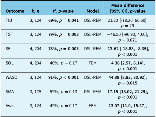
Only studies comparing patients with NT1 and HC were included in the meta-analysis, as this was the only comparison between CDH that included the same actigraphy outcome in at least three studies. Table 2 summarizes the meta-analysis data on the pooled absolute mean difference for the included outcomes. No significant differences were found between patients with NT1 and HC in terms of TIB and TST. We found a significantly lower SE in patients with NT1 compared to HC (absolute mean difference of -13.62; 95% CI -18.87 to -8.37; Figure 2); however, these patients showed a significantly higher SOL (absolute mean difference of 4.36; 95% CI 2.57–6.14; Figure 2), WASO (absolute mean difference of 44.88; 95% CI 8.83–80.92; Figure 2), SMA (absolute mean difference of 17.15; 95% CI 13.02–21.29; Figure 2) and Awk (absolute mean difference of 13.01; 95% CI 10.98–15.17; Figure 2).
Forest-plot on absolute mean difference on actigraphy outcomes between patients with narcolepsy type 1 and healthy controls. (A) - Sleep efficiency: a pooled absolute mean difference of -13.62 (95%CI of [−18.87, −8.37], p < .001) was observed, with significantly high heterogeneity between studies (I2 = 78%, p = .003). (B) - Sleep onset latency: a pooled absolute mean difference of 4.36 (95%CI of [2.57, 6.14], p < .001) was observed. No significant high heterogeneity between studies was obtained (I2 = 40%, p = .17). (C) - Wake after sleep onset: a pooled absolute mean difference of 44.88 (95%CI of [8.83, 80.92], p = .015) was observed, with significantly high heterogeneity between studies (I2 = 91%, p < .001). (D) - Sleep motor activity: a pooled absolute mean difference of 17.15 (95%CI of [13.02, 21.29], p < .001) was observed. No significant high heterogeneity between studies was obtained (I2 = 52%, p = .13). (E) – Awakenings: a pooled absolute mean difference of 13.07 (95%CI of [10.98, 15.17], p < .001) was observed, with no significantly heterogeneity between studies (I2 = 42%, p = .17).

Pooled main results for comparison of actigraphy outcomes between patients with narcolepsy type 1 and healthy controls

Values in bold are statistically significant. Difference expressed as the outcome for narcolepsy type 1 patients minus the outcome for healthy controls. k = number of studies included in the analysis; n = total number of participants included in the analysis; 95% CI = confidence interval at 95%; I 2 = heterogeneity; Awk = number of awake episodes; DSL-REM = DerSimonian–Laird random-effects model; FEM = fixed-effects model; SE = sleep efficiency; SMA = sleep motor activity; SOL = sleep onset latency; TIB = time in bed; TST = total sleep time; WASO = wake after sleep onset.
We found significantly high heterogeneity between studies (I 2 of 69%–91%) for all actigraphy outcomes assessed, with the exception of SOL (I2 of 40%, Figure 2), SMA (I 2 of 52%, Figure 2) and awakenings (I 2 of 42%, Figure 2). Sensitivity analysis for SE showed that removing the Leger et al. study reduced heterogeneity to 27% (p = 0.26). Using a fixed-effects model, we observed significantly lower SE in patients with NT1 compared to HC, with an absolute mean difference of -16.6 (95% CI -19.2 to -14.0; p < 0.001), similar to the results observed when all studies were included. As none of the meta-analyses included 10 or more studies, publication bias was not analysed using statistical tests. Funnel plots for each meta-analysis are shown in Figure S1 in the supplementary material.
Discussion
Summary of the findings
To our knowledge, this is the first systematic review evaluating the role of actigraphy in various CDH, and we have summarized the results of the studies on this topic. The actigraphy device used varied from study to study. The equipment used and the analysis software are crucial, as the methods of data acquisition, calculation and integration differ, leading to different actigraphy results.Reference Harneet, Walia and KHLaP22 Cook et al. support this claim by pointing out that estimates of sleep-wake parameters in actigraphy may vary between different scoring algorithms,Reference Cook and Plante23 and co-authors addressed this issue in their study and came to different conclusions than Filardi et al., with the actigraphy model being one of the causes for the results obtained.Reference Leger, Gauriau and Tonetti18 In all studies included in the review, the TST was the most commonly analysed actigraphy outcome and was found to be useful for discriminating between CDH.Reference Filardi, Pizza, Martoni, Vandi, Plazzi and Natale14–Reference Cook, Goldstein and Plante16,Reference Leger, Gauriau and Tonetti18–Reference Torstensen, Pickering, Kornum, Wanscher, Baandrup and Jennum21 Alakuijala et al. concluded that actigraphy is reliable in ruling out insufficient sleep in patients with narcolepsy and provides a more accurate TST assessment than PSG.Reference Alakuijala, Sarkanen, Jokela and Partinen24 As for the actigraphy outcomes, TST, SE and WASO were consistent when PSG was compared with actigraphy. However, the SOL result was not consistent.Reference Harneet, Walia and KHLaP22 Actigraphy has been shown to be useful in differentiating CDH compared to HC.Reference Filardi, Pizza, Martoni, Vandi, Plazzi and Natale14,Reference Torstensen, Pickering, Kornum, Wanscher, Baandrup and Jennum21 The notable differences between NT1 and HC were a decrease in TST, SE and DMA and an increase in WASO, Awk, SMA and LNAP.Reference Filardi, Pizza, Martoni, Vandi, Plazzi and Natale14,Reference Tonetti, Filardi, Plazzi and Natale17–Reference Leger, Gauriau and Tonetti18,Reference Torstensen, Pickering, Kornum, Wanscher, Baandrup and Jennum21 These results are similar to those of a recent systematic review evaluating the role of quantitative actigraphy in clinical sleep medicine.Reference Liguori, Mombelli and Fernandes25 Regarding DMA and LNAP, the decrease in DMA may be due to the increase in naps during the day in patients with NT1. In their new index DS, Filardi et al. combined actigraphic parameters from nighttime and daytime. This index has been shown to be effective in the diagnosis of NT1 as it reflects the disrupted nocturnal sleep and hypersomnolence that characterize this disorder.Reference Filardi, Pizza, Martoni, Vandi, Plazzi and Natale14 Subsequently, Leger and colleagues calculated an NDS that included LNAP and FI and distinguished NT1 patients from HC and PI patients. However, patients were not drug-free at the time of actigraphy recording.Reference Leger, Gauriau and Tonetti18 The main finding related to IH was increased LNAP compared to HC.14 These results are consistent with the literature: One criterion for IH diagnosis is a 24-hour total sleep time of ≥ 660 minutes as measured by 24-hour PSG or actigraphy. Here, actigraphy can be a useful tool due to its simple technique and recording.1 With regard to KLS, the study included in this review found no differences in actigraphy findings between HC and KLS patients during the asymptomatic period. However, DMA decreased at the onset of the hypersomnia attack, then increased in the late phase of the attack and recovered thereafter. Lin et al. found that decreased IS can be explained by a loss of stability of the circadian diurnal profile and may be a sign of incomplete recovery.Reference Lin, Chin and Huang20 The most important observation in patients with TBI was an increased TST. The need for neuronal plasticity may explain the increased need for sleep.Reference Imbach, Valko and Li15 In MD patients, TST was lower than in IH patients.Reference Chen, Trotti and Rye19 Increased TST was also observed in MDD patients.Reference Cook, Goldstein and Plante16
Other considerations
Actigraphy may also be used as a confirmatory tool for the evaluation of patients with hypersomnolence. Kretzschmara et al. have shown that actigraphy plays a more important role in the diagnosis of insufficient sleep, as these patients tend to overestimate their sleep in their medical history and sleep diaries. In the case of IH, the authors found that these patients described their sleep habits more accurately, with actigraphy serving as a confirmatory tool. Although no study of ISS was included in this review, it is very common, and actigraphy helps to distinguish it from other CDH.Reference Kretzschmar, Werth, Sturzenegger, Khatami, Bassetti and Baumann26 There is a lack of knowledge about the best actigraphy parameters for use in CDH. To fill this gap, Leger et al. found that seven days of actigraphic monitoring is sufficient for patients with NT1.Reference Leger, Tonetti and Gauriau27 Cook et al. also investigated the setting configuration for TST estimation in IH patients. Their results showed that a 25-second epoch parameter configuration was superior to the 30-second configuration for estimating the TST in IH patients.Reference Cook, Eftekari, Leavitt, Prairie and Plante28
Future directions
Since Filardi et al. and Leger et al. have presented DS and NDS, it is likely that functions of this type will be developed in the future. DS and NDS need further investigation to determine their diagnostic capabilities in CDH and whether the results can be generalized to all actigraphy models. NDS testing in drug-naïve patients with NT1 should also be performed. According to a review of actigraphy published by the AASM, actigraphy settings are fundamental to accurate data collection.Reference Smith, McCrae and Cheung6 Future studies should focus on closing this gap for all CDH. Currently, several devices on the market must be used with extreme caution, as they may lack a validated sleep assessment algorithm and fail to ensure accurate data acquisition. As a result, these devices have not been validated for clinical use and are not recommended for such applications. However, with ongoing advancements in digital health technologies, it is anticipated that many of these devices will undergo rigorous validation processes and eventually receive approval for CDH assessment in the future.Reference Ancoli-Israel, Martin and Blackwell29
According to Lammers et al., a new classification for CDH is needed. In fact, CDH encompasses a wide range of disorders for which there is neither a central cause nor a known causal or comorbid relationship.Reference Lammers, Bassetti and Dolenc-Groselj3 Actigraphy could play a more significant role in the assessment of CDH if the classification of sleep into three distinct categories were streamlined, as has been suggested in existing studies of narcolepsy and IH patients. Simplifying the categorization could improve the accuracy and clinical utility of actigraphy in assessing sleep patterns and differentiating between these disorders.
Limitations
Limitations of our review include the lack of studies on narcolepsy type 2 (NT2), IH with short sleep time (ISS) and hypersomnia due to medication or substance misuse, as well as the limited number of publications on the included cases of chronic hypersomnia disorders (CHD). In addition, our review revealed considerable variability between studies, particularly in the actigraphy data recorded. This variability limited the inclusion of actigraphy results in the quantitative synthesis, so that only comparisons between NT1 patients and HC could be analysed – and even then only for a limited number of parameters. The inclusion of 3–4 studies for each outcome further limits the interpretation of our pooled data, namely, when a high heterogeneity was observed, and should be regarded with caution. In addition, most of our meta-analyses showed significant heterogeneity, which could be due to differences in the actigraphs used and variability in data acquisition procedures across studies. This could introduce a potential source of bias. However, we believe that this is an acceptable limitation given the current state of the art. The establishment of standardized guidelines for actigraphy data collection could help to reduce these inconsistencies, improve comparability and ensure greater reliability of results in future research.
Conclusion
This review highlights the value of actigraphy as an objective tool to assess sleep in patients with CHD. In combination with clinical evaluation, actigraphy can help to distinguish between different CHD subtypes, in particular NT1, IH and ISS. However, the absence of standardized protocols for data collection and interpretation limits the systematic integration of this accessible technique into clinical practice.
Supplementary material
The supplementary material for this article can be found at https://doi.org/10.1017/cjn.2025.10114
Author contributions
SM: Conceptualization, data curation, formal analysis, interpretation of data, writing original draft. JIS: Conceptualization, data curation, formal analysis, interpretation of data, writing original draft. DFB: Formal analysis, interpretation of data, writing review and editing. JCL: Conceptualization, formal analysis, interpretation of data, writing review and editing. MG: Conceptualization, writing review and editing.
Funding statement
The authors received no financial support for the research, authorship or publication of this article.
Competing interests
None.