INTRODUCTION
The family Patellidae has been recognized as a complex taxonomic group for many years (Fisher-Piette, Reference Fisher-Piette1938), and its poor fossil record has complicated the study of the group (Ridgway et al., Reference Ridgway, Reid, Taylor, Branch and Hodgson1998). Nevertheless, there are many recent studies on taxonomic and phylogenetic topics (Corte-Real et al., Reference Corte-Real, Hawkins and Thorpe1996; Ridgway et al., Reference Ridgway, Reid, Taylor, Branch and Hodgson1998; Koufopanou et al., Reference Koufopanou, Reid, Ridgway and Thomas1999; Weber & Hawkins, Reference Weber and Hawkins2002, Reference Weber and Hawkins2003, Reference Weber and Hawkins2005; Nakano & Ozawa, Reference Nakano and Ozawa2004, Reference Nakano and Ozawa2007; Sá-Pinto et al., Reference Sá-Pinto, Branco, Harris and Alexandrino2005; Espinosa & Ozawa, Reference Espinosa and Ozawa2006; Nakano et al., Reference Nakano, Yazaki, Kurokawa, Yamaguchi and Kuwasawa2009). Although limpet fauna has been extensively studied in the north-east Atlantic, South Africa, North America, Australia–New Zealand and the Far East, poor attention has been conferred to West Africa. There is a lack of knowledge on ecological, taxonomic and genetic studies of West African limpets. One of these limpet species, Cymbula nigra (da Costa, 1771), has its distributional range from Namibia to the Mediterranean (Hodgson et al., Reference Hodgson, Ridgway, Branch and Hawkins1996; Koufopanou et al., Reference Koufopanou, Reid, Ridgway and Thomas1999). Christiaens (Reference Christiaens1973) mentioned two varieties of Cymbula nigra (formerly named Patella nigra): Patella nigra plumbea (typical form from Senegal) and Patella nigra ghananis (variety from Ghana). This author synonymized Patella plumbea Lamarck, 1819 and P. safiana Lamarck, 1819 into P. nigra da Costa, 1777, although Powell (Reference Powell1973) considered both taxa as a different species. The pictures included in Christiaens (Reference Christiaens1973) are clearly different, with a typical shell form of C. nigra in the former variety (identical to those observed in Morocco and the Mediterranean, personal observation), and a smaller shell in the latter. Morphological (Hodgson et al., Reference Hodgson, Ridgway, Branch and Hawkins1996) and genetic studies (Koufopanou et al., Reference Koufopanou, Reid, Ridgway and Thomas1999; Nakano & Ozawa, Reference Nakano and Ozawa2007) have pointed out that specimens from Angola and the Mediterranean are the same species (named Cymbula safiana by these authors) and belong to the genus Cymbula instead of the genus Patella. However, following the International Code of Zoological Nomenclature (Article 23) the specific name has to be the oldest name nigra (Moreno & Arroyo, Reference Moreno, Arroyo, Barea-Azcón, Ballesteros-Duperón and Moreno2008), therefore, the valid name for the taxon would be Cymbula nigra. The aim of the present study was to confirm if the varieties from West Africa belong to the taxon Cymbula nigra.
MATERIALS AND METHODS
Sampling collection
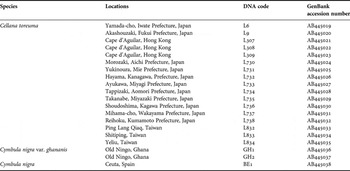
The species collected in the present study are shown in Table 1. Several specimens of Cymbula nigra from different localities of the Strait of Gibraltar were collected and their morphological characters were studied. After these observations were done, one specimen from Ceuta (North Africa, Strait of Gibraltar) was selected taking into account that all the specimens were morphologically similar. On the other hand, two specimens labelled as Cymbula nigra from Old Ningo, Ghana (West Africa) were collected by Dr David Reid (Natural History Museum, London). Additionally, several specimens of Cymbula nigra (collected in Ghana by Dr José Templado and deposited in the National Museum of Natural Sciences, Madrid, Spain) were studied morphologically. The genetic study on those samples was not possible due to their formalin preservation.
Sampling sites and species collected.

DNA extraction, PCR amplification and DNA sequencing
Living specimens were preserved in 95% ethanol. Total DNA was extracted from a fragment of the mantle or foot muscle tissue, either using High Pure PCR Template Preparation Kit (Roche) or a standard phenol/chloroform extraction method. In the latter procedure, a small amount of tissue was treated with 200 µl of TEN buffer (10 mM Tris, pH 8.0, 10 mM EDTA and 10 mM NaCl), 20 µl of 10% SDS, and 40 µl of proteinase K (20 mg/ml) at 37°C for 1–2 hours with gentle rotation, followed by phenol/chloroform and chloroform extractions, and ethanol precipitation.
Amplification of a part of the cytochrome-c oxidase I (COI) was achieved with the LCO1490 (5′–GGTCAACAAATCATAAAGATATTGG–3′) and HCO2198 (5′–TAAACTTCAGGGTGACCAAAAAATCA–3′) primer pair described by Folmer et al. (Reference Folmer, Black, Hoeh, Lutz and Vrijenhoek1994). PCR amplification was performed in 25 µl of reaction volume containing 10 mM Tris–HCl pH 8.3, 50 mM KCL, 1.5 mM MgCl2, 200 µM dNTPs, 0.2 µM each primer, 0.5 mg/ml BSA (Sigma), 2 units of Taq polymerase (Takara), and 1 µl of template DNA solution. The cycling parameters for amplification consisted of an initial denaturation for 3 minutes at 94°C; followed by 30 cycles of denaturation for 45 seconds at 94°C, annealing for 90 seconds at 54°C, and extension for 120 seconds at 72°C; and ended with a 5 minutes extension at 72°C. Amplification products were purified using High Pure PCR Product Purification Kit (Roche). Direct double-stranded cycle sequencing of 25 to 30 ng of COI PCR product was performed in both directions using the Applied Biosystems BigDye vr.3 dye terminator cycle sequencing kit. Cycle sequencing was performed using an Applied Biosystems GeneAmp PCR System 9700. The cycling parameters were 25 cycles of 10 seconds at 96°C, 5 seconds at 50°C, and 4 minutes at 60°C. Sequencing reaction products were purified using ethanol precipitation and analysed on an ABI PRISM 377 DNA sequencer.
Sequence analysis and phylogeny reconstruction
We determined the nucleotide sequences of a fragment of COI exactly 658bp. Sequence of COI was aligned using MacClade 4.03 (Maddison & Maddison, Reference Maddison and Maddison2002) referring to translated amino acid sequence. Third codon positions of COI sequences were retained. Distance analysis was carried out using the neighbour-joinning (NJ) algorithm (Saito & Nei, Reference Saito and Nei1987) with Kimura 2-p model and 1000 bootstrap replicates to assess the stability of each node.
Haplotype analysis based on the sequence of a fragment of COI was undertaken. The haplotypes were defined using the ‘Redundant Taxa’ option in MacClade 4.03 (Maddison & Maddison, Reference Maddison and Maddison2002), and the minimum spanning tree (MST) (Kruskal, Reference Kruskal1956; Prim, Reference Prim1957) was constructed using ARLEQUIN version 2.0 (Schneider et al., Reference Schneider, Roessli and Excoffier2000). The MST is computed from the matrix of pairwise distances calculated between all pairs of haplotypes using a modification of the algorithm described in Rohlf (Reference Rohlf1973). The MST is convertible to a minimum spanning network (MSN).
RESULTS
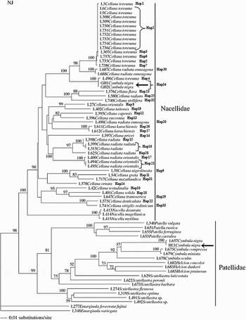
The specimens of Cymbula nigra from Ghana showed the same haplotype (Figures 1 & 2) but both belong to a different species than those specimens from Ceuta because of the great genetic distance found (186 substitutions). To establish the species identity of these sequences, a NJ tree (Figure 1) was constructed with some sequences from Nakano & Ozawa (Reference Nakano and Ozawa2007) and Nakano et al. (Reference Nakano, Yazaki, Kurokawa, Yamaguchi and Kuwasawa2009) (the codes used by these authors have been maintained. Note that individuals L3, L687, L688 and L496 had been previously sequenced in Nakano & Ozawa, Reference Nakano and Ozawa2004, Reference Nakano and Ozawa2007). We have included three sequences of C. nigra in the NJ tree: the individual BE1 (from Ceuta, Spain) and the individuals GH1–GH2 (from Old Ningo, Ghana), showing a very different sequence of the COI. The sample from Benzú (Ceuta, Spain) was grouped within the genus Cymbula as it was expected. Furthermore, the specimen was grouped with a sample of Cymbula nigra also from Ceuta sequenced previously by Nakano & Ozawa (Reference Nakano and Ozawa2007). However, the sample from Old Ningo (Ghana) was grouped closely with the species Cellana toreuma, indicating that both specimens from Ghana belong to the genus Cellana instead of Cymbula.
Neighbour-joining tree based on sequences of 658 bp obtained by Nakano & Ozawa (Reference Nakano and Ozawa2007) for COI fragment (the codes for each sequence have been maintained like in the original paper) with the inclusion of new sequences obtained for Cellana species and C. nigra from Benzú (Ceuta, Spain) and Old Ningo (Ghana) of individuals BE1 and GH1–GH2 respectively. Arrows indicate the position of each sequence in the tree. Note the position of the specimens GH1 and GH2 from Ghana. Corresponding haplotypes are indicated (see also Figure 2). Numbers on each node indicate bootstrap values.

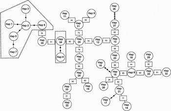
Minimum spanning network of the haplotypes found for Cellana species (same sequences as for phylogenetic tree of Figure 1). Dotted lines indicate the specimens identified as Cellana toreuma. Haplotypes 1–3 and 5–7 come from Japan, Taiwan and Hong Kong, whereas haplotype 4 comes from Java, Indonesia and haplotype 34 are the specimens GH1 and GH2 from Ghana.

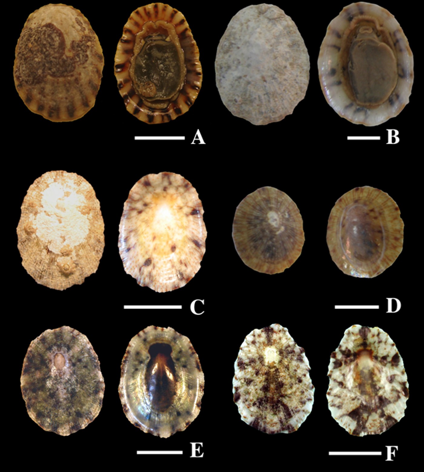
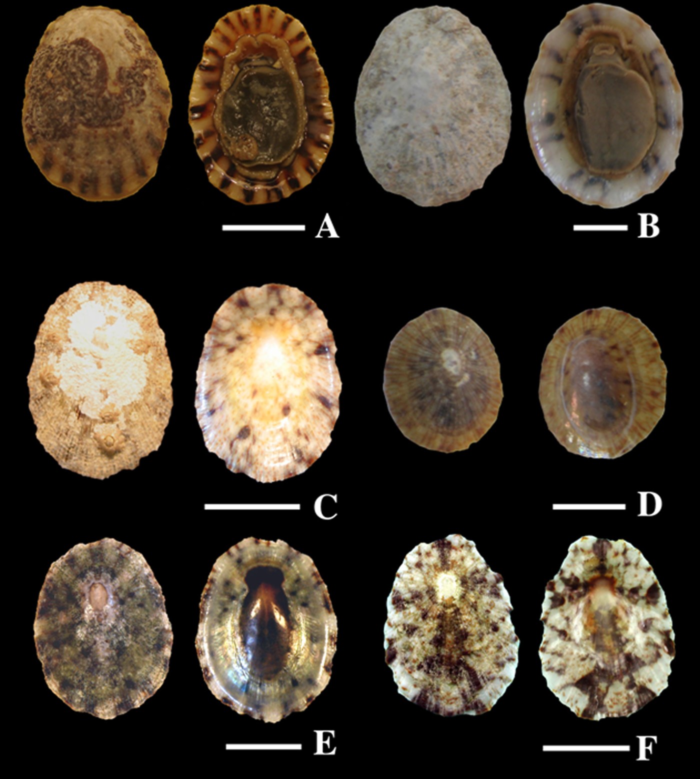
In order to clarify the taxonomic status of these individuals, a MST analysis was carried out with several sequences of Cellana species (from Nakano & Ozawa, Reference Nakano and Ozawa2004, Reference Nakano and Ozawa2007, Nakano et al., 2009), including those of Cellana toreuma sequenced during the present study (Figures 1 & 2). Specimens GH1 and GH2 from Ghana were grouped closely with C. toreuma from Java (Indonesia) and, subsequently, the MSN showed that specimens from Ghana are the same species as those from Java (only two substitutions of difference between haplotypes 4 and 34; see Figure 2). Conversely, C. toreuma from Japan–Taiwan–Hong Kong belong to a different species. Morphological characters of different specimens studied in the present study are shown in Figure 3.
(A) Cymbula nigra, specimen BE1 (Ceuta); (B) C. nigra, specimen from the National Museum of Natural Sciences, Madrid, Spain (Ghana); (C & D) Cellana sp., specimens from the Natural History Museum, London, UK (Ghana); (E) Cellana sp. (Java, Indonesia); (F) Cellana toreuma (Japan). Scale bars 1 cm.
DISCUSSION
The samples from Ghana belong to the genus Cellana, and would represent the first record of this genus in the Atlantic. Morphologically, the specimens are similar to C. toreuma species according to the pictures and description included in Powell (Reference Powell1973). However, this species is distributed in the Far East (Japan, China, Taiwan and Philippines) according to this author. In the same way, we have also identified the specimen from Java (Indonesia), which is closely related with the samples from Ghana, as C. toreuma based on morphological characters of the shell. Nevertheless, the genetic data obtained do not support this contention. The data indicate clearly that specimens from Java and Ghana belong to a different and cryptic species of C. toreuma. There are many works that have revealed cryptic species using molecular techniques within molluscan groups (Scutellastra, Paulay & Meyer, Reference Paulay and Meyer2002; Patelloida, Kirkendale & Meyer, Reference Kirkendale and Meyer2004, Nakano & Ozawa, Reference Nakano and Ozawa2005; Monodonta, Donald et al., Reference Donald, Kennedy and Spencer2005; Astraliun, Meyer et al., Reference Meyer, Geller and Paulay2005; Tricolia, Williams & Ozawa, Reference Williams and Ozawa2006 among others). In this context, it would be plausible to surmise, although our results are preliminary, that the two varieties mentioned by Christiaens (Reference Christiaens1973) of Cymbula nigra: Patella nigra plumbea (typical form from Senegal) and Patella nigra ghananis (variety from Ghana) were different species. The pictures included in this work are clearly different, with a typical shell form of C. nigra in the former variety (identical to those collected in the present study), and a smaller and more similar to Cellana toreuma shell in the latter (similar to both individuals sequenced from Ghana; see Figure 3). We cannot discard the fact that the variety from Ghana was really a Cellana species that could be broadly spread in West Africa, taking into account that Christiaens (Reference Christiaens1973) referred that he studied a great number of limpets from Ghana and all of those belong to the var. nov. Patella nigra ghananis. Additionally, the proximity between specimens GH1–GH2 from Ghana and L496 from Java (see Figures 1 & 2) revealed that this fact can be due to a recent introduction of this species in the Atlantic Ocean; otherwise, the genetic differences should be greater. In this sense, limpets with two different and distant distributional areas due to introduction events are not a new record (see Voss, Reference Voss1959; Baker et al., Reference Baker, Baker and Fajans2004 for the case of Siphonaria pectinata in the Gulf of Mexico/Florida and Southern Europe/Western Africa). Nakano & Ozawa (Reference Nakano and Ozawa2007) reported the specimens of C. toreuma from Japan and Indonesia as very close in their phylogenetic analyses and, apparently, belonging to the same species. This could be due to the fact that those analyses did not include the new sequence of Cellana radiata enneagona from Ogasawara (Bonin) Island, south-eastern Japan (see Figures 1 & 2, specimen L687; haplotype 30) that is inserted between the two C. toreuma groups (Nakano et al., Reference Nakano, Yazaki, Kurokawa, Yamaguchi and Kuwasawa2009). The similar tropical distribution of the specimens from Indonesia and Ghana could have facilitated the introduction of the species in West Africa from Indonesia as fouling or as larval stage in ballast waters. Further studies are required on samples from Ghana to clarify the presence of Cellana sp. and its relative abundance in this area to establish whether it is the consequence of a recent introduction or, on the contrary, this area has been its natural distributional area. Additionally, comparative studies need to be undertaken with samples from Indonesia to establish the taxonomic status of this species.
ACKNOWLEDGEMENTS
We express our thanks to Dr David G. Reid (Natural History Museum of London, UK) and Dr José Templado (Museo Nacional de Ciencias Naturales, Spain) for contributing specimens studied in the context of this work. Our gratitude to Autoridad Portuaria de Ceuta and Grant-in-Aid for JSPS Fellows no. 207024 to T. Nakano from the Japan Society for Promotion of Science for financial support. All material has been collected under appropriate collection and approved ethics guidelines.