Introduction
Studies of the mechanical properties of nanoporous (NP) metals have a twofold motivation. First, their performance as functional or structural materials requires strength, plastic deformability, and high values of either stiffness or compliance, depending on the application. Documenting and understanding the mechanical behavior is mandatory for establishing the required mechanical performance. Second, and of equal relevance, dealloying-made NP metals offer exciting opportunities for exploring and unraveling the mechanical behavior of small-scale systems in general. High sample-to-sample reproducibility and the availability of samples in the size regime from millimeter to centimeter, suitable for reliable macroscopic testing schemes, enable particularly meaningful investigations of small-scale plasticity. Furthermore, in-electrolyte tests afford an in situ variation of the surface state, providing unique access to the signatures of surface contributions to the elastic, plastic, and fracture behavior.
Fundamental observations of stiffness, strength, and fracture
Compression tests of nanoporous gold
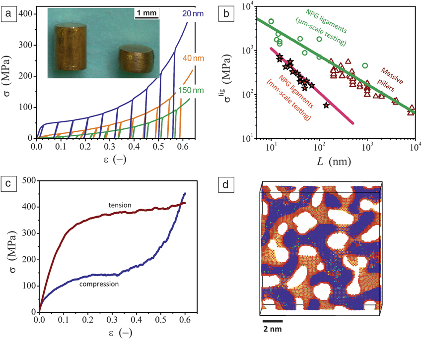
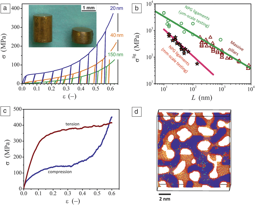
Figure 1 summarizes the plastic deformation behavior of nanoporous gold (NPG). As seen in Figure 1a (inset), the material can be deformed continuously to large plastic strain in compression without the formation of cracks.Reference Mameka, Wang, Markmann, Lilleodden and Weissmüller1 The experimental compression stress–strain curves show the systematic strengthening at small size and the pronounced strain hardening.Reference Mameka, Wang, Markmann, Lilleodden and Weissmüller1 This particular data set also exhibits unload–load sections that allow a precise measurement of Young’s modulus in each deformation state. The yield strength is also typically extracted from such data.
Stress–strain behavior and strength of nanoporous gold (NPG). (a) Experimental compressive stress–strain curves for millimeter-sized samples with various ligament sizes. Inset: sample before and after compression.Reference Mameka, Wang, Markmann, Lilleodden and Weissmüller1,Reference Mameka, Markmann, Jin and Weissmüller4 (b) Experimental results for apparent local ligament strength σlig of NPG ligaments versus ligament size L. Separate data sets from micrometer-scale and millimeter-scale tests on NPG. Also shown are results from compression tests on focused-ion-beam-machined gold pillars.Reference Mameka, Wang, Markmann, Lilleodden and Weissmüller1 (c) Molecular dynamics results comparing stress–strain behavior of NPG in compression and tension.Reference Farkas, Caro, Bringa and Crowson17 (d) Slice through a simulated NPG sample of 2-nm ligament diameter showing the effects of surface stress. Atoms in red are under tensile stresses greater than 2 GPa, mostly located at the surface of the ligaments. Atoms in blue are under compressive stress greater than 2 GPa and are located inside ligaments.Reference Crowson, Farkas and Corcoran20
Uniform deformation, no-crush bands
The compression stress–strain curve of conventional low-density open-cell metal foams typically shows a linear elasticity region, followed by a long plateau that is associated with the propagation of localized collapse or crush bands, and finally, a densification region where almost all pores have collapsed and the stress rises steeply.Reference Gibson and Ashby2 Figure 1a illustrates that NPG exhibits a different compression behavior:Reference Mameka, Wang, Markmann, Lilleodden and Weissmüller1,Reference Jin, Kurmanaeva, Schmauch, Rosner, Ivanisenko and Weissmüller3–Reference Ngô, Stukowski, Mameka, Markmann, Albe and Weissmüller5 the “elasticity” region is often nonlinear and involves pre-yielding; the stress plateau is replaced by a hardening region in which the stress increases gradually with strain; the onset of densification is also ill-defined.
The continuous hardening in NPG is associated with uniform densification in compression, as observed in electron backscatter diffraction (EBSD) images of compressed NPG samples. Reference Jin, Kurmanaeva, Schmauch, Rosner, Ivanisenko and Weissmüller3 Localized crush bands are not observed.Reference Jin, Kurmanaeva, Schmauch, Rosner, Ivanisenko and Weissmüller3 Most pores in NPG shrink simultaneously along the compression direction, but do not completely close, even at high compressive strains.
Dislocation accumulation
The deformation behavior of idealized individual nano-objects is often discussed in terms of dislocation starvation,Reference Greer and Nix6 where dislocations form by nucleation and are removed by egress after crossing the crystal. Jin et al.Reference Jin, Kurmanaeva, Schmauch, Rosner, Ivanisenko and Weissmüller3 provide EBSD images that suggest dislocation accumulation in NPG. Dislocation accumulation is also directly observed by molecular dynamics (MD) simulationsReference Ngô, Stukowski, Mameka, Markmann, Albe and Weissmüller5 and by transmission electron microscopy,Reference Dou and Derby7,Reference Dou and Derby8 and it is indirectly evidenced by studies of the strain-hardening behavior in finite element modeling (FEM).Reference Huber, Viswanath, Mameka, Markmann and Weissmueller9
Smaller is stronger
Figure 1b shows a compilation of experimental strength data from several studies of NPG with different ligament size, L.Reference Mameka, Wang, Markmann, Lilleodden and Weissmüller1,Reference Jin, Kurmanaeva, Schmauch, Rosner, Ivanisenko and Weissmüller3,Reference Biener, Hodge, Hayes, Volkert, Zepeda-Ruiz, Hamza and Abraham10–Reference Volkert and Lilleodden12 Note that the data refer to the local yield stress at the ligament level. This is obtained by factoring out the effect of the network geometry with the help of the uncorrected Gibson–Ashby scaling equation (see the “Strength of nanoporous gold: Scaling behavior and size effects” section in this article). The results fall in two distinct groups, one suggesting weaker behavior and a larger size exponent than the other. Remarkably, the data within each group are highly consistent. The figure also shows that the group exhibiting higher strength shows good agreement with the size-dependent strength of single-focused-ion-beam (FIB)-cut micropillars.Reference Greer and Nix6,Reference Volkert and Lilleodden12 The stronger group relates to early work using micrometer-scale testing schemes such as nanoindentation,Reference Biener, Hodge, Hayes, Volkert, Zepeda-Ruiz, Hamza and Abraham10–Reference Hakamada and Mabuchi14 whereas the second group utilizes more recent and arguably more reliable conventional tests on millimeter-sized samples.Reference Mameka, Wang, Markmann, Lilleodden and Weissmüller1,Reference Jin, Kurmanaeva, Schmauch, Rosner, Ivanisenko and Weissmüller3,Reference Buerckert, Briot and Balk15,Reference Balk, Eberl, Sun, Hemker and Gianola16 The low solid fraction in the latter experiments may be significant.
Tension–compression asymmetry
The MD simulation results (Figure 1c) emphasize another important aspect of the mechanical behavior of NPG—the material is significantly stronger in tension than in compression,Reference Farkas, Caro, Bringa and Crowson17,Reference Crowson, Farkas and Corcoran18 reflecting a tension–compression asymmetry that has been found in MD studies on plasticity of individual nanowires.Reference Diao, Gall, Dunn and Zimmerman19 For nanoscale face-centered-cubic (fcc) metals, the asymmetry is often attributed to the large tensile stress in the surface atomic layer and to the ensuing compressive stress in the interior of nano-objects (Figure 1d).Reference Diao, Gall, Dunn and Zimmerman19–Reference Weinberger and Cai23
Most plastic deformation results to date for NPG in tension come from computer simulations. Experimental tension tests show brittle failure in the elastic regime,Reference Balk, Eberl, Sun, Hemker and Gianola16,Reference Badwe, Chen and Sieradzki24 except for ligament sizes of a few hundred nanometers.Reference Badwe, Chen and Sieradzki24 The early failure in tension can be understood in terms of classic fracture mechanics concepts for brittle materials.Reference Badwe, Chen and Sieradzki24 However, inspection of the fracture surfaces reveals large plastic tensile deformation at the scale of individual ligaments.Reference Hodge, Hayes, Caro, Biener and Hamza13 Furthermore, compression of random networks such as NPG under uniaxial load involves a distribution of strain states in individual ligaments, including ligaments that are deformed plastically (and locally ductile) in tension.Reference Lührs, Zandersons, Huber and Weissmüller25
A benchmark for effective elasticity: Foam scaling equations
Figure 2 summarizes experimental data for the networks’ effective macroscopic Young’s modulus, Y eff, complementing the previously discussed results. A natural starting point for discussing these data is the Gibson–Ashby scaling relation for the effective Young’s modulus of open cell foams:Reference Gibson and Ashby2
 $${Y^{{\rm{eff}}}} = {Y^{{\rm{lig}}}}{{\rm{\varphi }}^2}$$,
where Y lig is the Young’s modulus of individual ligaments and φ is the solid volume fraction. The theoretical basis for the Gibson–Ashby laws holds for porous aggregates with φ < 0.1.Reference Gibson and Ashby26 Empirically, however, good agreement is found at any volume fraction for various microscale foams.Reference Gibson and Ashby26 As a matter of routine, studies of the elasticity of NPG with φ on the order of 0.3 use the scaling Equation 1 or its equivalent for the strength as a benchmark.Reference Jin, Kurmanaeva, Schmauch, Rosner, Ivanisenko and Weissmüller3,Reference Huber, Viswanath, Mameka, Markmann and Weissmueller9,Reference Biener, Hodge, Hayes, Volkert, Zepeda-Ruiz, Hamza and Abraham10,Reference Volkert and Lilleodden12,Reference Biener, Hodge, Hamza, Hsiung and Satcher27–Reference Briot and Balk29
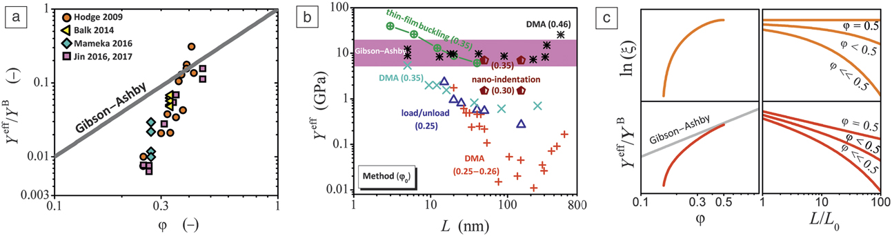
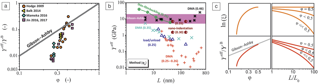
Macroscopic Young’s modulus of nanoporous gold (NPG). (a) Effective Young’s modulus Y eff (normalized to bulk Au, Y B) of as-prepared NPG Reference Mameka, Wang, Markmann, Lilleodden and Weissmüller1,Reference Briot, Kennerknecht, Eberl and Balk30–Reference Liu and Jin33 versus solid volume fraction φ. Note the systematic deviation from the Gibson–Ashby scaling law at small φ.Reference Mameka, Wang, Markmann, Lilleodden and Weissmüller1,Reference Briot, Kennerknecht, Eberl and Balk30–Reference Liu and Jin33 (b) Variation of Y eff when the ligament size, L, varies with coarsening by thermal annealing. Data for various samples with initial solid fraction, φ0, estimated from the Au atom fraction in the master alloy.Reference Mameka, Wang, Markmann, Lilleodden and Weissmüller1,Reference Hodge, Doucette, Biener, Biener, Cervantes and Hamza31–Reference Mathur and Erlebacher34 Colored region indicates Gibson–Ashby prediction with φ varying in the range of 0.25∼0.50. DMA: Dynamic mechanical analysis. (c) Schematic variation of connectivity density in NPG with φ, and with L during coarsening. Top row shows variation of scaled connectivity density ξ with φ in as-prepared NPG and with ligament size, L, which represents an increase over L 0 of the as-prepared network during coarsening. Bottom row shows impact on the effective Young’s modulus Y eff/Y B.
Anomalous variation of compliance with solid fraction
Figure 2a compares the scaling law to data for Y eff of NPG samples from different studiesReference Mameka, Wang, Markmann, Lilleodden and Weissmüller1,Reference Briot, Kennerknecht, Eberl and Balk30–Reference Liu and Jin33 plotted versus φ. The data were selected to include only as-prepared samples, ignoring samples where the ligament size was varied by annealing. At higher solid fractions, the data were compatible with the Gibson–Ashby scaling equation.Reference Hodge, Doucette, Biener, Biener, Cervantes and Hamza31,Reference Mathur and Erlebacher34 Remarkably, NPG with φ ≤ 0.3 is much more compliant than Gibson–Ashby networksReference Hodge, Doucette, Biener, Biener, Cervantes and Hamza31,Reference Mathur and Erlebacher34 and Y eff decays significantly faster when the density of as-dealloyed NPG is reduced.Reference Hodge, Doucette, Biener, Biener, Cervantes and Hamza31 For φ ≤ 0.3, Y eff is found to be up to 10× lower than predicted by the scaling relations (see Figure 2b).Reference Mameka, Wang, Markmann, Lilleodden and Weissmüller1,Reference Ngô, Stukowski, Mameka, Markmann, Albe and Weissmüller5,Reference Huber, Viswanath, Mameka, Markmann and Weissmueller9 Furthermore, increasing L of samples with φ ≈ 0.3 by annealing leads to a further drop in Y by up to another factor of 10.Reference Mameka, Wang, Markmann, Lilleodden and Weissmüller1,Reference Liu, Ye and Jin32,Reference Liu and Jin33 MD simulations also suggest anomalously low compliance.Reference Ngô, Stukowski, Mameka, Markmann, Albe and Weissmüller5 Clearly, the Gibson–Ashby scaling law does not appropriately catch all the relevant physics of dealloying-made nanoporous metals. Other structural parameters, such as the contribution of network connectivity, needs to be considered (see discussion next).
In situ experiments in electrolyte reveal a strong contribution of the surface to the mechanical behavior
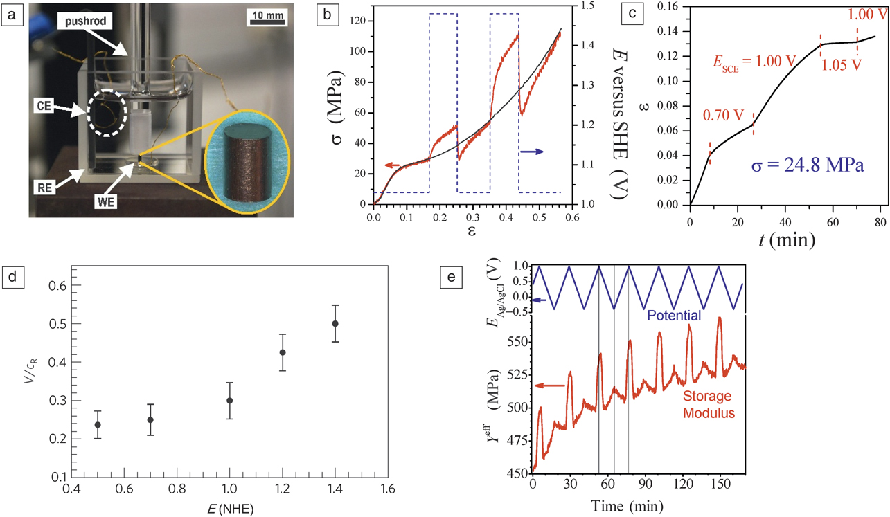
The pronounced size effects on strength and stiffness of NPG point to the vital role of the surface for the deformation of small-scale solids. This notion is best illustrated by the electrochemical modulation of mechanical response of NPG. Figure 3 compiles relevant results. Figure 3a shows a typical experimental setup, where NPG is plastically or elastically deformed in electrolyte and under potential control. In such experiments, the entire internal pore surface is wetted and the electric potential control affords precise and reversible adjustment of the superficial charge density or of the adsorbate coverage. The experiments exemplify the in operando property tuning discussed in the Introductory article in this issue.Reference Weissmüller and Sieradzki35 Jin and WeissmüllerReference Jin and Weissmüller36 were able to demonstrate this effect by controlling the yield strength and flow stress of NPG (Figure 3b). The largest recoverable change of strength (by a factor of two) was induced by the adsorption/desorption of a single atomic monolayer of OH on the ligament surfaces. Pinning of dislocations at surfaces, which is sensitive to the surface states, was suggested as the atomic-scale mechanism.Reference Jin and Weissmüller36 The deposition of self-assembled monolayers can also affect the hardness of NPG.Reference Miyazawa, Ishimoto, Hakamada and Mabuchi37,Reference Corcoran, Brankovic, Dimitrov, Sieradzki, Cammarata, Nastasi, Busso and Oliver38
Surface effects on the plastic, elastic, and fracture behavior of nanoporous gold (NPG) probed by exploiting electrochemical modulation of the surface state. (a) Representative experimental setup showing sample (enlarged in inset) immersed in electrolyte in a glass cuvette for laser-based optical strain measurements. Working electrode (sample, WE), reference electrode (RE), and counter electrode (CE) are designated; gold wires are for electrical control. The setup is installed in a conventional mechanical testing rig and load applied via a fused silica pushrod.Reference Mameka, Markmann and Weissmüller67 (b) The macroscopic flow stress of NPG, σ, varies as the surface state is switched between a clean surface (potential 1.03 V) and a surface covered by one molecular monolayer of OH (1.48 V) (L = 20 nm).Reference Jin and Weissmüller36 (c) Creep of NPG varies with potential (L = 20 nm).Reference Duan, Yi, Huang and Wang45 (d) The terminal crack velocity, V (normalized by the Rayleigh wave speed c R) as a function of the electrochemical potential for NPG (L = 40 nm). Reprinted with permission from Reference Sun, Chen, Badwe and SieradzkiReference 40. © 2015 Macmillan Publishers Ltd. The σ represents the stress applied during the creep test. (e) The effective macroscopic Young’s modulus Y eff, varies during cycles of the electrode potential, E, with respect to a pseudo Ag/AgCl reference electrode.Reference Mameka, Markmann, Jin and Weissmüller4 (L = 40 nm). Note: SHE, standard hydrogen electrode; SCE, saturated calomel electrode; NHE, normal hydrogen electrode.
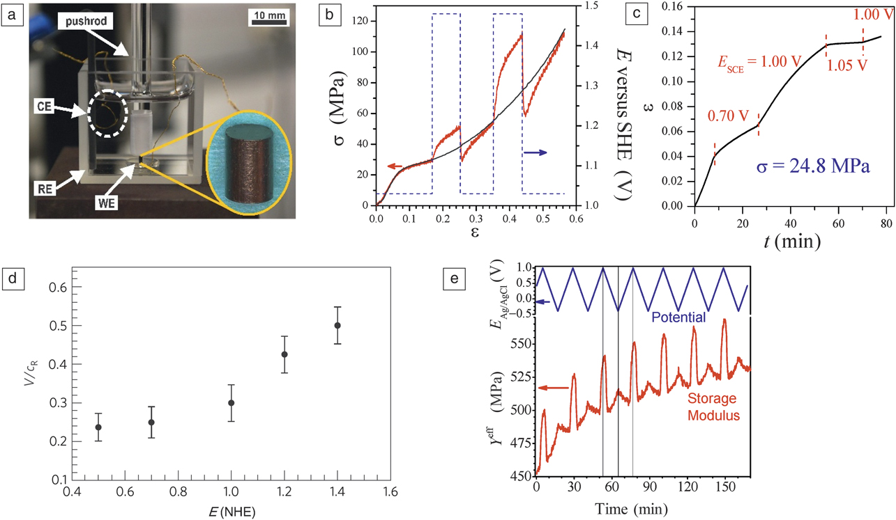
The creep behavior of NPG is even more sensitive to the surface modification. Ye et al.Reference Ye and Jin39 reported that the creep rate of NPG varies with applied potential (Figure 3c) even in the double-layer potential region where surface sorption is insignificant. Surprisingly, the highest creep rate was observed at a potential where the coarsening rate was the highest. This may connect the creep deformation to the surface tension of the solid, which is the major driving force for NPG coarsening.
Sieradzki et al.Reference Sun, Chen, Badwe and Sieradzki40 showed that fracture processes are also sensitive to the surface adsorbate coverage; specifically, the crack propagation speed increases significantly in NPG when the surface is covered by OH (see Figure 3d). Mameka et al.Reference Mameka, Markmann, Jin and Weissmüller4 also demonstrated that besides plastic deformation and fracture, the surface state affects the elastic behavior. Stiffening is observed during capacitive electron enrichment in the surface. Most notably, the stiffness reproducibly increases by 8% when a single monolayer of OH absorbs (see Figure 3e).
In the previously discussed instances, the mechanical behavior reacts to changes exclusively in the surface state. This observation establishes that surface effects strongly affect the plastic and elastic materials response of nanostructures such as NPG. Exploring surface effects on the mechanical response of NP metals provides new opportunities for adding novel functions to metals, and for understanding the governing mechanisms of deformation of small-scale solids.
Surface excess elasticity and nonlinear bulk elasticity do not explain the size-dependent stiffness
We now explore three issues that may be related to the anomalous compliance of NPG, namely surface excess elasticity, bulk nonlinear elasticity, and systematic changes in the topology with φ and with L.
Surface-induced strain and surface excess elasticity
Since the bonding near the surface of a crystal is different from that in the bulk, the surface regions experience a stress in their unstrained state; this is parameterized by the surface stress, f. The elastic response near the surface differs from the bulk; this phenomenon is quantified by surface elastic parameters, which are derivatives of the surface stress with respect to the tangential strain.Reference Gurtin and Murdoch41,Reference Gurtin, Weissmüller and Larché42 Next, we summarize the continuum description of these phenomena for a long circular nanowire, represented as a ligament in nanoporous metal.
The surface stress requires compensating stresses S A (axial) and S R (radial) in the bulk of the nanowire, which obey:Reference Weissmüller and Cahn43
 $${S_{\rm{A}}} = - {\rm{\alpha }}f\,{\rm{and}}\,{S_{\rm{R}}} = - {1 \over 2}{\rm{\alpha }}f$$,
along with the axial (εA) and radial (εR) strains:Reference Weissmüller, Duan and Farkas44
 $${{\rm{\varepsilon }}_{\rm{A}}} = - {\rm{\alpha }}f{{1 - {\nu ^{\rm{B}}}} \over {{Y^{\rm{B}}}}}{\rm{and}}\,{{\rm{\varepsilon }}_{\rm{R}}} = - {\rm{\alpha }}f{{1 - 3{\nu ^{\rm{B}}}} \over {2{Y^{\rm{B}}}}}$$,
where Y B and v B denote Young’s modulus and the Poisson’s ratio for the bulk metal, respectively. The ligament size L enters Equations 2 and 3 through the specific (per volume of the solid phase) surface area α; here α = 4/L. The consequences of surface excess elasticity are exemplified by the effective values for Young’s modulus for axial tension or compression of a nanowire:Reference Duan, Yi, Huang and Wang45,Reference Elsner, Müller, Bargmann and Weissmüller46
 $${Y^{{\rm{lig}}}} = {Y^{\rm{B}}} + {\rm{\alpha }}\left( {{{\rm{\lambda }}^{\rm{S}}}\left( {1 - {\nu ^{\rm{B}}}^2} \right) + 2{{\rm{\mu }}^{\rm{S}}}\left( {1 + {\nu ^{\rm{B}}}^2} \right)} \right)$$.
The surface Lamé constants λS and μS quantify the surface excess elasticity.Reference Gurtin and Murdoch41 Effective values of Y lig for use with the bending stiffness and for the shear modulus for use with torsion stiffness have also been derived.Reference Elsner, Müller, Bargmann and Weissmüller46 The surface Lamé constants can be obtained from calculations within electronic density functional theory (DFT).Reference Elsner, Müller, Bargmann and Weissmüller46 For ligaments of NPG with clean surfaces, the results imply elastic softening in tension, bending, and torsion.Reference Elsner, Müller, Bargmann and Weissmüller46 This agrees with the observation of an anomalously low experimental stiffness in NPG, yet the predicted magnitude of the softening is less than 10% for ligament sizes larger than 10 nm. Apparently, surface excess elasticity cannot explain the drastic reduction in stiffness observed by experiments on NPG, nor can the effect be resolved in small-scale tension studies with individual gold nanowires.Reference Wu, Heidelberg and Boland47
Note that the previous discussion refers to clean surfaces; adsorbate-covered surfaces can be drastically stiffer than the bulk, as demonstrated in Figure 3e.Reference Mameka, Markmann, Jin and Weissmüller4
Nonlinear elasticity in the bulk
Several studies, on either NPGReference Mameka, Wang, Markmann, Lilleodden and Weissmüller1,Reference Ngô, Stukowski, Mameka, Markmann, Albe and Weissmüller5,Reference Ngô, Roschning, Albe, Weissmüller and Markmann48 or individual nanowires,Reference Liang, Upmanyu and Huang49,Reference Chen, Richter, Sullivan and Gianola50 have emphasized the relevance of nonlinear elasticity in the bulk of the metal nanostructures for their effective elastic response. The surface-induced strains, Equation 3, can be large enough for higher-order elastic parameters to become relevant. These parameters can imply either stiffening or elastic softening, depending on the stress state and the crystallography. Ngô et al. have pointed out that the nonhydrostatic nature of the stress and strain in Equations 2 and 3 implies shear, which generally brings a trend for elastic softening or even for shear instability at small ligament size.Reference Ngô, Stukowski, Mameka, Markmann, Albe and Weissmüller5
The impact of nonlinear elasticity on the effective elastic response of NPG has been probed by simulations comparing MD with the full nonlinear response of an embedded atom potential for gold, along with surface-stress effects, to FEM simulation exploring linear elastic behavior and no surface stress.Reference Ngô, Roschning, Albe, Weissmüller and Markmann48 The latter provides threefold stiffer behavior, supporting the relevance of shear softening at the lower nanoscale. Yet, at L = 3mm, the MD simulation considered extremely small ligaments. The experiments in Figure 2b refer to samples with at least tenfold larger L. Here, the strains of Equation 3 are too small to allow for substantial bulk elastic nonlinearity. This emphasizes the role of additional factors other than surface excess elasticity or bulk nonlinear elasticity.
Topology varies systematically with solid fraction and ligament size
Several recent studiesReference Mameka, Wang, Markmann, Lilleodden and Weissmüller1,Reference Liu, Ye and Jin32,Reference Hu, Ziehmer, Wang and Lilleodden51,Reference Mangipudi, Epler and Volkert52 point out the decisive role of the topology of dealloying-made metal networks on mechanical properties. In fact, there are good reasons for expecting the nanoscale self-organization processes of dealloying to create networks with systematically different connectivity, depending on φ and on the acting processes during dealloying and coarsening.
The concept of network connectivity is discussed in the article by Lilleodden and Voorhees in this issue.Reference Lilleodden and Voorhees53 Here, we simply recall that there are various ways to define a scaled connectivity parameter, ξ, which measures the number of connections in a characteristic volume that scales with the third power of a characteristic structure size, for instance, the mean spacing between neighboring struts.
Reducing the solid fraction in a network while maintaining all its struts and their connections (constant ξ) requires increasing the aspect ratio of each strut. In other words, small φ requires long, thin ligaments. Yet, such ligaments are liable to pinch off due to the Plateau–Rayleigh instability:Reference Rayleigh54–Reference McCue, Karma and Erlebacher56 thin cylinders with surface tension can lower their energy by necking and subsequent disintegration. Thus, ξ of NPG is expected to decrease with decreasing φ. Eventually, at sufficiently low φ (low fraction of gold in the master alloy), dealloying will result in disconnected clusters, ξ = 0. Figure 2c (top left) illustrates this schematically. The consequences for the mechanical behavior appear evident: Y eff may be expected to vanish at the finite value of φ, which corresponds to the network’s percolation-to-cluster transition. As this value is approached, Y eff must vary more steeply with φ than the quadratic law of Equation 1 (Figure 2c, bottom left). This reasoning is consistent with the experimental observations in Figure 2a.
Mameka et al.Reference Mameka, Wang, Markmann, Lilleodden and Weissmüller1 have pointed out another link between microstructure evolution and connectivity, which is illustrated in Figure 2c (top right). Coarsening treatments, which underlie the adjustment of the ligament size of NPG in experiments, again rely on the pinch-off of ligaments. For networks with φ = 0.5, symmetry requires the coarsening to be self-similar, maintaining ξ constant. For NPG, this implies that samples with larger φ are liable to maintain constant stiffness during coarsening, so that Y eff depends only weakly on L.Reference Liu and Jin33 In samples with lesser φ, in contrast, annealing will simultaneously increase L and decrease ξ; such samples then exhibit a steeper apparent decrease of Y eff with L (Figure 2c, bottom right). The reasoning is reflected in the data of Figure 2b. The samples with lowest φ exhibit the steepest Y eff(L) decrease.Reference Mameka, Wang, Markmann, Lilleodden and Weissmüller1,Reference Liu, Ye and Jin32,Reference Liu and Jin33
This interpretation, however, does not apply to the “thin-film buckling” dataReference Mathur and Erlebacher34 in Figure 2b , where a steep Y eff(L) decrease occurs despite a high φ. This is because thin films have large external surface area, where microstructure evolution during coarsening differs from the bulk.
The insights into reduced stiffness and strength during coarsening of NPG, especially at lower φ, suggest that coarsening and solid fractions φ < 0.3 may need to be avoided when large strength and stiffness are sought in NPG. This strategy has been used to fabricate (Pt-doped) NPG with yield strength up to ∼200 MPa, which is ∼5× stronger than the fully solid parent alloy prior to dealloying.Reference Ye and Jin57,Reference Ye, Lu, Li, Du, Tan and Jin58
Strength of nanoporous gold: Scaling behavior and size effects
Scaling behavior of strength
Compared with conventional foams, the scaling behavior of strength in NP metals is more complicated because the ligament strength, σlig , depends on L.Reference Biener, Hodge, Hayes, Volkert, Zepeda-Ruiz, Hamza and Abraham10,Reference Volkert and Lilleodden12 Incorporating the ligament size effect into a scaling equation—for instance, as σlig ∝ L −n, where n is the power-law exponent —provides a description for NP metals.Reference Hodge, Biener, Hayes, Bythrow, Volkert and Hamza11,Reference Briot and Balk29 The Gibson–Ashby scaling equation for the effective macroscopic strength:
 $${{\rm{\sigma }}^{{\rm{eff}}}} = 0.3\,{{\rm{\sigma }}^{{\rm{lig}}}}\,{{\rm{\varphi }}^{3/2}}$$,
can also be inverted to extract σlig based on experimental data for σeff. This results in plots such as Figure 1b. Yet, the results are not always consistent. CompressionReference Mameka, Wang, Markmann, Lilleodden and Weissmüller1,Reference Jin, Kurmanaeva, Schmauch, Rosner, Ivanisenko and Weissmüller3,Reference Buerckert, Briot and Balk15,Reference Weissmüller, Newman, Jin, Hodge and Kysar28,Reference Jin and Weissmüller36 and tensionReference Balk, Eberl, Sun, Hemker and Gianola16,Reference Briot, Kennerknecht, Eberl and Balk30 tests on millimeter-scale NPG samples reveal systematically lower strength than nanoindentation tests or extrapolation from data for FIB-cut nano- and micropillars.Reference Diao, Gall, Dunn and Zimmerman19,Reference Wu, Heidelberg and Boland47,Reference Dou and Derby59
In view of the discussion of topology, one may assume that Equation 5, which, similar to Equation 1, ignores the possibility of systematic changes in connectivity with φ, requires corrections. Empirically, several modified scaling equations have been proposed for NPG. They introduce new scaling exponents,Reference Buerckert, Briot and Balk15,Reference Briot and Balk29 compare with gyroid or spinodal structures,Reference Mangipudi, Epler and Volkert52,Reference Pia, Brun, Aymerich and Delogu60 consider structure disorder,Reference Huber, Viswanath, Mameka, Markmann and Weissmueller9,Reference Roschning and Huber61 joint or node geometry,Reference Liu and Antoniou62–Reference Jiao and Huber64 and other simulatedReference Sun, Xu, Li, Feng and Gao65 or reconstructedReference Hu, Ziehmer, Wang and Lilleodden51 geometries. Each expression matches selected experiments on NPG. Yet, a generally applicable model has yet to be found.
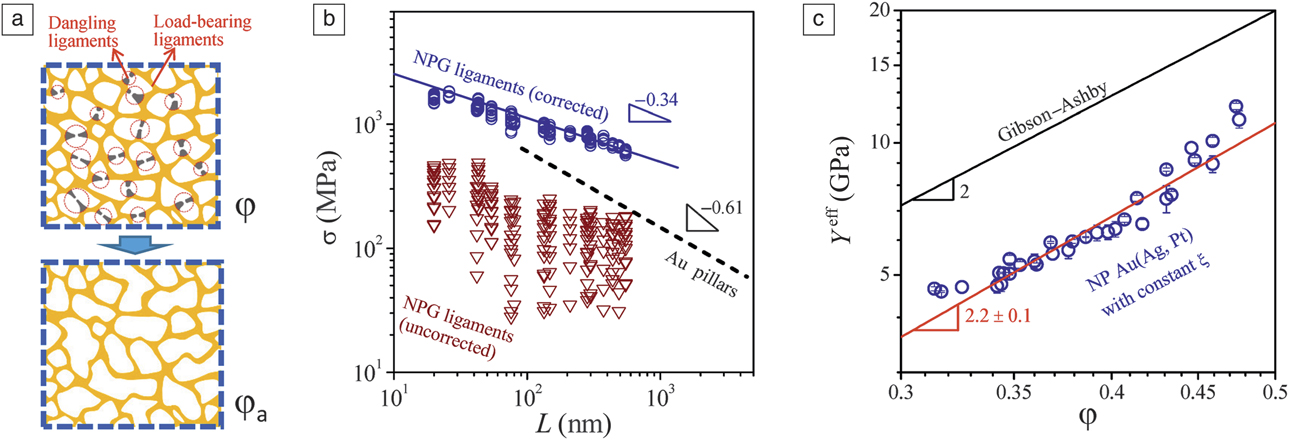
An empirical correction to the scaling equation is again associated with the network connectivity.Reference Mameka, Wang, Markmann, Lilleodden and Weissmüller1,Reference Liu, Ye and Jin32 Ligaments that are “broken” (not connected, lowering ξ) will not bear load (Figure 4a). Equations 1 and 5 can then be corrected by simply replacing the relative density φ with an apparent relative density, φa,Reference Liu, Ye and Jin32 which represents the volume fraction of load-bearing ligaments. Jin et al. have pointed out that φa is readily estimated from experimental stiffness data of NPG if one accounts for the fact—previously discussed—that the local Young’s modulus of the ligaments is essentially independent of their size. Deviations of the experimental Y eff from Equation 1 can be traced back to the broken ligaments. Equation 1 may be inverted to determine φa as:
 $${{\rm{\varphi }}_{\rm{a}}} = \sqrt {{{{Y^{{\rm{eff}}}}} \over {{Y^{\rm{B}}}}}}$$.
One can then write the corrected scaling equation for the strength as:
 $${{\rm{\sigma }}^{{\rm{eff}}}} = 0.3\,{{\rm{\sigma }}^{{\rm{lig}}}}\,{\rm{\varphi }}_{\rm{a}}^{3/2}$$.
Correction of Gibson–Ashby relations by considering unconnected ligaments. (a) Schematic illustration of dangling ligaments in a nanoporous structure with relative density φ, and a mechanically equivalent structure with apparent relative density φa. (b) Corrected and uncorrected ligament strengths as a function of ligament diameter, and comparison with Au pillars.Reference Liu, Ye and Jin32 (c) Variation of Young’s modulus of Pt-doped NP Au(Ag) with φ, while ξ was nearly unchanged.Reference Liu and Jin33
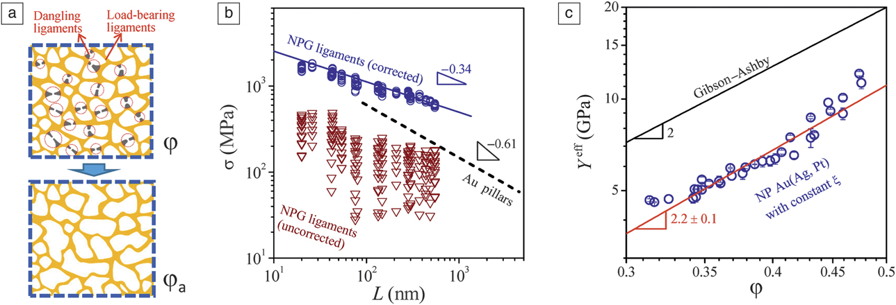
Figure 4b shows results for σlig, estimated based on experimental data for σeff using Equation 5 (triangles); the large scatter is obvious and casts doubt on the applicability of the scaling equation for NPG. When the same data set is evaluated by means of Equation 7 (circles), then the scatter is removed and the data coincide with a clear trend line. This finding supports the analysis based on the effective, load-bearing solid fraction φa, and emphasizes substantial variations in connectivity in NPG.Reference Liu, Ye and Jin32
Two observations further strengthen the previously discussed notion: (1) the corrected σlig in Figure 4b is comparable to the strength of Au nanowires (which is much higher than the uncorrected value derived using Equation 5); and (2) Y eff of NPG scales roughly with φ2 when the connectivity is maintained nearly constant by avoiding structural coarsening (Figure 4c).Reference Liu and Jin33
Size effect on strength
Early studies using small-scale testsReference Biener, Hodge, Hayes, Volkert, Zepeda-Ruiz, Hamza and Abraham10–Reference Volkert and Lilleodden12,Reference Briot and Balk29 on NPG reported the exponent n in σlig ∝ L n between –0.5 and –0.6, which compares favorably with data for Au pillars and nanowires.Reference Volkert and Lilleodden12,Reference Dou and Derby59 Macroscale testsReference Mameka, Wang, Markmann, Lilleodden and Weissmüller1,Reference Jin, Kurmanaeva, Schmauch, Rosner, Ivanisenko and Weissmüller3 suggesting the more negative n ≈ –1.1 were confirmed by in situ x-ray diffraction experiments of the strength.Reference Wang, Hartig, Blankenburg, Mueller, Guenther and Weissmüller66 Yet, these studies used Equation 5, ignoring the connectivity issue. When taking disconnected ligaments into account via Equation 7, the significantly more positive exponent n ∼ –0.34 is obtained (see the “corrected” σlig in Figure 4b).Reference Liu, Ye and Jin32 Presently, the size exponent of the local strength in NPG is not conclusively established and there is a need for further experiments.
Tension–compression asymmetry: A signature of surface stress or surface tension?
The large surface-to-volume area in nanoporous materials means that capillary terms can also significantly affect the strength. This notion has been invoked to explain the observation in atomistic computer simulations that the tensile yield strength exceeds the compressive strength (Figure 1c).Reference Farkas, Caro, Bringa and Crowson17,Reference Crowson, Farkas and Corcoran18 In fact, tension–compression asymmetry ratios of about 10 were observed in simulations of NPG with ligament sizes of about 2 nm.Reference Farkas, Caro, Bringa and Crowson17
One possible explanation for the tension–compression asymmetry is the surface-induced stress of Equation 2. Figure 1d shows MD simulation results illustrating the stress state:Reference Crowson, Farkas and Corcoran18 the local stresses in the surface atomic layers are generally tensile (positive surface stress f), and the compensating surface-induced stress in the interior of the ligaments compressive. The surface-induced compressive stress in the bulk of the ligament superimposes over the stresses in reaction to external load. Thus, the local stress state in the bulk favors plastic compression over plastic tensile elongation. The disparity increases at small size as ∝ f/L.
A different explanation of the tension–compression asymmetry focuses on energy as opposed to stress.Reference Mameka, Markmann and Weissmüller67 The plastic tensile elongation of a ligament increases its net surface area A, while compression reduces A. The net energy change is γδA, where γ denotes the surface tension, an energy excess per area of surface. For macroscopic metal wires, the zero creep experiments by H. Udin in the mid-20th century demonstrate a tension-compression asymmetry of the creep due to this energy-related effect:Reference Udin, Schaler and Wulff68,Reference Buttner, Udin and Wulff69 Wires contract spontaneously, unless a tensile stress of magnitude 2γ/L is applied to arrest the contraction. Nanoscale structures may be expected to reflect the same phenomenon, just much stronger.
The 2γ/L contribution to the flow stress results from a simple energy balance at equilibrium, which makes it forceful. Yet, the effect may be concealed if yielding is governed by dissipative processes that involve barriers of much higher magnitude. Specifically, the surface-induced bulk stress of Equation 2 may affect the nucleation of dislocations in the interior of ligaments. The magnitude of the observed yield stress will then increase under tension and decrease under compression.Reference Diao, Gall, Dunn and Zimmerman19 However, it is known that dislocations indeed nucleate at the surface of nanowires.Reference Gall, Diao and Dunn21,Reference Diao, Gall and Dunn70–Reference Hyde, Espinosa and Farkas72 The very origin of the compressive stress in the bulk is an opposing tensile stress in the surface regions. If this stress affects nucleation, as it should, then the opposite sign of the anisotropy could be expected, strengthening in compression and weakening in tension. Furthermore, a dislocation sweeping a complete glide plane would probe the area-average stress, over surface regions and bulk. As pointed out in Reference Mameka, Markmann and WeissmüllerReference 67, the contribution of surface-related stresses to this average is zero, so those stresses might not affect the strength at all.
The arguments linking the tension–compression asymmetry in small-scale plasticity to capillary effects are supported and clarified by two experiments reported very recently. Both exploit the strategy of in situ electrochemical modulation of the surface state during plastic deformation of NPG for identifying surface effects on strength. Lührs et al. investigated the plastic Poisson’s ratio of NPG and provide experimental confirmation of the tension–compression asymmetry.Reference Lührs, Zandersons, Huber and Weissmüller25 Mameka et al. provide compelling evidence for surface tension and not surface stress as the decisive capillary parameter by comparing jumps in the flow stress to the known variations of γ and f during potential modulation.Reference Mameka, Markmann and Weissmüller67
Conclusion
This article exemplifies how recent research on dealloying-made nanoporous metal has contributed at a fundamental level to documenting and unraveling the mechanics of small-scale plasticity. In the future, advances in preparation could extend the materials portfolio to cost-efficient, lightweight metals and composites.Reference McCue, Karma and Erlebacher56 This emphasizes the vision and promise of dealloying-made structural materials. Exciting insights can be expected when future studies exploit more systematically the opportunities for ligaments sizes well below 10 nmReference Ye and Jin57 and for materials with organized structural hierarchy on different size scales.Reference Juarez, Biener, Weissmüller and Hodge73
Acknowledgments
H.J. is supported by the National Key R&D Program of China (Grant No. 2017YFA0204401) and NSFC (Grant No. 51571206) is acknowledged. J.W. would like to thank Deutsche Forschungsgemeinschaft, SFB 986, Subproject B2. D.F. acknowledges the National Science Foundation, IRD and DMREF programs. H.J. and J.W. thank the “Helmholtz-CAS Joint Research Groups” Project (Grant No. HCJRG-315).

Hai-Jun Jin is a professor and group leader in the Shenyang National Laboratory for Materials Science at the Institute of Metal Research (IMR), Chinese Academy of Sciences (CAS), China. He received his PhD degree in materials science from IMR-CAS in 2004. He was then appointed assistant professor at IMR. He was an Alexander von Humboldt postdoctoral researcher and research scientist at the Institute of Nanotechnology, Karlsruhe Institute of Technology, Germany, from 2005 to 2010. His current research focuses on the fabrication, mechanical properties, and surface-controlled functionalities of nanoporous metals. Jin can be reached by email at hjjin@imr.ac.cn.

Jörg Weissmüller is a full professor and head of the Institute of Materials Physics and Technology at Hamburg University of Technology. He is also the leader of the Hybrid Materials Systems Group at Helmholtz-Zentrum Geesthacht, Germany. He studied materials science at Saarland University, Germany, and the University of Dundee, UK. He received his PhD degree in engineering and Habilitation degree in experimental physics from Saarland University. He has worked at the Institute of New Materials, Germany; the National Institute of Standards and Technology, and the Institute of Nanomaterials at Karlsruhe Institute of Technology, Germany, and is presently a visiting professor at the Technion Haifa, Israel. His current research focuses on interfaces and nanomaterials with interface-controlled behavior, with emphasis on the coupling between mechanics and chemical or electrochemical processes. Weissmüller can be reached by email at weissmueller@tuhh.de.

Diana Farkas is a professor in the Department of Materials Science and Engineering at Virginia Tech. She received her PhD degree in metallurgy in 1980 from the University of Delaware. She is a Fellow of MRS and ASM. She was a Jefferson Science Fellow at the US Department of State in 2010 and program director at the National Science Foundation, Division of Materials Research, until 2017. Her research focuses on atomistic computer simulation of mechanical behavior of metallic materials at the nanoscale. She has co-authored more than 200 publications. Farkas can be reached by email at diana@vt.edu.