Introduction
γδ T-cells
γδ T-cells are an evolutionarily conserved T-cell population, distinguished by the genes that encode their antigen receptor T-cell receptor (TCR). Though γδ T-cells remain an enigma and their overall importance to the immune system is still debated, they clearly have the capacity, if properly stimulated, to mediate a large array of effector cell activities (Hayday, Reference Hayday2000). γδ T-cells are potent cytolytic cells (Ciccone et al., Reference Ciccone, Viale, Bottino, Pende, Migone, Casorati, Tambussi, Moretta and Moretta1988; Rivas et al., Reference Rhodes, Hewinson and Vordermeier1989), produce an array of cytokines that enhance the activities of macrophages and neutrophils, as well as other lymphocytes (Ferrick et al., Reference Ferrick, Schrenzel, Mulvania, Hsieh, Ferlin and Lepper1995; Mak and Ferrick, Reference Mak and Ferrick1998; Born et al., Reference Born, Cady, Jones-Carson, Mukasa, Lahn and O'Brien1999), can present antigen (Collins et al., Reference Collins, Werling, Duggan, Bland, Parsons and Howard1998; Brandes et al., Reference Brandes, Willimann and Moser2005), and induce as well as suppress inflammation (Zuany-Amorim et al., Reference Zuany-Amorim, Ruffie, Haile, Vargaftig, Pereira and Pretolani1998; Egan and Carding, Reference Egan and Carding2000; O'Brien et al., Reference O'Brien, Yin, Huber, Ikuta and Born2000). They are found in virtually all portals of entry into the body prior to infection and are particularly well represented at the gut mucosal surface, constituting a large fraction of the intraepithelial lymphocyte (IEL) population (Komano et al., Reference Komano, Fujiura, Kawaguchi, Matsumoto, Hashimoto, Obana, Mombaerts, Tonegawa, Yammamoto, Itohara, Nanno and Ishikawa1995; Boismenu and Havran, Reference Boismenu and Havran1998). After epithelial cells, γδ T-cells are one of the first cell populations of the innate immune system to encounter pathogens that invade through the gut epithelial lining (Ferrick et al., Reference Ferrick, King, Jackson, Braun, Tam, Hyde and Beaman2000). γδ T-cells also respond to inflammatory stimuli; thus they can be recruited to sites of infection within the gut (Wilson et al., Reference Wilson, Aydintug and Jutila1999). Through these activities, γδ T-cells have been shown to respond to and participate in host defense responses in a variety of pathogen-induced diseases including: HSV-1 encephalitis, malaria, toxoplasmosis, leishmaniasis, cryptosporidiosis, tuberculosis, listeriosis, salmonellosis, tularemia, brucellosis, erlichiosis and AIDS (Hayday, Reference Hayday2000). These cells have also been shown to participate in tissue functions, particularly in maintaining the health of epithelial cells lining mucosal surfaces and in wound repair (Jameson and Havran, Reference Jameson and Havran2007).
Bovine γδ T-cells
γδ T-cells represent a minor percentage of the peripheral lymphocyte pool in most animals. In contrast, they represent a major lymphocyte subset in cattle and can constitute up to 60–70% of the circulating T-cell pool in calves (Davis et al., Reference Davis, Brown, Hamilton, Wyatt, Orden, Khalid and Naessens1996; and M. A. Jutila, unpublished observations). As such, γδ T-cells are likely critical to bovine immunity, perhaps more so than in other animals. As in humans, the percentage of γδ T-cells in the peripheral blood decreases in calves as the animal ages, implicating them as important to immunity in neonates (Hayday, Reference Hayday2000).
Studies on bovine γδ T-cells have elucidated: (i) potential antigens that drive their responses (Abrahamsen, Reference Abrahamsen1998; Fikri et al., Reference Fikri, Denis, Pastoret and Nyabenda2001, Reference Fikri, Pastoret and Nyabenda2002; Naiman et al., Reference Naiman, Alt, Bolin, Zuerner and Baldwin2001; Rhodes et al., Reference Rhodes, Hewinson and Vordermeier2001; Smyth et al., Reference Smyth, Welsh, Girvin and Pollock2001; Baldwin et al., Reference Baldwin, Sathiyaseelan, Naiman, White, Brown, Blumerman, Rogers and Black2002; Mwangi et al., Reference Mwangi, McKeever, Nyanjui, Barbet and Mahan2002), (ii) their potential role in responses against Mycobacterium spp. (Rhodes et al., Reference Rhodes, Hewinson and Vordermeier2001; Smyth et al., Reference Smyth, Welsh, Girvin and Pollock2001), Cryptosporidium parvum (Abrahamsen, Reference Abrahamsen1998), Cowdria ruminantium (Mwangi et al., Reference Mwangi, McKeever, Nyanjui, Barbet and Mahan2002), Leptospira borgpetersenii (Naiman et al., Reference Naiman, Alt, Bolin, Zuerner and Baldwin2001), Staphylococcus spp. (Fikri et al., Reference Fikri, Denis, Pastoret and Nyabenda2001), Theileria parva (Daubenberger et al., Reference Daubenberger, Taracha, Gaidulis, Davis and McKeever1999), Anaplasma marginale (Lahmers et al., Reference Lahmers, Hedges, Jutila, Deng, Abrahamsen and Brown2006) and Salmonella enterica serovar Typhimurium (Hedges et al., Reference Hedges, Buckner, Rask, Kerns, Jackiw, Trunkle, Pascual and Jutila2007) infection in cattle, (iii) ligands and counter-receptors important in their activation (Sopp and Howard, Reference Sopp and Howard2001; Ahn et al., Reference Ahn, Konno, Gebe, Aruffo, Hamilton, Park and Davis2002; Fikri et al., Reference Fikri, Pastoret and Nyabenda2002; Sathiyaseelan et al., Reference Sathiyaseelan, Naiman, Welte, Machugh, Black and Baldwin2002), (iv) subset-specific responses (Hedges et al., Reference Hedges, Cockrell, Jackiw, Meissner and Jutila2003a; Meissner et al., Reference Meissner, Radke, Hedges, White, Behnke, Bertolino, Abrahamsen and Jutila2003), and (v) molecular basis for their trafficking behavior (Walcheck and Jutila, Reference Walcheck and Jutila1994; Jutila and Kurk, Reference Jutila and Kurk1996; Jones et al., Reference Jones, Watts, Robinson, Vestweber and Jutila1997; Jutila et al., Reference Jutila, Wilson and Kurk1997; Wilson et al., Reference Wilson, Aydintug and Jutila1999, Reference Wilson, Hedges, Butcher and Jutila2002). Though many functions of bovine γδ T-cells have been elucidated, it is likely that much remains to be discovered concerning their importance and roles within the immune system and participation in tissue homeostasis. Most early studies examined bovine γδ T-cells within accepted paradigms of T-cell biology based on years of study of αβ T-cells, mainly in humans and rodents. Recently, multiple groups, including ours, have applied functional genomics approaches to begin to gain an unbiased view of bovine γδ T-cells, which have revealed interesting and novel functional responses in these cells.
Our first studies analyzed global gene expression patterns in circulating bovine γδ T-cell subsets based on cell surface markers. In addition to the γδ TCR, bovine γδ T-cells express lineage-specific surface antigens grouped together into a family called WC1 (Wijngaard et al., Reference Wijngaard, MacHugh, Metzelaar, Romberg, Bensaid, Pepin, Davis and Cleavers1994; MacHugh et al., Reference MacHugh, Mburu, Carol, Wyatt, Orden and Davis1997), which are not found on rodent or human cells. Other surface antigens useful in the study of these cells include CD8 and CD2, which, along with the WC1 family members, define functionally distinct subsets (Tuo et al., Reference Tuo, Bazer, Davis, Zhu and Brown1999). Gene expression profiles of γδ T-cell subsets defined by expression or lack of expression of CD8, regardless of specific TCR usage, were compared using microarrays and serial analysis of gene expression (SAGE) (Hedges et al., Reference Hedges, Cockrell, Jackiw, Meissner and Jutila2003a, Reference Hedges, Graff and Jutilab; Meissner et al., Reference Meissner, Radke, Hedges, White, Behnke, Bertolino, Abrahamsen and Jutila2003). These studies concluded that while CD8− γδ T-cells were activated, proliferative and inflammatory, the CD8+ subset expressed anti-inflammatory genes and genes consistent with quiescence and trafficking to the mucosa. These early studies also suggested that bovine γδ T-cells express a number of myeloid cell genes.
The intent of this review is to summarize our recent functional gene expression work, which sets γδ T-cells apart from αβ T-cells, increases the known similarities of γδ T-cells to myeloid cells and has led to a new innate-like antigen-independent priming model of γδ T-cell responses to infection. Using this model, assays have been developed and used to screen natural compound libraries for novel bovine γδ T-cell agonists, and the results of some of these screens will be summarized. A number of other recent reviews (Born et al., Reference Born, Reardon and O'Brien2006, Reference Born, Jin, Aydintug, Wands, French, Roark and O'Brien2007; Moser and Brandes, Reference Moser and Brandes2006) provide a broader overview of the studies of this enigmatic T-cell population, which will not be summarized here.
Gene expression in αβ and γδ T-cells from blood, spleen and mucosal lymphatics
Since our original reports, we have performed additional functional gene expression analyses using SAGE in an extensive comparison of gene expression in sorted bovine γδ and αβ T-cells isolated from the blood and spleen. These studies showed (i) that these two subsets respond to global mitogen signals, such as Con-A and PMA/ionomycin, in distinct fashions, (ii) differences in the gene expression patterns of blood- and spleen-derived T-cells and (iii) the impact of different sorting approaches (magnetic bead and FACS) on basal gene expression (Graff et al., Reference Graff, Behnke, Radke, White and Jutila2006). Briefly, consistent with other global gene expression analyses in γδ and αβ T-cells (Fahrer et al., Reference Fahrer, Konigshofer, Kerr, Ghandour, Mack, Davis and Chien2001; Shires et al., Reference Shires, Theodoridis and Hayday2001), nearly all (95%) of the genes expressed in the resting T-cell populations were the same. However, following stimulation with Con-A/IL-2, there was an approximately 5-fold increase in the number of genes selectively expressed in γδ T-cells. Baseline gene transcription was more robust in spleen versus blood γδ T-cells, consistent with expression of a potent transcriptional repressor [B-lymphocyte induced maturation protein-1 (BLIMP-1); see below] in the cells from blood. Finally, both methods of cell sorting impacted gene transcription in bovine γδ T-cells, but FACS appeared to have the least effect. In total, 16 SAGE libraries were constructed and analyzed in this work. These libraries can be accessed on a Web site (http://vmbmod10.msu.montana3.edu/vmb/jutila-lab/sagebov.htm) that contains a number of different bioinformatics tools to facilitate searches of the extensive datasets.
We also participated in a study done by Dr Wendy Brown's group at Washington State University who compared γδ and αβ T-cell clone responses against peptides from Anaplasma marginale, an intraerythrocytic rickettsial pathogen of cattle. The T-cell clones were previously shown to respond to peptide P10 of a conserved region of the major surface protein 2 (MSP2) of A. marginale. Gene expression profiles of activated T-cell clones were compared using two different microarray platforms (cDNA and oligonucleotide arrays), and the results of differentially expressed genes were confirmed by real-time RT-PCR and protein analyses (Lahmers et al., Reference Lahmers, Hedges, Jutila, Deng, Abrahamsen and Brown2006). These studies demonstrated that while αβ and γδ T-cells possess some conserved functions, such as production of IFN-γ, TNF-α and T-cell-associated chemokines, there are dramatic differences between these two cell types. Consistent with our analyses of gene expression in γδ T-cell subsets, WC1+ γδ T-cell clones preferentially expressed genes generally associated with myeloid cells, which included CD11b, macrophage scavenger and mannose receptors, CD68 and Toll-like receptor 4 (TLR4).
Also relevant to animal health, we have examined gene expression patterns in mesenteric lymphatic γδ and αβ T-cells prior to and during enterocolitis caused by Salmonella serovar Typhimurium (Hedges et al., Reference Hedges, Buckner, Rask, Kerns, Jackiw, Trunkle, Pascual and Jutila2007). Among many gene expression patterns detected in these studies were patterns indicative of early innate immune response and function by γδ T-cells. In this investigation, the early transcriptional activities of mucosal lymphatic T-lymphocyte subsets during Salmonella serovar Typhimurium-induced enterocolitis revealed substantial differences in how naive γδ T-cells and αβ T-cells respond to this infection. We found that γδ T-cells were subtly activated, or primed, 48 h after Salmonella serovar Typhimurium infection in calves, as evidenced by the increase in IL-2Rα on cells derived from the intestinal lymphatics. Infection did not increase gene expression in αβ T-cells over that of the negative control; rather, Salmonella serovar Typhimurium infection appeared to have a dampening effect on this T-cell subset. Minimal changes in γδ T-cell phenotype were observed in the blood of the infected calves, which were consistent with responses seen in human Salmonella serovar Typhimurium-induced enterocolitis. In this study, the functional proliferative response to various pathogen-associated molecular patterns (PAMPs) was elucidated and the PAMP-induced priming model began to emerge.
Myeloid gene expression in bovine γδ T-cells
Our genomic analyses of bovine γδ T-cell subsets underscored the relationship of γδ T-cells to myeloid cells by their expression of genes such as CD11b, CD14, CD68, scavenger receptor 1, mannose-binding protein, multiple TLRs and BLIMP-1, a master regulator of B- and myeloid cell differentiation. Extensive analyses to confirm the predictions from these early gene expression studies have been performed, which underscored the validity of the microarray and SAGE data (Hedges et al., Reference Hedges, Lubick and Jutila2005, Reference Hedges, Buckner, Rask, Kerns, Jackiw, Trunkle, Pascual and Jutila2007; Kress et al., Reference Kress, Hedges and Jutila2006; Lahmers et al., Reference Lahmers, Hedges, Jutila, Deng, Abrahamsen and Brown2006). Of the myeloid genes defined to date, BLIMP-1 is one of particular interest because it is a potent transcriptional repressor, which has been shown in other systems to control B-cell and myeloid cell differentiation (Lin et al., Reference Lin, Wong and Calame1997). BLIMP-1 is expressed in both CD8+ and CD8− γδ T-cells, though the latter expresses higher levels (Meissner et al., Reference Meissner, Radke, Hedges, White, Behnke, Bertolino, Abrahamsen and Jutila2003). Because of the potential significance of BLIMP-1, its function in bovine γδ T-cells was further investigated.
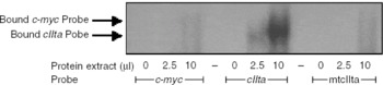
BLIMP-1 expression in bovine γδ T-cells was originally confirmed in RNase protection assays (Meissner et al., Reference Meissner, Radke, Hedges, White, Behnke, Bertolino, Abrahamsen and Jutila2003) and its expression was also demonstrated in human γδ T-cells, (J. C. Graff and M. A. Jutila, unpublished observations). BLIMP-1 is a repressor of c-myc and, therefore, proliferation in B- and myeloid cells (Lin et al., Reference Lin, Wong and Calame1997), but its function in γδ T-cells is unknown. To determine whether the BLIMP-1 protein expressed by bovine γδ T-cells is functional, we tested its ability to bind to specific DNA sequences of B-cell cIIta and c-myc promoters (Lin et al., Reference Lin, Wong and Calame1997; Piskurich et al., Reference Piskurich, Lin, Lin, Wang, Ting and Calame2000). Electrophoretic mobility shift assays (EMSA) were performed using nuclear protein lysates from bovine γδ T-cells and the known BLIMP-1 binding sites in the cIIta and c-myc promoter sequences (Fig. 1). BLIMP-1 from bovine γδ T-cells bound to both promoter sequences, though to a lesser extent to the c-myc promoter. BLIMP-1 had a much weaker interaction with the mutated cIIta promoter sequence, demonstrating specificity (Fig. 1). Nuclear proteins from human γδ T-cells also bound the cIIta and c-myc promoter sequences in the same fashion (data not shown). Thus, γδ T-cells express functional BLIMP-1 protein, confirming the predictions of the original SAGE studies (Meissner et al., Reference Meissner, Radke, Hedges, White, Behnke, Bertolino, Abrahamsen and Jutila2003). We have also found that BLIMP-1 is regulated by mitogen activation in γδ T-cells (J. C. Graff and M. A. Jutila, unpublished observations). Current experiments are focused on determining if BLIMP-1 represses proliferation in bovine γδ T-cells, as it does in B- and myeloid cells.
Bovine γδ T-cell BLIMP-1 binds known promoter elements in vitro. As seen by EMSA, nuclear proteins isolated from purified bovine γδ T-cells (>97% pure) bind the known BLIMP-1 binding sites in both the c-myc promoter (Lin et al., Reference Lin, Wong and Calame1997) and cIIta promoter III region (Piskurich et al., Reference Piskurich, Lin, Lin, Wang, Ting and Calame2000). Mutation of the cIIta promoter (mtcIIta) greatly weakened the protein–DNA complex.

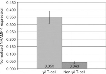
Another surprising differentially expressed myeloid gene detected in resting and activated blood γδ T-cells (9 tags) and not αβ T-cells (0 tags) identified by SAGE was natural resistance-associated macrophage protein-1 (NRAMP-1; SLC11A1; http://vmbmod10.msu.montana.edu/vmb/jutila-lab/sagebov.htm). NRAMP-1, a metal ion transporter across phagosomal membranes in macrophages, is involved in susceptibility to intracellular pathogens (Nevo and Nelson, Reference Nevo and Nelson2006). We used real-time RT-PCR to examine the expression of NRAMP-1 transcripts in sorted bovine γδ T-cells and the remaining PBMCs after the sort (predominantly αβ T-cells and B-cells). As predicted by the SAGE analyses, we found in these preliminary studies that γδ T-cells expressed high levels of NRAMP-1 transcripts, whereas non-γδ T-lymphocytes expressed low levels (Fig. 2). This finding is particularly intriguing, since NRAMP-1 function has previously been thought to be restricted to monocytes and dendritic cells. This observation suggests alternative explanations involving γδ T-cells for recent findings that loss or alteration of NRAMP-1 increases susceptibility of the host to numerous infectious agents, as well as autoimmune disorders (Blackwell et al., Reference Blackwell, Goswami, Evans, Sibthorpe, Papo, White, Searle, Miller, Peacock, Mohammed and Ibrahim2001). Investigations of the role of NRAMP-1 in γδ T-cells are currently under way.
Selective expression of NRAMP-1 in bovine γδ T-cells. Bovine γδ T-cells and non-γδ T-cells (predominantly αβ T-cells, NK cells and B-cells) were sorted by FACS and NRAMP-1 transcripts analyzed by real-time RT-PCR. Values were normalized to 18S and the data reflect means and SEM from triplicate samples.

Bovine γδ T-cells express PAMP receptors and respond to microbial PAMPS: defining a PAMP primed γδ T-cell
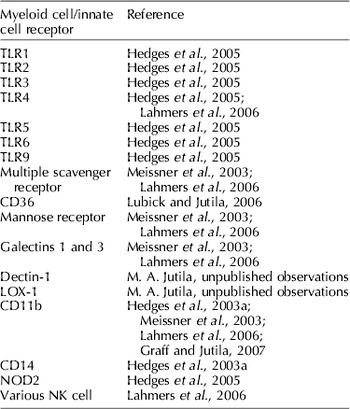
A consistent finding in our gene expression work on bovine γδ T-cells has been their expression of a number of myeloid cell-associated genes, which include numerous receptors for PAMPs (Table 1). Confirmation studies have been performed for each of these receptors at the RNA level (Hedges et al., Reference Hedges, Lubick and Jutila2005). There are a few antibody reagents available to analyze some of these receptors in bovine cells, and using them, we have confirmed Dectin-1 (E. Kress and M. A. Jutila, unpublished observations), CD36 (Lubick and Jutila, Reference Lubick and Jutila2006) and CD11b (Graff and Jutila, Reference Graff and Jutila2007) at the protein level, as well.
List of innate receptors detected in bovine γδ T-cells

In addition to expressing a wide array of genes encoding PAMP receptors, bovine γδ T-cells also directly respond to PAMPs. As another approach to confirm the expression of PAMP receptors on bovine γδ T-cells, we examined the response of these cells to various PAMPs, including peptidoglycan (PGN; signals through TLR2 and NOD2), lipoteichoic acid (LTA; signals through TLR2 and CD36), muramyl dipeptide (signals through NOD2) and lipopolysaccharide (LPS; signals through TLR4/CD14, CD11b and/or scavenger receptors). Using the limited number of mAbs against bovine PAMP receptors and RNA-interference assays, we have confirmed a role for CD36 in regulating responses to LTA (Lubick and Jutila, Reference Lubick and Jutila2006) and intracellular NOD2 receptors in the response to muramyl dipetide. These PAMPs activated sorted bovine γδ T-cells, but the response was quite subtle. Modest increases in transcription of genes encoding IL-2Rα and chemokines, such as MIP1α and MIP1β, were detected (Hedges et al., Reference Hedges, Lubick and Jutila2005; Lubick and Jutila, Reference Lubick and Jutila2006). Strikingly, genes for the prototypic markers of activated γδ T-cells, TNF-α and IFN-γ, were minimally affected by the PAMP treatments. The subtle response by bovine γδ T-cells to PAMPs is functionally relevant, as sufficient levels of chemokines were induced, which directed migration of specific target cells in in vitro assays (Hedges et al., Reference Hedges, Lubick and Jutila2005). This subtle response is termed ‘antigen-independent priming’ to differentiate between the canonical overt activation of γδ T-cells.
Another characteristic of a PAMP primed γδ T-cell is its increased responsiveness to secondary signals such as antigen or cytokines. Based on these early PAMP studies, we hypothesized that the rapid in vivo response of γδ T-cells early in infection with Salmonella was a response to LPS generated by bacterial infection in the gut. In support of that hypothesis, increase in IL-2Rα protein expression on γδ T-cells was apparent in vivo and its function was demonstrated in vitro with Salmonella serovar Typhimurium LPS alone. Specifically, pretreating largely naïve sorted bovine γδ T-cells with Salmonella serovar Typhimurium LPS for 48 h greatly increased the downstream proliferative response to IL-2 or IL-15 (Hedges et al., Reference Hedges, Buckner, Rask, Kerns, Jackiw, Trunkle, Pascual and Jutila2007). This response is similar, in part, to an antigen-driven response, except that it occurs with a large fraction of the naïve γδ T-cell population. The effect was not restricted to Salmonella LPS in that crude Escherichiacoli LPS, ultra-pure E. coli LPS, muramyl dipeptide and β-glucan, among other PAMPs, also primed γδ T-cells to proliferate in response to IL-2 (Hedges et al., Reference Hedges, Buckner, Rask, Kerns, Jackiw, Trunkle, Pascual and Jutila2007). While we have not precisely determined the mechanism of LPS detection, transcripts encoding many TLRs and other pattern recognition proteins are readily detected in γδ T-cells (Table 1) (Mokuno et al., Reference Mokuno, Matsuguchi, Takano, Nishimura, Washizu, Ogawa, Takeuchi, Akira, Nimura and Yoshikai2000; Hedges et al., Reference Hedges, Lubick and Jutila2005; Deetz et al., Reference Deetz, Hebbeler, Propp, Cairo, Tikhonov and Pauza2006; Kress et al., Reference Kress, Hedges and Jutila2006; Lubick and Jutila, Reference Lubick and Jutila2006; Wesch et al., Reference Wesch, Beetz, Oberg, Marget, Krengel and Kabelitz2006). Co-stimulatory effects of TLR agonists and TCR engagement on γδ T-cells have been shown, which are similar in many respects to the priming effect described here (Deetz et al., Reference Deetz, Hebbeler, Propp, Cairo, Tikhonov and Pauza2006; Wesch et al., Reference Wesch, Beetz, Oberg, Marget, Krengel and Kabelitz2006). Similarly, in regulatory T-cells, the effect of the combination of TLR2 agonist and TCR engagement that is enhanced by IL-2 is consistent with our results (Liu et al., Reference Liu, Komai-Koma, Xu and Liew2006). Also on regulatory T-cells, an additive response of Salmonella serovar Typhimurium LPS and IL-2 in the absence of TCR engagement has been observed (Caramalho et al., Reference Caramalho, Lopes-Carvalho, Ostler, Zelenay, Haury and Demengeot2003). Priming of γδ T-cells is reminiscent of that of innate cells and is well defined for macrophages, where prior exposure to LPS dramatically increases subsequent responses to secondary signals (Aderem et al., Reference Aderem, Cohen, Wright and Cohn1986).
Priming model
Our studies of PAMP responses in bovine γδ T-cells have led to a new functional model that describes early responses of γδ T-cells to infection (Fig. 3). Early in the course of infection, γδ T-cells sense pathogens leading to alterations in production of cytokine transcripts and some surface proteins, such as increased IL-2Rα. The γδ T-cell is now primed for enhanced downstream responses to antigen and/or cytokines that may be associated with infection. The cell activated by the secondary antigen/cytokine represents the prototypic effector γδ T-cell, characterized by increased TNF-α and IFN-γ production (Wang et al., Reference Wang, Das, Kamath and Bukowski2001).
Priming model for bovine γδ T-cells.

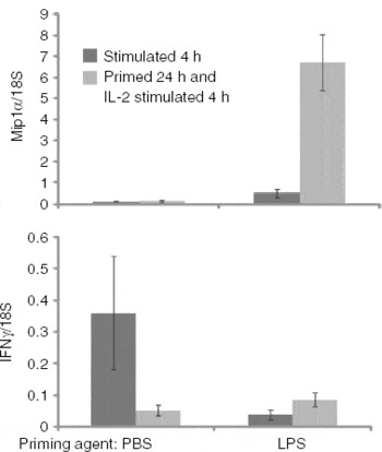
PAMP primed γδ T-cells are defined by up-regulation of IL-2Rα transcripts and usually protein, rendering them highly responsive to IL-2 and IL-15, suggesting a change in IL-15Rα as well. The primed γδ T-cell, though not overtly activated, is triggered to immediately participate in early myeloid cell responses against infection by production of chemokines, such as MIP1α, MIP1β, RANTES and possibly IL-8 (Hedges et al., Reference Hedges, Lubick and Jutila2005; and M. A. Jutila, unpublished observations). In our model, the initial, localized myeloid cell response to cytokines from primed γδ T-cells may be sufficient to control infection and the primed γδ T-cell would then return to a resting state. We have started preliminary experiments to investigate changes in gene expression patterns during the transitional primed stage and after interactions with downstream signals. PAMP priming for 4 h usually yields a subtle increase in several key cytokines, while, depending on the priming agent, priming for 24 h followed by IL-2 stimulation results in a substantial increase in the same cytokines and a slight increase in additional activation markers, such as IFN-γ (Fig. 4). Our current assumption is that a PAMP primed γδ T-cell has a short-term immunological advantage in responses to additional secondary signals. It is likely that addition of TCR antigen stimulation greatly enhances the differences between PAMP primed and resting γδ T-cells, consistent with the observations of others (Deetz et al., Reference Deetz, Hebbeler, Propp, Cairo, Tikhonov and Pauza2006; Wesch et al., Reference Wesch, Beetz, Oberg, Marget, Krengel and Kabelitz2006). Experiments designed to determine responses of PAMP primed γδ T-cells to antigen, the length of time a γδ T-cell remains primed and if activated/memory γδ T-cells also respond to PAMPs in a similar fashion are currently under way.
Enhanced response to secondary signals following priming. After 4 h of stimulation (dark grey) with a microbial PAMP (LPS), bovine γδ T-cells increased expression of several cytokines, such as Mip1α, and usually demonstrate no change or decreased expression in IFN-γ compared to resting cells. After priming for 24 h and addition of IL-2 for 4 h, there is a much greater increase in Mip1α and a slight increase in IFN-γ in primed cells compared to negative controls (PBS then IL-2 stimulated).

Plant condensed tannins are potent priming agents for bovine γδ T-cells
We have used the model described above to develop two semi-high-throughput screening assays to identify novel priming agents, aside from known PAMPs. Both assays use two-color FACS to follow naïve γδ T-cell responses in a mixed PBMC preparation. The first assay measures cell activation (measured by increased expression of IL-2Rα or CD69) and the second uses CFSE-labeled cells to follow cell proliferation. Using these assays, we tested numerous plant and microbial extracts for novel agonists that induce selective up-regulation of IL-2Rα and/or cell division in bovine γδ T-cells. Interestingly, water-soluble extracts of many common herbal supplements were identified as sources of γδ T-cell priming agents. Fractionation of these extracts identified γδ T-cell agonist activity in the polyphenol fraction, specifically the condensed tannin fraction. As described by Holderness et al. (Holderness et al., Reference Holderness, Jackiw, Kimmel, Kerns, Radke, Hedges, Petrie, McCurley, Glee, Palecanda and Jutila2007), extracts of non-ripe Malus domestica fruit peel [apple polyphenol (APP) in Apple Poly™ (from Apple Poly LLC, applepoly.com)] and Uncaria tomentosa bark (Cat's Claw, Nature's Way) induced IL-2Rα up-regulation selectively on bovine γδ T-cells and not other lymphocytes. Further analysis of the plant tannins showed that they act in a manner similar to PAMP-induced priming by subtly activating the γδ T-cell population, but requiring additional mitogenic signaling in the form of IL-2 to achieve optimal proliferation. This priming activity is limited to select tannin species and appears to act directly on the γδ T-cell via a receptor-mediated process.
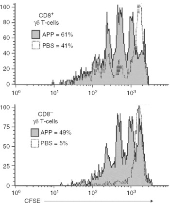
The plant-derived tannins are the most potent innate priming agents for bovine γδ T-cells that we have defined to date (Holderness et al., Reference Holderness, Jackiw, Kimmel, Kerns, Radke, Hedges, Petrie, McCurley, Glee, Palecanda and Jutila2007). Within 4 h after treatment of sorted γδ T-cells, transcripts for the PAMP-associated chemokines MIP1α and MIP1β are induced (Graff and Jutila, Reference Graff and Jutila2007; and M. A. Jutila, unpublished observations), within 24 h IL-2Rα protein can be detected by FACS, and even in the absence of any exogenous growth factor, such as IL-2, the plant tannins induce a low level of proliferation in bovine γδ T-cells. Treatment of highly pure bovine γδ T-cells (>96% purity) with plant tannins makes nearly all γδ T-cells hyper-responsive to IL-2 (Holderness et al., Reference Holderness, Jackiw, Kimmel, Kerns, Radke, Hedges, Petrie, McCurley, Glee, Palecanda and Jutila2007), demonstrating that tannins induce their priming effect on γδ T-cells directly, and not through an accessory cell. Furthermore, there is no subset specificity to the γδ T-cell response, in that both CD8+ and CD8− γδ T-cells are primed by the plant tannins (Fig. 5).
Plant tannins prime bovine CD8+ and CD8− (WC1+) γδ T-cells to proliferate in response to IL-2. CFSE-labeled bovine PBLs were treated with oligomeric tannins from apple extract (apple polyphenol, APP) for 48 h and washed and culture medium was replaced with IL-2 (1 ng ml−1)-containing medium. After 5 days in culture, three-color FACS was done and the proliferation of CD8+ and CD8− γδ T-cells was compared. Shaded histograms represent APP-treated cells, whereas the open histograms represent PBS-treated cells. The percentage values represent the percentage of γδ T-cells that divided at least once, as determined by loss of CFSE intensity.

The response to plant tannins also occurs with mouse and human γδ T-cells, indicating that this mechanism is evolutionarily well conserved. The specificity of the response in the mouse is strikingly similar to what is seen in bovine cells, in that γδ T-cells are selectively primed to respond to IL-2 (B. A. Freedman and M. A. Jutila, unpublished observations). This identifies one of the first conserved functional responses in rodent and ruminant γδ T-cells, which we normally define as being quite divergent. Plant tannins also effectively prime human γδ T-cells, but whereas γδ T-cells are the only bovine cell type affected by plant tannins, human NK cells and subsets of αβ T-cells additionally respond in a manner similar to γδ T-cells. Another disparity between human and bovine γδ T-cell responses is that whereas bovine γδ T-cells proliferate to a small degree in the absence of secondary stimulation (IL-2), tannin-treated human γδ T-cells require secondary stimuli to achieve significant proliferation. Although the priming event induced by plant tannins induces only small phenotypic changes in the γδ T-cell, similar to γδ T-cells treated with LPS, the full consequence of priming is realized when further treated with the human γδ T-cell agonist, HDMAPP. In the case of APP, this tannin enhances human γδ T-cell proliferative responses to HDMAPP by >300-fold (Holderness et al., Reference Holderness, Jackiw, Kimmel, Kerns, Radke, Hedges, Petrie, McCurley, Glee, Palecanda and Jutila2007). Due to the similarities of the γδ T-cell response to LPS, we propose this plant tannin augmentation is due to a change in the γδ T-cell priming state and that this priming event is required for optimal γδ T-cell response to mitogenic signaling.
The induction of an antigen-independent priming state in γδ T-cells treated with tannins suggests that this is a conserved, host-developed response to the environment. Cattle certainly consume large amounts of various plant tannins and other polyphenols; thus we speculate that diet may contribute to the maintenance of the large pool of γδ T-cells in these animals. In support of this possibility, studies have shown that feeding of similar condensed tannin preparations to mice in their water leads to an expansion of their γδ T-cell pool within the gut mucosa (Akiyama et al., Reference Akiyama, Sato, Watanabe, Nagaoka, Yoshioka, Shoji, Kanda, Yamada, Totsuka and Teshima2005). This suggests the possibility that tannin-based agonist preparations might be used to augment γδ T-cell function and immunity in general in vivo, which we are currently investigating.
The mechanism of plant tannin action on γδ T-cells is unclear. Common themes in the study of tannins include their potent antioxidant and apoptosis-inducing properties, which are a conserved characteristic of all tannins. It is unlikely, however, that the γδ T-cell response can be attributed directly to antioxidant properties since smaller tannins (monomers) have little impact on γδ T-cell proliferation (Holderness et al., Reference Holderness, Jackiw, Kimmel, Kerns, Radke, Hedges, Petrie, McCurley, Glee, Palecanda and Jutila2007; and J. Holderness and M. A. Jutila, unpublished observations) yet possess an increased antioxidant potential compared to oligomeric tannins (Osakabe et al., Reference Osakabe, Yasuda, Natsume, Takizawa, Terao and Kondo2002). Furthermore, many tannin preparations do not induce γδ T-cell priming (Holderness et al., Reference Holderness, Jackiw, Kimmel, Kerns, Radke, Hedges, Petrie, McCurley, Glee, Palecanda and Jutila2007), which rules out non-specific activities of tannins as a whole, such as antioxidant properties, as the mode of γδ T-cell priming.
Tannins often demonstrate individual properties not shared by all tannins. Historically, tannin-based preparations have been used as natural remedies to modify the immune response, which varies greatly depending on the type of tannin used. Some studies demonstrate that tannins induce pro-inflammatory cytokine (IL-1β) production (Miyamoto et al., Reference Miyamoto, Murayama, Nomura, Hatano, Yoshida, Furukawa, Koshiura and Okuda1993), though many other reports illustrate tannins that elicit a more anti-inflammatory response (Zhang et al., Reference Zhang, Liu, Xie, Yin, Li, Kwik-Uribe and Zhu2006; Hou et al., Reference Hou, Masuzaki, Hashimoto, Uto, Tanigawa, Fujii and Sakata2007). The differences observed in pro- or anti-inflammatory responses are associated with different tannin species. A comparison of these contrasting immune responses is found in studies by Mao et al., who have performed a number of experiments treating human PBMCs with tannin fractions from cocoa. The authors observe increased anti-inflammatory responses [IL-5 (Mao et al., Reference Mao, van de, Keen, Schmitz and Gershwin2002a) and TGFβ (Mao et al., Reference Mao, van de, Keen, Schmitz and Gershwin2003)] from smaller procyanidins and pro-inflammatory responses from larger, oligomeric procyanidins [IL-1β (Mao et al., Reference Mao, Powel, van de, Keen, Schmitz, Hammerstone and Gershwin2000)] and TNFα (Mao et al., Reference Mao, van de, Keen, Schmitz and Gershwin2002b)]. Although the tannins tested by Mao et al. were from cocoa and have not been tested on γδ T-cells, these data emphasize the conflicting responses to different tannin species even within condensed tannins from the same plant source.
The differences in immune response to various tannin preparations can be explained by tannin binding affinities for different proteins. Originally defined as low-affinity and non-specific protein-binding complexes with antioxidant activity, tannins are increasingly portrayed as additionally having high-affinity counter-receptors (Hagerman and Butler, Reference Hagerman and Butler1981; Frazier et al., Reference Frazier, Papadopoulou, Mueller-Harvey, Kissoon and Green2003). Immunologically relevant examples of tannins binding to specific proteins include: (i) tannic acid binding to CXCL12, preventing engagement with its receptor, CXCR4, and thereby preventing chemotaxis (Chen et al., Reference Chen, Beutler, McCloud, Loehfelm, Yang, Dong, Chertov, Salcedo, Oppenheim and Howard2003); (ii) Apple tannins blocking FcεR1/IgE binding (Tokura et al., Reference Tokura, Nakano, Ito, Matsuda, Nagasako-Akazome, Kanda, Ikeda, Okumura, Ogawa and Nishiyama2005) and preventing epidermal growth factor signaling by blocking the receptor (Kern et al., Reference Kern, Tjaden, Ngiewih, Puppel, Will, Dietrich, Pahlke and Marko2005); and (iii) epigallocatechin gallate binding to and suppressing CD11b, an adhesion molecule important for leukocyte migration to sites of inflammation, expression and function (Kawai et al., Reference Kawai, Tsuno, Kitayama, Okaji, Yazawa, Asakage, Hori, Watanabe, Takahashi and Nagawa2004).
Based on the observation that tannins affect monocytes by suppressing and down-regulating CD11b (Kawai et al., Reference Kawai, Tsuno, Kitayama, Okaji, Yazawa, Asakage, Hori, Watanabe, Takahashi and Nagawa2004), we tested the apple tannin preparation (APP) used to stimulate γδ T-cells to determine if CD11b regulation could be the cause of γδ T-cell priming. Although APP binds to and suppresses CD11b expression on monocytes in a manner similar to the tannin tested by Kawai et al., APP interestingly had the opposite effect on bovine γδ T-cells, and instead induced CD11b expression on a subset of cells. Furthermore, unlike with monocytes, this regulation of CD11b on γδ T-cells does not occur through tannin interaction with CD11b (Graff and Jutila, Reference Graff and Jutila2007). This suggests that there is a select group of tannins responsible for the γδ T-cell response, which differ from the tannins that bind to CD11b and affect monocytes. Therefore, to observe the direct effects of the γδ T-cell tannin agonist, the identification and isolation of the optimal tannin complex for γδ T-cell agonist activity are currently a top priority in our laboratory.
Another priority is the identification of the cellular receptor(s) for the bovine γδ T-cell tannin agonist. Due to the selective γδ T-cell response with low concentrations (1–40 μg ml−1) of the crude tannin preparation, our data to date are consistent with active tannin(s) acting through one or, perhaps, a restricted number of receptors on the bovine γδ T-cell and not through a non-specific mechanism, such as antioxidant activity. The first information in support of this comes from the restrictive pattern of gene regulation induced by plant tannins in γδ T-cells. Selective up-regulation of surface markers [IL-2Rα and CD69 (Holderness et al., Reference Holderness, Jackiw, Kimmel, Kerns, Radke, Hedges, Petrie, McCurley, Glee, Palecanda and Jutila2007)] and gene transcripts [MIP1α (Graff and Jutila, Reference Graff and Jutila2007)] is similar to PAMP-associated γδ T-cell responses and therefore consistent with a receptor-mediated event. Additionally, studies on other tannin/cell receptor studies suggest that the concentration of tannin extract (APP) used to prime γδ T-cells correlates with characterized tannin–protein interactions. For example, the specific binding of tannic acid to CXCR4 shows that this interaction competitively inhibits CXCL12 binding at concentrations (IC50=360 ng ml−1; Chen et al., Reference Chen, Beutler, McCloud, Loehfelm, Yang, Dong, Chertov, Salcedo, Oppenheim and Howard2003) similar to those we predict for the active component of APP.
The effective dose range of crude plant tannin preparations required to induce IL-2Rα on γδ T-cells is quite limited (1–40 μg ml−1 for bovine cells), due to toxicity of the preparations at the higher concentrations, which is consistent with other tannin preparations (Chen et al., Reference Chen, Beutler, McCloud, Loehfelm, Yang, Dong, Chertov, Salcedo, Oppenheim and Howard2003). We predict that isolation of the active tannin(s) from the crude extract will likely reduce the toxic effects of harmful tannin species. However, isolation of the active tannin component may be unnecessary for sub-toxic, biologic effects in vivo since the gut regulates tannin concentrations to both bioactive and safe concentrations. Both rats given a single oral dose of APP at 2000 mg kg−1 (Shoji et al., Reference Shoji, Akazome, Kanda and Ikeda2004) and mice receiving a prolonged exposure by replacing their drinking water with 1·0% w/v) APP for up to 9 weeks (Akiyama et al., Reference Akiyama, Sato, Watanabe, Nagaoka, Yoshioka, Shoji, Kanda, Yamada, Totsuka and Teshima2005) do not demonstrate an obvious toxic response. This increased in vivo resistance to the toxic effects can be explained by studies of Shoji et al., who demonstrate that plasma uptake of tannins plateaus at 10·2 μg ml−1 (1000 mg kg−1 oral dose) and does not increase when dosed up to 2000 mg kg−1 (Shoji et al., Reference Shoji, Masumoto, Moriichi, Akiyama, Kanda, Ohtake and Goda2006). This tannin concentration is optimal for γδ T-cell activation in vitro (Holderness et al., Reference Holderness, Jackiw, Kimmel, Kerns, Radke, Hedges, Petrie, McCurley, Glee, Palecanda and Jutila2007) and, furthermore, effectiveness of these in vivo treatments is confirmed by γδ T-cell expansion in animals treated with 1·0% (w/v) APP after 2 weeks (Akiyama et al., Reference Akiyama, Sato, Watanabe, Nagaoka, Yoshioka, Shoji, Kanda, Yamada, Totsuka and Teshima2005). This suggests that oral administration may naturally prevent overdose, and regulate tannin absorption into the plasma at optimal priming concentrations. Moreover, APP is currently marketed and sold as a nutritional supplement without anecdotal evidence of adverse effects, supporting its safety and underscoring the need for further characterization of this tannin-based supplement.
Summary
We have used a variety of approaches to study global gene expression in bovine T-cell subsets, including following various stimuli and during enterocolitis induced by Salmonella serovar Typhimurium. These studies suggest that bovine γδ T-cells express many genes associated with innate immunity, including many myeloid cell-associated genes, and rapidly respond to infection after a priming state induced by recognition of PAMPs. Follow-up studies confirmed the microarray and SAGE analyses and provided functional evidence for a new model of γδ T-cell responses to infection. Specifically, γδ T-cells rapidly respond to PAMPs, leading to a subtle response we define as antigen-independent priming. PAMP primed γδ T-cells produce cytokines that attract and activate myeloid cells and respond more robustly to secondary signals that include growth factors, such as IL-2, and specific antigen. Semi-high-throughput screening assays based on this model were used to identify novel γδ T-cell priming agents. A number of plant extracts were identified as containing potent priming agonists. Oligomeric tannins in some of these extracts represent the most potent priming agonists defined to date. These latter results expand the PAMP-induced priming model to include components of diet, which can prime γδ T-cells, similar to PAMPs, and potentially enhance innate immune responses in the intestinal mucosa.
Acknowledgments
Funding from a number of sources contributed to studies summarized in this review. These sources include: NIH (contract no. HHSN266200400009/N01-AI40009), USDA-IFAFS, USDA-NRI, multiple USDA and NIH equipment grants, Animal Health Formula Funds, Montana Agricultural Experiment Station, NIH COBRE (RR020185) and the M. J. Murdock Charitable Trust.