Serum proteins have multiple functions. Albumin is the most osmotically active serum protein and is a carrier of many substances. Globulins are a heterogeneous group of proteins which includes antibodies and other inflammatory molecules, haemostatic and fibrinolytic proteins, carriers of lipids, vitamins and hormones. Physiological or pathologic conditions can cause shifts in albumin and globulin concentrations and their evaluation by electrophoresis is a valuable diagnostic tool (Alberghina et al. Reference Alberghina, Casella, Vazzana, Ferrantelli, Giannetto and Piccione2010). In dairy cows the period of transition from late gestation to early lactation is recognized as inducing considerable metabolic adaptation (Van Dorland et al. Reference Van Dorland, Richter, Morel, Doherr, Castro and Bruckmaier2009; Polakova et al. Reference Polakova, Kudrna, Kodes, Hucko and Mudrik2010). Alterations in defence mechanisms occurring during the periparturient period are associated with changes in hormone profiles and the metabolic and physical stress of parturition (Mallard et al. Reference Mallard, Dekkers, Ireland, Leslie, Sharif, Lacey-Vankampen, Wagter and Wilkie1998). It seems that an alteration of immune and innate resistance mechanisms of the dairy cow normally begins approximately 3 weeks prior to calving, it is maximal at parturition and until 3 weeks after calving (Mallard et al. Reference Mallard, Dekkers, Ireland, Leslie, Sharif, Lacey-Vankampen, Wagter and Wilkie1998). These periods are all characterized by mobilization of body fat, protein and mineral stores to satisfy the requirements for milk production and maintenance (Van Dorland et al. Reference Van Dorland, Richter, Morel, Doherr, Castro and Bruckmaier2009). Serum proteins have been often used to evaluate the infections that could occur during the post-partum period and that contribute to a lengthening of the calving-to-conception interval (Mallard et al. Reference Mallard, Dekkers, Ireland, Leslie, Sharif, Lacey-Vankampen, Wagter and Wilkie1998). Most of these problems manifest after calving and, usually, establish as early as at the end of previous lactation or at the end of the dry-up period (Polakova et al. Reference Polakova, Kudrna, Kodes, Hucko and Mudrik2010). Cow milk protein fractions during phases of lactation have been extensively studied (Ontsouka et al. Reference Ontsouka, Bruckmaier and Blum2003; Schopen et al. 2009; Bonfatti et al. Reference Bonfatti, Di Martino, Cecchinato, Vicario and Carnier2010) but little attention has been addressed to the serum protein pattern of dairy cow during different stages of gestation and lactation. Therefore the aim of this study was to analyse the modifications of serum protein fractions during these physiological phases in healthy Holstein cows of high average milk production.
Materials and Methods
The experiment was carried out in Sicily (Italy), in a farm site in Ragusa (36°55′N; 14°43′E), on five multiparous clinically healthy Holstein Friesian cows, in good nutritional condition (5 years old, mean body weight 650±50 kg). Dairy cows were in their 5th pregnancy and with an average 305-d production of about 7600 kg. They were enrolled 60 d prior to expected calving date. All housing and care conditions conformed to the standards recommended by the Guide for the Care and Use of Laboratory Animals and Directive 86/609 CEE. Their health status was evaluated based on rectal temperature, heart rate, respiratory rate, appetite, faecal consistency and haematologic profile. All animals were free from internal and external parasites and were negative to the coprologic examination, carried out using the flotation method, before the experimental period. During the trial, all animals were kept under natural photoperiod and ambient temperature. Ambient temperature and relative humidity for all experimental periods were continuously recorded with a data logger (Gemini, Chichester, West Sussex, UK). No treatments were administered before the start of the experiment.
Cows were milked twice daily at 5·30 and 16·30 by means of a milking machine, and fed twice daily at 7·00 and 18·00. Cows were fed two diets containing different ratios of forage (on a dry matter basis, 64% triticosecale silage and 36% alfalfa hay) and concentrate, with a crude protein content of 15·5% of dry matter. Lactation diet was formulated to contain 53% forage and 47% concentrate. Concentrate composition was: corn meal (17%), soy protein flour (16%), beet pulp (15%), barley flour (13%), carob bean sprouts (7%), wheat bran (7%), fat-extracted sunflower meal (6%), carobs (5%), molasses (4%), extruded soy (4%), rumen by-pass fats Megalac (5%), calcium carbonate (1%), rock salt (0·6%). Dry matter intake averaged 21 kg/d. Dry period diet was formulated to contain 82% forage and 18% concentrate. Concentrate composition was: barley flour (16%), fat-extracted sunflower meal (18%), wheat bran (5%), soy protein flour (32%), carob bean sprouts (8%), corn meal (16%), minerals and salts (‘Dairy dry’) (5%). Dry matter intake averaged 12 kg/d. Water was available ad libitum.
All the experimental subjects were selected on the basis of their pregnancy status and blood samples were collected at different physiological phases: dry period (at −60 d, −30 d to expected calving), transition period (almost 7 d to calving, 7 d after calving), and lactation (in weeks 2, 5 and 15 after calving) giving a total of eight blood samples for each cow. Sampling time 7 d before expected calving was equivalent to 5±2 d before actual calving. Each sample, collected at the same hour in the morning from a jugular vein into vacuum glass tubes containing no additive, consisted of 10 ml of blood. After standing at room temperature for 20 min, the tubes were centrifuged at 1785 g for 10 min and harvested plasma was stored at –20°C until analysed. At the time of analysis serum samples were thawed at 20°C for 30 min before assessing protein concentrations. Total protein concentrations were determined by the biuret method, using an automated analyser (Konelab 20, Dasit, Helsinki, Finland). The protein standard was albumin (5·00 g/dl; Dasit, Milano, Italy). Electrophoresis was performed using a semiautomated AGE system (Helena Laboratories, Helena Biosciences, Gateshead, UK) according to the procedure described by the manufacturer and reported in detail by Alberghina et al. (Reference Alberghina, Casella, Vazzana, Ferrantelli, Giannetto and Piccione2010).
Using the computer software Phoresis (Helena Biosciences), electrophoretic curves plus related quantitative specific protein concentrations, for each sample, were displayed. Relative protein concentrations within each fraction were determined as the optical absorbance percentage, and absolute concentrations (g/dl) were calculated using the total serum protein concentration. All the results were expressed as means±sd. On all data, normally distributed (P<0·05, Kolmogorov-Smirnov's test), one-way Repeated Measure Analysis of Variance (ANOVA), was applied to evaluate the influence of time on serum protein fractions. Whenever the F test was signficant (P<0·05), Bonferroni's test was applied for post hoc comparison. Data were analysed using Statistica 7·5 software package (Statsoft Inc., Tulsa OK, USA).
Results
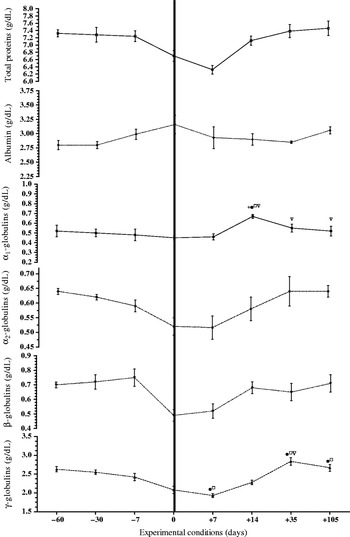
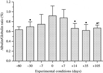
Mean values (±sd), together with the statistical significances, of total proteins and protein fractions obtained in the five cows during the experimental period, are shown in Fig. 1. A significant effect of the different physiological stages was observed on total proteins (P<0·05), α1-globulins (P<0·005), β-globulins (P<0·05), γ-globulins (P<0·005) and albumin/globulin (A/G) ratio (P<0·001). Albumin was the only protein fraction to show a slight increase in post-partum periods. Significant declines in concentrations around parturition were observed for: α1-globulins, β-globulins and γ-globulins, whereas albumin and α2-globulins showed non-significant changes in serum concentration. A/G ratio increased post partum compared with late gestation and following phases of lactation. Moreover during week 1 of lactation A/G ration was significantly higher than in week 5. The pattern of A/G ratio is shown in Fig. 2.
Pattern of serum protein levels in five Holstein Friesian cows, monitored from dry to lactation period (−60 d to calving; −30 d to calving; −7 d to calving; 0, calving (vertical dashed line); +7 days after calving; +14, week 2 of lactation; +35, week 5 of lactation; +105, week 15 of lactation). Statistical significance: * v. −60 d to calving (P<0·001); ●v. −30 d to calving (P<0·0001); □v. –7 d to calving (P<0·01); ◊v. calving (P<0·001). ∇v. 7 d after calving

The pattern of albumin/globulin ratio, analysed in five Holstein Friesian cows, monitored from dry to lactation period (–60 d to calving; –30 d to calving; −7 d to calving; 0, calving; +7 d after calving; +14, week 2 of lactation; +35, week 5 of lactation; +105, week 15 of lactation). Statistical significance: ●v. −30 d to calving (P<0·0001); □v. –7 d to calving (P<0·01)

Discussion
Serum protein concentrations were within the normal range (Alberghina et al. Reference Alberghina, Giannetto, Vazzana, Ferrantelli and Piccione2011). The post-partum period, compared with previous phases, was characterized by a decrease of total proteins due to the decrease of γ-globulins and to a consequent increase of A/G ratio. Total proteins continued to decrease at week 1 of lactation, whereas these slightly increase during the following phases. In agreement with other reports (Larson & Kendall, Reference Larson and Kendall1957; McLennan & Willoughby, Reference McLennan and Willoughby1973; Grünberg et al. Reference Grünberg, Donkin and Constable2011) total protein concentrations at calving were lower than concentrations outside the parturient period. The decline of total protein concentration has been indicated as effect of a decreased globulin fraction whereas changes in plasma albumin were marginal (Larson & Kendall, Reference Larson and Kendall1957). In the present study, albumin concentration was similar thorough these physiological phases with a small increase in concentration at calving. The slight increase of albumin at calving could be due to higher albumin synthesis by liver or to a decrease of plasma volume masked by hypoglobulinaemia. Pregnancy is accompanied by a substantial and progressive increase in plasma volume followed by its decrease 6–24 h after parturition (Jansakul et al. Reference Jansakul, King, Boura, Brennecke and Handberg1989). Albumin, with A/G normal, usually increases as an effect of decreased plasma volume because albumin and globulins are affected to the same extent. In this case A/G ratio was increased due to translocation of immunoglobulins into the mammary gland, making it more difficult to interpret the albumin concentration increase. Regarding the concentration of albumin at calving, some authors found a decrease in some but not in all cows studied (Rowlands et al. Reference Rowlands, Manston, Stark, Russel, Collis and Collis1980) while others found a general decrease (Little, Reference Little1974; Rowlands & Manston, Reference Rowlands and Manston1983; Park et al. Reference Park, Shirley, Titgemeyer, Cochran, DeFrain, Wickersham and Johnson2010; Grünberg et al. Reference Grünberg, Donkin and Constable2011). These fundamental differences in albumin pattern could be reflective of different post-partum time samplings or to differences in methods used for albumin measurement. The absolute albumin levels derived from a combination of total protein estimation and the relative proportions obtained from an electrophoretic trace is more accurate than the Bromocresol green (BCG) method (Bush, Reference Bush1991). The increase of bilirubin levels at calving (Rowlands & Manston, Reference Rowlands and Manston1983) can give falsely low readings with the hydroxyazobenzene benzoic acid (HABA) dye-binding method (Rowlands & Manston, Reference Rowlands and Manston1983) but also BCG readings are influenced by interference with bilirubin levels (Bush, Reference Bush1991). The α1-globulin fraction includes the acute phase proteins (APP), α1-antichymotrypsin and orosomucoid, and their increase post partum in dairy cows was reported also by others (Cairoli et al. Reference Cairoli, Battocchio, Veronesi, Brambilla, Conserva, Eberini, Wait and Gianazza2006). The slight decrease of β-globulins, observed at calving and at week 1 of lactation, should be interpreted as the passage of tranferrin, plasminogen and lipoproteins from plasma to colostrum and milk. Sánchez et al. (1988) found the highest concentration of transferrin in cows’ colostrum and first milk, suggesting the existence of a specific transport of transferrin from blood to milk. γ-Globulins represent one of the most important colostrum and milk constituents: the immunoglobulins, necessary to provide the young animal with passive immunization (Stelwagen et al. Reference Stelwagen, Carpenter, Haigh, Hodgkinson and Wheeler2009). Immunoglobulins of milk may be blood-derived or produced in situ by intramammary plasma cells (Stelwagen et al. Reference Stelwagen, Carpenter, Haigh, Hodgkinson and Wheeler2009). The decrease observed in late gestation in this study, according to results from others (Larson & Kendall, Reference Larson and Kendall1957) could indicate that immunoglobulins leave the plasma during the last month of gestation when cholostrum is formed in the mammary gland. After the decrease of γ-globulins in the post-partum period, levels increase from week 5 of lactation. This decrease of γ-globulins should be considered as one of the factors associated with the higher predisposition of dairy cows to infection after partum. The pattern of A/G showed an increase post partum and in early lactation as a result of the decrease of γ-globulins. This study provides information concerning the high-producing dairy cow and shows the modifications of the serum proteins occurring during different physiological phases from dry period, until calving and lactation. Most of the detected modifications were related to the transition from gestation to lactation, and show that it is a period of great metabolic stress for cows. On the basis of the results obtained we can affirm that the pattern of serum protein fractions could give information about dehydration, plasma volume expansion and hepatic function occurring during the peripartum period in dairy cows.