Heterotaxy syndromes have been known to be associated with various disorders of the cardiac conduction system. There is a relatively high incidence of complete atrioventricular block in patients with left isomerism, which often starts early in fetal life. The prognosis for fetuses with high grade atrioventricular block in association with major cardiac anomalies has historically been dismal, and has remained unchanged over the past few decades in spite of advances in medical field. Recent advances in fetal echocardiography have enabled us to identify these anomalies of viscerocardiac heterotaxy early in gestation, and to assess more carefully the type and severity of the prenatal cardiac arrhythmias. In the light of these advances, we report a fetus with left isomerism with atrioventricular septal defect and common atrioventricular valvar orifice, in whom we were able to identify, and follow with Doppler echocardiography, the progression of the conduction disturbance from low grade second degree to complete atrioventricular block. Although cardiac anomalies and atrioventricular block have previously been well described in fetuses with left isomerism, as far as we are aware, ours is the first case in which the Wenckebach phenomenon, also known as Mobitz type I atrioventricular block, and its gradual progression to complete atrioventricular block, has been recorded in a fetus with spectral Doppler. We also discuss briefly the various cardiac and extracardiac anomalies found in children with left isomerism.
Case report
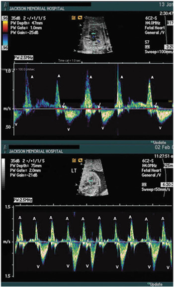
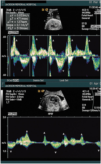
A pregnant lady at 21 weeks of gestation was referred to us for fetal echocardiography, an abnormal cardiac 4-chamber view having previously been seen on ultrasonic scanning. The routine ultrasound also showed a midline liver, and the spleen was not visualized. The fetal echocardiogram showed the heart to be left sided, with isomerism of the left atrial appendages. There was a common atrium, bilateral superior caval veins, each draining to the roofs of the common atrium on each side, and interruption of the inferior caval vein with azygous continuation. There was a balanced atrioventricular septal defect, with mild regurgitation through the common atrioventricular valvar orifice. The great arteries were normally related, with no outflow obstruction, and ventricular function was normal. The atrial rhythm was regular at 120 to 130 beats per minute, and the ventricular rhythm was regularly irregular. Spectral Doppler tracings of the inflows to and outflows from the ventricles indicated second degree atrioventricular block of the Wenckebach type, or Mobitz type I (Fig. 1). In the follow-up echocardiogram at 24 weeks gestation, the atrial rhythm was still regular at 130 to 140 beats per minute. By this stage, however, the atrioventricular block had progressed to high grade atrioventricular block of the Mobitz type I, with alternating 3:2 and 2:1 conduction (Fig. 1). At 28 weeks gestation, there was Mobitz II atrioventricular block with consistently 2:1 conduction with the atrial rate at 120 to 130 beats per minute, and the ventricular rate at 60 to 65 beats per minute (Fig. 2). The ventricular function and the regurgitation across the common atrioventricular valve remained unchanged, without any evidence of fetal hydrops. At 32 week of gestation, there was complete atrioventricular block, with bradycardia (Fig. 2) and severe hydrops, requiring emergency delivery of the fetus by Caesarian section. The fetal echocardiographic diagnoses were confirmed postnatally. The infant had complete atrioventricular block with resultant bradycardia, ventricular noncompaction, and severe ventricular dysfunction, requiring implantation of a VVI pacemaker. The patient also had severe lung disease related to prematurity, and died at 3 weeks of age in spite of all supportive measures. Autopsy confirmed the diagnosis made echocardiographically.
Top panel: Spectral Doppler mapping of the left ventricular inflow showing progressive prolongation of the atrioventricular interval (white arrows) leading on to a blocked atrial beat. (Mobitz type I – Wenckebach block). Bottom panel: Spectral Doppler showing alternating 3:2 and 2:1 block. (High grade Wenckebach atrioventricular block). A – Atrial Doppler wave; V – Ventricular systolic Doppler wave.

Top panel: Spectral Doppler mapping of the left ventricular inflow showing Mobitz type II, 2nd degree atrioventricular block. Bottom panel: Spectral Doppler showing complete atrioventricular block with dissociation between the atrial (A) and the ventricular (V) beats and severe ventricular bradycardia.

Discussion
Heterotaxy is strictly defined as any disturbance from the usual arrangement, but in the practice of paediatric cardiology, it is usually interpreted to mean an abnormal arrangement of the thoraco-abdominal organs across the right–left axis. Other terms used include cardiosplenic syndrome, and isomerism. The spectrum of cardiac and extra-cardiac anomalies associated with left isomerism has been described in many series.Reference Peoples, Moller and Edwards1, Reference Ho, Cook, Anderson, Allan and Fagg2 In general, patients with left isomerism present with less severe cardiac defects when compared to those with right isomerism. In a recent large series reported by Gilljam et al.,Reference Gilljam, McCrindle, Smallhorn and Williams3 one-sixth of a total of 163 patients had normal hearts, nine-tenths had an interrupted inferior caval vein, and half had atrioventricular septal defects with common atrioventricular junction. Only three-fifths of the patients, however, had multiple spleens, the spleen being normal in one-quarter, and hypoplastic or absent in the remainder. Left atrial, or coronary sinus, rhythm, wandering atrial pacemaker, bradycardia and congenital complete atrioventricular block are well known to occur in this condition.
Diagnosis of these syndromes can now be made with high accuracy by fetal echocardiography. The diagnosis of left isomerism should be strongly entertained in the presence of a combination of at least 2 of the following features: atrioventricular septal defect with common atrioventricular junction, interruption of inferior caval vein, fetal atrioventricular block, and viscerocardiac heterotaxy.Reference Berg, Geipel and Smrcek4 The presence of interruption of the inferior caval vein is a reliable clue in the prenatal diagnosis of cardiosplenic syndromes.Reference Sheley, Nyberg and Kapur5 The prognosis remains dismal for the fetus with left isomerism in the presence of major structural heart disease and high grade atrioventricular block.Reference Chitayat, Lao, Wilson, Fagerstrom and Hayden6–Reference Shenker, Reed, Anderson, Marx, Sobonya and Graham9 Another recent study demonstrated atrioventricular block to be an independent risk factor for mortality in this group of patients.Reference Berg, Geipel and Smrcek4
As far as we are aware, our case is unique in that we were able to follow the progression from low to high grade atrioventricular block in this fetus. To our best knowledge, this is the first description of the Wenckebach phenomenon in the fetus as shown by Doppler echocardiography. In addition, our experience provides an excellent demonstration of the usefulness of spectral Doppler analysis in the detection of fetal arrhythmias. Studies have also shown that spectral Doppler analysis of the flows in the fetal heart can be very useful in the diagnosis of fetal arrhythmias.Reference Chan, Woo, Ghosh, Tang and Lam10 A prolonged mechanical atrioventricular interval measured by Doppler obtained from the left ventricular outflow, or simultaneous interrogation of the superior caval vein and ascending aorta, may be the first indication of disease of the conduction system, and should be made a part of the fetal echocardiographic study in these patients. Advances in fetal echocardiography have enabled us, over the past 2 decades, to identify these anomalies early in gestation and offer counseling to the parents to make informed decisions.