Significant outcomes
Different postnatal experiences have profound effects on adult neurochemistry, although the effects varied among strain, neurotransmitters and brain area examined.
The neurochemical phenotype of Lister Hooded (LH) rats suggests that this strain may be more vulnerable to develop schizophrenic-like manifestations following isolation.
The Wistar (W) rat strain could provide better information on several parameters resembling depressive-like behaviours in schizophrenia.
Limitations
Animal models of mental disorders are difficult to assess, especially for schizophrenia, which is a complex, multifactorial clinical syndrome.
The choice of a selected strain of animal to model a specific disease could be related to economical aspects and not to scientific evidence.
The indiscriminate use of strains could represent a potential source of conflicting results in scientific literature.
Introduction
Early adverse experiences and especially vulnerability to these events may ‘shape' a pre-existing genetic vulnerability to stress and disease. Thus, young animals raised under adverse-rearing conditions, such as social isolation or maternal separation, present with long-lasting behavioural and physiological changes in adulthood that resemble depression and/or schizophrenia, including increased anxiety, compromised cognitive function, poor social interaction as well as various depressive-like behaviours (Reference Harvey and Shahid1). From a clinical point of view, depression and schizophrenia lie on a continuum of symptom presentation and underlying biology. Indeed, schizophrenic patients often show a depressive state as prodrome of the psychiatric disorder (Reference Yung, Buckby and Cosgrave2). Rearing rats in social isolation from weaning results in long-term effects on brain structure, neurotransmitter function and behaviour and many of the effects mimic those seen in schizophrenia and depression (Reference Muchimapura and Marsden3). Specifically, isolation rearing of rats has been regarded as an ‘environmental model' of schizophrenia (Reference Weiss and Feldon4) since the long-term alterations induced by this non-pharmacological method resemble symptoms seen in schizophrenic patients (Reference King, Seeman, Marsden and Fone5), such as hyper-reactivity to novel environments and cognitive impairment (Reference Geyer and Ellenbroek6,Reference Lapiz, Fulford, Muchimapura, Mason, Parker and Marsden7). Impairments in prepulse inhibition (PPI) are often reported in isolated rats (Reference Geyer, Wilkinson, Humby and Robbins8) and in patients with schizophrenia (Reference Braff, Geyer and Light9). However, this may also be observed in other psychiatric disorders such as depression, where it may reflect stimulus overload-induced cognitive fragmentation. Moreover, loss of social contact and behavioural withdrawal are associated with both schizophrenia and depression in humans (Reference Heim, Plotsky and Nemeroff10).
The isolation rearing model is based on the observation that rats reared in single housing from weaning throughout adulthood exhibit schizophrenia- and depression-like behavioural, morphological and neurochemical abnormalities (Reference Fone and Porkess11). Among the most common behavioural and morphological changes observed in isolated rats are deficits in sensorimotor gating, increased locomotor activity (Reference Cilia, Reavill, Hagan and Jones12), anxiety-related behaviours (Reference Lukkes, Watt, Lowry and Forster13) and decreased cortical and hippocampal synaptic plasticity (Reference Fone and Porkess11). The dopaminergic hypothesis is one of the most widely accepted theories regarding the aetiology of schizophrenic symptoms, such that impairment in the dopaminergic system could represent the neurochemical basis of these behavioural alterations (Reference Harvey, Stein and Emsley14). Moreover, it also explains the common use of dopamine (DA) antagonists as clinically effective antipsychotic agents. Nonetheless, there is evidence that a key effect of social isolation may be loss of neuronal plasticity combined with change in the functionality of various cortical and hippocampal neurotransmitters, including glutamate and serotonin (5-HT) (Reference Harvey, Stein and Emsley14). Although reduced glutamate function may underlie the deficits in memory function, this can be reversed by administration of a 5-HT6 receptor antagonist (Reference Marsden, King and Fone15), emphasising the importance of cross-talk between these two transmitters. Moreover, many new generation antipsychotics target 5-HT in their pharmacological profile (Reference Harvey, Stein and Emsley14) and are effective not only for schizophrenia but also for treatment-resistant depression (Reference Shelton, Osuntokun, Heinloth and Corya16). However, there is a wealth of evidence showing that gender and age can affect both the degree and direction of the resultant long-term changes in behaviour and neurochemistry produced by exposure to early life adversity in the rat (Reference Ferdman, Murmu, Bock, Braun and Leshem17).
An additional variable in isolation experiments that has not been extensively characterised is the extent to which the strain of rat used in the experiments may affect the results. Indeed social behaviour differs by strain, making it a significant variable when considering the influence of social-rearing conditions on later behaviour (Reference Hall, Huang, Fong, Sundstrom and Pert18). Few published studies have compared two different rat strains, such as Wistar (W) and Lister Hooded (LH), with respect to their response to an adverse condition. However, there is a surprising lack of comparative studies on LH and W rats of both genders, which motivated us to use these two strains. In particular, we have chosen the LH strain for our studies on isolation rearing as it has been found to produce consistent and reproducible behavioural effects with the isolation procedure (Reference Marsden, King and Fone15). Specifically, LH and W rats have revealed considerable behavioural differences in kindling responses, anxiety (Reference Ennaceur, Michalikova, Bradford and Ahmed19), attention tasks, open field and food-hoarding behaviour (Reference Broersen and Uylings20). The results of these studies seem to support increased anxiety and lower cognitive performance in W compared to LH rats.
The above reported scenario raises the intriguing question as to whether difference in these rat strains may have a neurochemical basis and whether a different genetic background could be responsible for possible neurochemical alterations in isolated rats (ISO). Thus, the first aim of this study was to evaluate the neurochemical profile of W and LH rat strains by examining tissue content of DA, 5-HT and relative metabolism, in several brain regions involved in mental disorders, such as prefrontal cortex (PFC), nucleus accumbens (NAC), hippocampus (Hipp) and striatum (St) of group housed (GRP) and ISO LH and W rats. Moreover, besides alterations in several brain areas, functional imaging and neuropsychological studies have provided convincing evidence for the critical involvement of the PFC in schizophrenia (Reference Martins-de-Souza, Gattaz and Schmitt21). Hence, in a second experiment, in vivo microdialysis was performed in the PFC of W rats as well as LH animals, either in GRP or ISO conditions to determine basal in vivo concentrations of the above transmitters.
Materials and methods
Animals
The present study was conducted in W and LH adult male rats. They were housed at constant room temperature (22 ± 1 °C) and relative humidity (55 ± 5%) under a 12-h light/dark cycle (lights on from 07:00 to 19:00 h). Food and water were freely available.
Procedures involving animals and their care were conducted in conformity with the institutional guidelines that are in compliance with national (D. L. No. 116, G. U., Suppl. 40, 18 Febbraio 1992, Circolare No. 8, G. U., 14 Luglio 1994) and international laws and policies (EEC Council Directive 86/609, OJ L 358, 1, December 12, 1987; Guide for the Care and Use of Laboratory Animals, US National Research Council, 1996). All efforts were made to minimise the number of animals used and their suffering.
Social isolation protocol
For the social isolation procedure (Reference Leng, Feldon and Ferger22), rats were mated such that all the mothers gave birth within 1 week. At weaning (postnatal day 21), the pups were separated from their mothers and divided and reared either in ISO (one rat per cage) or in GRP (three or four rats per cage) such that each litter contributed only one subject to the GRP and one subject to the ISO condition to avoid a litter effect (Reference Lehmann and Feldon23). Animals were disturbed only for cleaning purposes, which consisted of changing the cage once a week for ISO and twice a week for GRP animals. ISO and GRP rats maintained visual, auditory and olfactory contact with the other animals throughout the studies. All experiments were conducted at the end of 6–8 weeks of isolation rearing after body weight assessment.
Post-mortem tissue preparation
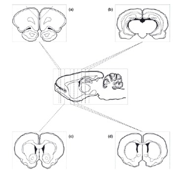
Rats were killed by decapitation and brains immediately removed. For dissection, the brains were placed dorsal side up in an ice-chilled rat brain matrix (World Precision Instruments Inc., Sarasota, FL, USA) with slits spaced at 1 mm using an ice-chilled razor blade, brain slices containing the target regions were dissected according to the atlas of Paxinos and Watson (Reference Paxinos and Watson24) (Fig. 1). Then PFC, NAC, St and Hipp were collected by tissue dissection and immediately frozen on dry ice. For the biogenic amine extraction, samples were homogenised in 10 volumes (w/v) of perchloric acid 0.1 M. The homogenates were stored on ice for 30 min and then centrifuged at 10000 × g for 10 min at 4 °C. The supernatants were then filtered (0.45 µm syringe filters, Millipore, Vimodrone, Milan, Italy) and diluted (PFC and NAC not diluted, St 1:4, Hipp 1:2) with 0.1 M perchloric acid before high-performance liquid chromatography (HPLC) analysis. DA turnover was expressed as (HVA+DOPAC)/DA and 5-HT turnover as 5-HIAA/5-HT.
Regions of the post-mortem tissue preparation. The locations of the razor blades used to cut the adjacent coronal slices are indicated in a sagittal view of the rat brain with the ventral surface down. Brain regions of interest were dissected on the superior face of the coronal slices as indicated (a: PFC; b: Hipp; c: NAC; d: St).

In vivo microdialysis
Rats were anaesthetised with 3.6 ml/kg Equithesin intraperitoneally (1.2 g sodium pentobarbital; 5.3 g chloral hydrate; 2.7 g MgSO4; 49.5 ml propylene glycol; 12.5 ml ethanol and 58 ml distilled water) and secured in a stereotaxic frame (David Kopf Instruments, Tujunga, CA, USA). A hole was drilled in the frontal bone and a small incision was made in the dura with a bent needle tip. The probe (4 mm) was lowered slowly into the PFC (AP = +3.7, ML = +0.7, DV = −4.8) from bregma according to the Paxinos and Watson atlas (Reference Paxinos and Watson24). The dialysis probes (AN69 Hospal SpA,; 20 kDa cut-off) were prepared as described by Robinson and Whishaw (Reference Robinson and Whishaw25). The rats were allowed to recover from anaesthesia for at least 15 h before the neurotransmitter release study (Reference Trabace, Kendrick and Castrignano26).
On the day of the experiment, the fibres were perfused with an artificial cerebrospinal fluid containing: 145 mM NaCl, 3 mM KCl, 1.26 mM CaCl2, 1 mM MgCl2 and 1.4 mM Na2HPO4 in distilled water (1 l); the solution was buffered at pH 7.3 with a 0.6 mM NaH2PO4 and filtered (0.45 µm). The fibres were perfused at a constant flow rate of 2 µl/min using a CMA/100 microinjection pump (CMA Microdialysis, Stockholm, Sweden). The microdialysis membrane was allowed to stabilise for 2 h. Then, samples were collected at 20-min intervals. All samples of perfusate were immediately analysed by HPLC.
The position of the microdialysis probe was verified by histological procedures at the end of each experiment. DA and 5-HT concentrations were determined by HPLC coupled with an electrochemical detector (INTRO, Antec Leyden, The Netherlands). Amines separation was performed by an LC18 reversed phase cartridge column (15 cm × 2 mm, 3 µm; Phenomenex, Castel Maggiore, Bologna, Italy). The detection was accomplished by a Unijet cell (BASi) with a 6-mm diameter glassy carbon electrode at a working potential of 0.65 V versus Ag/AgCl. The mobile phase used was 85 mM CH3COONa, 0.8 mM octan sulfonic acid, 0.3 mM ethylenediaminetetraacetic acid, 15 mM NaCl, methanol 6%, solved in distilled water, buffered at pH 4.85 with CH3COOH and filtered. The flow rate used was 0.220 ml/min.
Statistical analysis
For the post-mortem tissues analysis, content of neurotransmitters were analysed by two-way analysis of variance (ANOVA), with main factors of housing condition (r), comparing ISO and GRP rats, and strain (st), comparing W and LH animals.
For the microdialysis analysis, four consecutive samples were collected (80 min). Since differences between rearing conditions and strain were time independent, individual comparisons between marginal mean values of neurotransmitters levels (pooled data of 4 samples for each factor) were made and analysed by ANOVA using housing condition and strain as between-subjects factors.
In the case of significant main effects, the differences between individual means were assessed with the post hoc Tukey's test. Differences were considered significant only when p-values were less than 0.05.
Results
Body weight
Animal body weight was evaluated at the end of the isolation-rearing period. This parameter was estimated in order to consider any possible gross sign of animal distress that could relate to the difference in brain neurochemistry, leading to data misinterpretation.
After 6–8 weeks of rearing, no significant difference in the body weight (GRP LH: 338.7 ± 8.7 g, ISO LH: 354 ± 7.3 g, GRP W: 339.4 ± 10.2 g, ISO W: 320.5 ± 21.7 g) of the two different rat strains [F(st)1,26 = 1.203, n.s.] or the two different rearing conditions [F(r)1,26 = 0.0121, n.s.] was observed (data not shown). Moreover, no interaction between rearing conditions and strain was found [F(r × st)1,26 = 1.312, n.s.].
Post-mortem analysis
In order to assess the neurochemical profile of W and LH rat strains, we examined tissue content of DA, 5-HT and metabolites in several brain regions involved in mental disorders, including the PFC, NAC, Hipp and St from GRP and ISO rats from both strains.
Prefrontal cortex
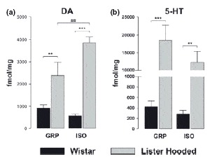
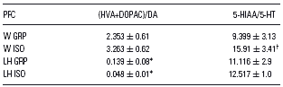
A significant difference in DA content between W and LH rats, with more DA in the PFC of LH than W rats, was observed (Fig. 2). Moreover, in LH strain, ISO rats showed higher DA content than GRP animals [F(st)1,23 = 49.070, p < 0.001; F(r)1,23 = 2.743, n.s.; F(r × st)1,23 = 7.173, p < 0.05] (Fig. 2). Moreover, DA turnover in LH rats was significantly lower compared to W, independently from rearing conditions (two-way ANOVA, p < 0.01, Table 1), while no difference was evident in W rats after isolation.
Effects of social isolation on PFC content of monoamines in W (black bar) and LH (grey bar) rats. **p < 0.01, ***p < 0.001 comparing W and LH rats in the same rearing condition. ##p < 0.01 comparing GRP and ISO rats of the same strain. All values are the mean ± SEM (n = 5–8 rats).

Effects of social isolation on tissue DA and 5-HT turnover in PFC of W and LH rats
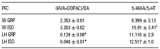
* All values are the mean ± SEM (n = 5–8 rats).
** p < 0.001 vs. W rats in the same rearing condition.
p < 0.05 vs. GRP rats of the same strain.
W and LH differed significantly in 5-HT content, being 5-HT content higher in LH rats, irrespective of housing conditions [F(st)1,22 = 28.061, p < 0.001; F(r)1,22 = 1.261, n.s.; F(r × st)1,22 = 1.154, n.s.] (Fig. 2). In addition, isolation caused an increase in 5-HT turnover only in W strain (two-way ANOVA, p < 0.05, Table 1).
Hippocampus
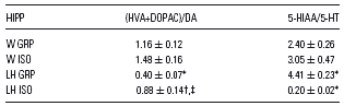
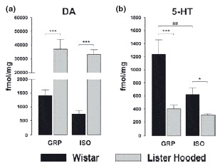
W and LH rats differed significantly in the content of DA [F(st)1,24 = 37.746, p < 0.001; F(r)1,24 = 0.327, n.s.; F(r × st)1,24 = 2.720, n.s.], either in GRP or in ISO rats, showing higher DA content in LH rats (Fig. 3). In regard to DA metabolism, LH strain showed a DA turnover significantly higher (two-way ANOVA, p < 0.05), however in this strain such parameter was increased by isolation (two-way ANOVA, p < 0.001) (Table 2). W rats exhibited higher 5-HT content only in GRP group. Moreover, social isolation significantly reduced 5-HT levels in W strain [F(st)1,23 = 15.665; p < 0.001; F(r)1,23 = 2.309, n.s.; F(r × st)1,23 = 3.904, p < 0.05] (Fig. 3). No difference in turnover was found in W, while in LH the isolation rearing significantly reduced, 5-HT metabolism (two-way ANOVA, p < 0.0001, Table 2).
Effects of social isolation on Hipp content of monoamines in W (black bar) and LH (grey bar) rats. **p < 0.01, ***p < 0.001 comparing W and LH rats in the same rearing condition. #p < 0.05 comparing GRP and ISO rats of the same strain. All values are the mean ± SEM (n = 5–8 rats).

Effects of social isolation on tissue DA and 5-HT turnover in Hipp of W and LH rats
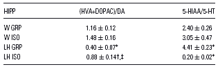
* All values are the mean ± SEM (n = 5–8 rats).
p < 0.001 vs. W rats in the same rearing condition.
p < 0.05 vs. W rats in the same rearing condition.
p < 0.05 vs. GRP rats of the same strain.
Nucleus accumbens
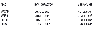
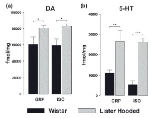
As shown in Fig. 4, a significant effect of strain was evident [F(st)1,23 = 72.062, p < 0.001; F(r)1,23 = 0.324, n.s.; F(r × st)1,23 = 0.160, n.s.] reflecting an increased DA content in LH rats in comparison with W animals, either in GRP or ISO animals. Moreover, DA turnover in LH rats was significantly lower compared with W, independently from rearing conditions (two-way ANOVA, p < 0.001, Table 3), while no difference was evident in W rats after isolation.
Effects of social isolation on NAC content of monoamines in W (black bar) and LH (grey bar) rats. *p < 0.05, ***p < 0.001 comparing W and LH rats in the same rearing condition. ##p < 0.01 comparing GRP and ISO rats of the same strain. All values are the mean ± SEM (n = 5–8 rats).

Effects of social isolation on tissue DA and 5-HT turnover in NAC of W and LH rats
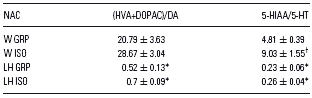
* All values are the mean ± SEM (n = 5–8 rats).
† p < 0.01 vs. W rats in the same rearing condition.
p < 0.05 vs. GRP rats of the same strain.
A strain-dependent effect was observed also when considering 5-HT content, either in GRP or in ISO rats. Indeed, LH animals showed decreased neurotransmitter levels. Moreover, the analysis revealed a significant decreased 5-HT content only in W rats, consequent to isolation rearing [F(st)1,23 = 25.091; p < 0.001; F(r)1,23 = 9.647, p < 0.01; F(r × st)1,23 = 5.200, p < 0.05] (Fig. 4). Finally, isolation caused an increase in 5-HT turnover in W strain (two-way ANOVA, p < 0.05 Table 3); however, independently from rearing conditions, LH rats showed lower 5-HT turnover (two-way ANOVA, p < 0.05, Table 3).
Striatum
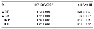
Irrespective of housing conditions, the analysis showed a significant increase in DA content in LH rats with respect to W strain [F(st)1,21 = 9.922, p < 0.01; F(r)1,21 = 0.00715, n.s.; F(r × st)1,21 = 0.0588, n.s.] (Fig. 5), while no difference was evident in DA turnover (Table 4). As far as 5-HT, a strain-dependent effect was evident either in GRP or ISO rats, resulting in increased 5-HT content in LH animals [F(st)1,21 = 37.693, p < 0.001; F(r)1,21 = 1.034, n.s.; F(r × st)1,21 = 0.818, n.s.] (Fig. 5). In addition, isolation caused an increase in 5-HT turnover in W strain (two-way ANOVA, p < 0.05, Table 4). In LH strain 5-HT turnover was lower compared to W, independently from rearing conditions (two-way ANOVA, p < 0.05, Table 4). In addition, isolation caused an increase in 5-HT turnover only in W strain (two-way ANOVA, p < 0.001, Table 4).
Effects of social isolation on St content of monoamines in W (black bar) and LH (grey bar) rats. *p < 0.05, **p < 0.01, ***p < 0.001 comparing W and LH rats in the same rearing condition. All values are the mean ± SEM (n = 5–8 rats).

Effects of social isolation on tissue DA and 5-HT turnover in St of W and LH rats
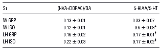
* All values are the mean ± SEM (n = 5–8 rats).
† p < 0.05 vs. GRP rats of the same strain.
‡ p < 0.05 vs. W rats in the same rearing condition.
p < 0.001 vs. W rats in the same rearing condition.
Overall differences in post-mortem tissue content of neurotransmitters
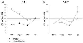
Fig. 6 summarises the overall differences in post-mortem tissue content of the neurotransmitters considered in this study (Fig. 6: DA; Fig. 6: 5-HT). Results showed the different strain-dependent responses of ISO rats in percent relative to normal GRP animals.
The different strain-dependent responses to ISO rats in percent relative to normal GRP animals for DA and 5-HT in PFC, Hipp, NAC and St of W(1) and LH (1) rats.

In vivo microdialysis
To understand if social isolation alters neurotransmitter release, we have performed microdialysis in freely moving animals to measure the extracellular concentration of DA and 5-HT in PFC of rats.
Since differences between rearing conditions and strain were time independent, individual comparisons between marginal mean values (pooled data of four samples for each factor) were made and a two-way ANOVA was performed.
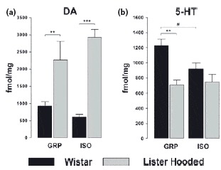
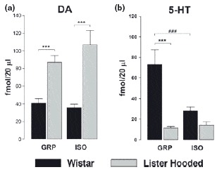
Results showed higher DA basal levels in LH rats compared with W rats, irrespective of housing conditions [F(st)1,91 = 45.96, p < 0.001; F(r)1,91 = 0.753, n.s.; F(r × st)1,91 = 2.104, n.s.] (Fig. 7).
Effects of social isolation on in vivo PFC levels of monoamines in W (black bar) and LH (grey bar) rats. ***p < 0.001 comparing W and LH rats in the same rearing condition. ###p < 0.001 comparing GRP and ISO rats of the same strain. All values are the mean ± SEM (n = 5–8 rats).

Basal levels of 5-HT were higher in W with respect to LH rats, but only in GRP conditions. Moreover, social isolation decreased 5-HT concentrations only in W rats [F(st)1,91 = 21.513, p < 0.001; F(r)1,91 = 6.546, p < 0.05; F(r × st)1,91 = 8.384, p < 0.01] (Fig. 7).
Discussion
This study suggests that the choice of rat strain can make a crucial difference when analysing the effects of isolation rearing on regional brain monoamine content. Clear and distinct neurochemical profiles in the brain areas examined were observed. In this regard, no extraneous experimental variables, such as body weight, could be forwarded as an explanation, as no significant difference in the body weight of the two different rat strains was observed.
Often, the rationale behind selecting one strain can be mainly speculative and affected by anthropomorphic inference (Reference Holmes27), although it can be addressed by economical evaluations of the experiments and by the surprising lack of references in comparative studies on behavioural and monoaminergic alterations following social isolation rearing in W and LH rats, as shown in the literature review by Lukkes and co-workers (Reference Lukkes, Watt, Lowry and Forster13).
Thus, the discussion of these results should consider two aspects: (a) strain-dependent differences in neurochemistry and (b) strain-related differences consequent to isolation. Furthermore, a fundamentally important question should be addressed: could strain-dependent differences explain differential effects of isolation, and as such be an important consideration for using the model in basic studies of schizophrenia or depression?
When analysing strain-dependent neurochemical differences, it appeared that brain regions in LH rats, at least those examined, contained higher DA levels with respect to similar brain areas in W rats. In line, in this strain DA metabolism resulted reduced with the exception of St.
Thus, considering that the dopaminergic hypothesis is one of the most widely accepted hypotheses regarding the aetiology of schizophrenic symptoms (Reference Jentsch and Roth28), these results seem to explain at least some of the behavioural contrasting results found in the isolation-based animal model of schizophrenia, where studies are performed in different rat strains, and why LH rats are a preferred strain.
In this regard, the PPI paradigm used in rodents has been proposed to model the attention impairments and deficits in psychomotor gating seen in psychiatric disorders such as schizophrenia (Reference Geyer, Moghaddam, Davies, Charney, Coyle and Nemeroff29). There is converging evidence for the important involvement of dopaminergic systems in the control of PPI. Specifically, neurotoxic lesion experiments (Reference Swerdlow, Geyer and Braff30) support the notion that increased DA transmission and DA receptor super- or sub-sensitivity underlie the disruption in PPI consequent to the above interventions. Indeed, it has been previously reported that LH rats demonstrate a greater reduction in startle reactivity relative to W animals (Reference Weiss, Di Iorio, Feldon and Domeney31).
Interestingly, the lower basal DA levels found in all brain areas examined in W rats with respect to LH animals could be consistent with the finding that the W rat strain appears to be more resistant to developing certain behaviours typical of animals subjected to ISO, specifically disruption of PPI. Indeed, raised accumbal DA, as it was particularly evident in LH rats, will imply an increase in salience culminating in an attenuation in sensorimotor gating and an associated decrease in startle reactivity, while reduced DA in W rats imply reduced salience and an increase in startle reactivity. These data seem to suggest that optimum levels of DA are necessary to observe schizophrenic-like behavioural alterations in specific rat strains (Reference Kinney, Wilkinson, Saywell and Tricklebank32).
Moreover, it has been demonstrated that LH rats display increased locomotor activity in comparison with W rats (Reference Weiss, Di Iorio, Feldon and Domeney31). The higher DA content found in the St of LH rats with respect to W rats could account for this behavioural evidence.
Strain-related differences were also observed in the serotonergic system. In particular, in vivo results showed decreased extracellular 5-HT concentrations in the PFC of LH rats, with respect to W animals. Prefrontal DA is under strong restraint via cortical serotonergic projections (Reference Harvey, Naciti, Brand and Stein33), such that reduced 5-HT may underlie the marked elevations in PFC DA observed in LH rats. Surprisingly, however, post-mortem tissue content of 5-HT were increased in the PFC and St of LH rats, while the same rats showed lower 5-HT content in the NAC and Hipp. Additionally, in these areas 5-HT metabolism was reduced compared to W rats, independently from isolation-rearing condition. In this strain, however, hippocampal 5-HT turnover was increased by isolation. The discrepancy between in vivo and ex vivo results found in the PFC may depend on the loss of neuronal connection in post-mortem analyses, as well as reflecting the whole neuronal pool of transmitter versus the synaptic levels.
The different effect found in microdialysis study with respect to ex vivo data may be due to the fact that chronic stress of isolation decreased 5-HT release (Reference Fone and Porkess11), thus increasing tissue content.
Rearing rats in isolation produced specific strain-dependent changes in the post-mortem tissue concentrations and in vivo basal levels of monoamines. In particular, LH-isolated animals had significantly higher concentrations of DA in the PFC when compared to GRP LH controls, but no alterations were observed in the NAC, St or Hipp. With regard to W rats, no variations of DA concentrations were found after isolation in all brain areas analysed. This re-emphasises the critical role of PFC DA in mediating the behavioural responses akin to schizophrenia, and the more limited role for DA in the latter regions.
Selective alterations in cortical DA function have been reported previously following isolation in LH rats (Reference Fone and Porkess11). As in this study, the effects on DA in the PFC were also specific, as no alterations in the NAC or St were found in the ISO LH rats.
The impact of social deprivation in rats is, however, not restricted to effects on dopaminergic transmission. In this study, post-mortem studies revealed that 5-HT content was unaffected in LH rats by rearing conditions in all brain areas considered. Conversely, 5-HT content was significantly decreased in the NAC and Hipp of W rats following isolation. It could be hypothesised that the decreased 5-HT levels in these brain regions following chronic isolation in W rats cannot be compensated by an increase in 5-HT synthesis, leading then to depressive-like behaviour. Indeed, reduced 5-HT content or 5-HT release has also been found in cortex and Hipp of ISO animals (Reference Miura, Qiao, Kitagami, Ohta and Ozaki34). Turning to the in vivo data, 5-HT was markedly reduced in the PFC in W rats following isolation, but no effects were observed in LH rats. Serotonergic changes subsequent to isolation are consistent with data describing the ability of some antidepressant compounds to reverse the effects of isolation rearing (Reference El Yacoubi, Bouali and Popa35).
Then, it is pertinent to note that neuropsychiatric disorders like depression have been considered a consequence of inadequate neurochemical adaptation in response to stressors (Reference Owens, Miller, Lawrie and Johnstone36). Interestingly in this context, it must be underlined that depression is a common symptom before (Reference an der Heiden, Konnecke, Maurer, Ropeter and Hafner37), during and after an episode of schizophrenia (Reference Brenes, Rodriguez and Fornaguera38). These results seem to support the developmental hypothesis that early adverse environmental life events could predispose to adult depressive-like response (Reference Brenes and Fornaguera39). However, further studies should be conducted to evaluate the involvement of other neurotransmitters. In particular, it is hypothesised that early life stressful events may lead to glutamate hyperfunction, thus preventing neuronal connections to the cortex and resulting in dopaminergic and serotonergic dysfunctions (Reference Powell, Sejnowski and Behrens40). In line with this, further investigations directed to assess glutamate involvement in different rat strains are warranted.
In conclusion, this data provide the first evidence that choice of strain and genetic background are of importance when considering an environmental animal model of schizophrenic disorders. From these results we could hypothesize that the neurochemical phenotype of the LH rat, particularly with respect to DA/5-HT changes in the PFC, may constitute a valuable genetic model of vulnerability for the development of schizophrenic manifestations in ISO animals, while the W rat strain could better provide information on several parameters associated with depression or in schizophrenia comorbid with depression.
Acknowledgements
G. W. serves as Editor-in-Chief for Acta Neuropsychiatrica, but was not involved in and actively withdrew from the review/decision process of this paper. No other conflicts of interest are to be declared.