Introduction
The genus Isoparorchis Southwell (Reference Southwell1913), belonging to the family Isoparorchiidae Travassos (Reference Travassos1922), are species of well-known parasites in the air bladder of freshwater catfishes (Gibson, Reference Gibson, Gibson, Jones and Bray2002; Shimazu et al., Reference Shimazu, Urabe and Grygier2011, Reference Shimazu, Cribb, Miller, Urabe, Van Ha, Binh and Shed'ko2014). Species of Isoparorchis are widely distributed, including in Vietnam (Moravec & Sey, Reference Moravec and Sey1989; Shimazu et al., Reference Shimazu, Cribb, Miller, Urabe, Van Ha, Binh and Shed'ko2014), India (Bhalerao, Reference Bhalerao1932, Reference Bhalerao1936; Pandey, Reference Pandey1969; Shimazu et al., Reference Shimazu, Cribb, Miller, Urabe, Van Ha, Binh and Shed'ko2014), Australia (Johnston, Reference Johnston1927; Cribb, Reference Cribb1988; Shimazu et al., Reference Shimazu, Cribb, Miller, Urabe, Van Ha, Binh and Shed'ko2014), Pakistan (Bilquees & Khatoon, Reference Bilquees, Khatoon, Bilquees, Saeed, Rehana, Khatoon and Kaikabad1972), Indonesia (Bovien, Reference Bovien1927), Bangladesh (Chandra, Reference Chandra2006), China (Wu, Reference Wu1938; Chen & Li, Reference Chen and Li1973), Russia (Shimazu et al., Reference Shimazu, Cribb, Miller, Urabe, Van Ha, Binh and Shed'ko2014), Japan (Kobayashi, Reference Kobayashi1921; Yamashita & Nishida, Reference Yamashita and Nishida1955; Sawada & Osako, Reference Sawada and Osako1969; Shimazu et al., Reference Shimazu, Urabe and Grygier2011, Reference Shimazu, Cribb, Miller, Urabe, Van Ha, Binh and Shed'ko2014), Korea (Sohn & Na, Reference Sohn and Na2018) and Thailand (Ejsmont, Reference Ejsmont1932). Currently, Isoparorchis comprises at least five species, including four valid species: Isoparorchis hypselobagri Billet, Reference Billet1898, Isoparorchis trisimilitubis Southwell, Reference Southwell1913, Isoparorchis eurytremum Kobayashi, Reference Kobayashi1915 and Isoparorchis tandani Johnston, 1927 (Shimazu et al., Reference Shimazu, Cribb, Miller, Urabe, Van Ha, Binh and Shed'ko2014). Generally, the Isoparorchis species (with the exception of I. tandani) are thought to rely on four hosts (mollusc, crustacean, fish and the fish definitive host) to develop to adult stages and complete their life cycle (Cribb, Reference Cribb1988; Shimazu et al., Reference Shimazu, Cribb, Miller, Urabe, Van Ha, Binh and Shed'ko2014). For instance, the first intermediate hosts reported were gastropod snails (Cribb, Reference Cribb1988; Besprozvannykh & Ermolenko, Reference Besprozvannykh and Ermolenko1989). In Australia, the metacercarial stage has been reported in freshwater shrimps Caridina indistincta and Paratya australiensis, which act as second intermediate hosts (Cribb, Reference Cribb1988; Shimazu et al., Reference Shimazu, Cribb, Miller, Urabe, Van Ha, Binh and Shed'ko2014). Elsewhere, a large number of families of freshwater fish have been implicated as third intermediate hosts (Bhalerao, Reference Bhalerao1932, Reference Bhalerao1936; Suzuki, Reference Suzuki1967; Pandey, Reference Pandey1969; Nagasawa et al., Reference Nagasawa, Katahira and Nitta2013; Shimazu et al., Reference Shimazu, Cribb, Miller, Urabe, Van Ha, Binh and Shed'ko2014; Shimazu, Reference Shimazu2015), whereas freshwater catfishes (Siluriformes) have been extensively reported to be the definitive hosts and naturally infected by Isoparorchis species (Bhalerao, Reference Bhalerao1932; Suzuki, Reference Suzuki1967; Pandey, Reference Pandey1969; Gibson, Reference Gibson, Gibson, Jones and Bray2002; Shimazu et al., Reference Shimazu, Cribb, Miller, Urabe, Van Ha, Binh and Shed'ko2014, Shimazu, Reference Shimazu2015). Adult flukes live in the air bladder, body cavity, musculature and internal organs of fish hosts – typically Siluriformes, and occasionally Anabantiformes (Yamashita & Nishida, Reference Yamashita and Nishida1955; Suzuki, Reference Suzuki1967; Pandey, Reference Pandey1969; Sawada & Osako, Reference Sawada and Osako1969; Gibson, Reference Gibson, Gibson, Jones and Bray2002; Shimazu et al., Reference Shimazu, Urabe and Grygier2011, Reference Shimazu, Cribb, Miller, Urabe, Van Ha, Binh and Shed'ko2014). Isoparorchis infection in the body muscles of fish hosts leads to ink spot disease, resulting in the surrounding tissues being swollen and a change in colour to black-brown (Yamashita & Nishida, Reference Yamashita and Nishida1955; Sawada & Osako, Reference Sawada and Osako1969). Furthermore, the pathogenesis also causes morphological, behavioural and haematological changes (Dollfus, Reference Dollfus1959; Mahajan et al., Reference Mahajan, Agrawal, John and Katta1979; Gibson, Reference Gibson, Gibson, Jones and Bray2002). For instance, Mahajan et al. (Reference Mahajan, Agrawal, John and Katta1979) found that the morphological changes in Channa punctatus caused by I. hypselobagri infection resulted in the necrosis of fin tissue, scale loss and loss of pigmentation with damage to the viscera, especially the gonads, and the abdominal muscles. Behavioural changes included loss of equilibrium, irritability and convulsions (Mahajan et al., Reference Mahajan, Agrawal, John and Katta1979). Haematological changes resulted in hypochromic macrocytic and normochromic normocytic anaemia (Mahajan et al., Reference Mahajan, Agrawal, John and Katta1979). In addition, reptiles and mammals, including humans, have also been reported to carry Isoparorchis infections (Chandler, Reference Chandler1926; Bhalerao, Reference Bhalerao1932, Reference Bhalerao1936; Cribb, Reference Cribb1988). Human cases may be accidental infections after consuming raw fish containing infective stages (Chandler, Reference Chandler1926; Bhalerao, Reference Bhalerao1932, Reference Bhalerao1936). Regarding the prevalence and mean intensity, the infection of I. hypselobagri in Channa argus has been reported as 70% and 3.1, respectively, in Japan (Yamashita & Nishida, Reference Yamashita and Nishida1955). In India, the prevalence and mean intensity of the infection of I. hypselobagri in Wallago attu have been reported as 83.33% (25/30) and 1.4, respectively (Choudhary et al., Reference Choudhary, Ray and Agrawal2019). In Korea, the overall prevalence of Isoparorchis sp. infection of four freshwater fish species is 8.4% (Sohn & Na, Reference Sohn and Na2018).
Recently, the taxonomic key of Isoparorchis species was created by considering the morphological characters together with molecular analysis based on the internal transcribed spacer 2 (ITS2) (Shimazu et al., Reference Shimazu, Cribb, Miller, Urabe, Van Ha, Binh and Shed'ko2014). This approach recognized at least five distinct species, including four valid species (Shimazu et al., Reference Shimazu, Cribb, Miller, Urabe, Van Ha, Binh and Shed'ko2014). Furthermore, the 28S ribosomal RNA (rRNA) gene has also been applied to identify I. hypselobagri collected from the freshwater fish W. attu in India (Choudhary et al., Reference Choudhary, Ray and Agrawal2019). In Thailand, adults of I. hypselobagri were reported in freshwater fish from the Chao Phraya River (Ejsmont, Reference Ejsmont1932). For almost 90 years, however, there has been no further report from Thailand. Moreover, there have been few surveys on parasitic infections in freshwater shrimp from Thailand. For these reasons, we investigated the prevalence and intensity of parasite specimens in freshwater shrimp by integrating morphological and molecular features based on ITS2 sequence data.
Materials and methods
Sample collection and parasitological examination
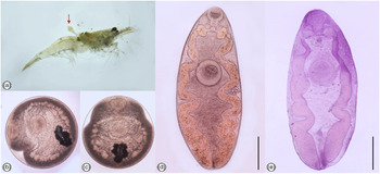
A total of 300 specimens of freshwater shrimp (Macrobrachium lanchesteri and Caridina sp.) were obtained from the Suan Phueng district, Ratchaburi province, Thailand (N13°34.385′E, 099°15.150′). The living specimens were transported to the Department of Biology at Srinakharinwirot University. These specimens were examined using the press with glass slide method (Habe et al., Reference Habe, Lai, Agatsuma, Ow-Yang and Kawashima1993). The metacercariae were isolated from the cephalothorax and the muscle of freshwater shrimp (fig. 1a). After that, the metacercariae were washed with distilled water and observed under a stereomicroscope, while the excysted metacercariae specimens were fixed with 4% formalin. Some specimens were preserved in 95% ethanol and stored at −20°C for DNA analysis. After two weeks, specimens in 4% formalin were washed with distilled water, stained with Haematoxylin solution, dehydrated in an alcohol series (10%, 20%, 30%, 50%, 70%, 80% and 95% ethanol; ethanol:n-butanol [1:1]; butanol; and n-butanol:xylene [1:1]), cleared in xylene and mounted in Permount. All fluke specimens were examined and identified as relating to the genus Isoparorchis by morphological characteristics (Shimazu et al., Reference Shimazu, Cribb, Miller, Urabe, Van Ha, Binh and Shed'ko2014). After that, various organs of the specimens were measured in micrometres, including the minimum (min), maximum (max) and mean, and compared with previously reported morphological descriptions of Isoparorchis species (Vankara et al., Reference Vankara, Mani and Vijayalakshmi2011; Sen, Reference Sen2014; Sohn & Na, Reference Sohn and Na2018).
Morphological features of the metacercarial stages of Isoparorchis sp. from freshwater shrimps (Macrobrachium lanchesteri and Caridina sp.). (A) Freshwater shrimps were naturally infected by Isoparorchis sp. (red arrow shows the excysted metacercaria); (B) Metacercarial stages show well-developed sucker, observed under stereomicroscope; (C) Other metacercariae showing the same organs; (D) Body of the excysted metacercariae; (E) Excysted metacercariae were stained by Haematoxylin. Scale bars: (d, e) 500 μm.

DNA extraction, amplification, purification and sequencing
The genomic DNA of each sample was extracted with the GF-1 Tissue kit (Vivantis, Shah Alam, Selangor Darul Ehsan, Malaysia) following the manufacturer's protocol and eluted in 50 μl of elution buffer. The genomic DNA concentration was then measured using a Nanodrop (Thermo Fisher Scientific, 3rd Avenue, Waltham, USA). The DNA concentration was adjusted to 6.0 ng/μl before use in molecular analysis. The ITS2 region including partial 5.8S and 28S rRNA genes of each sample was amplified by using the primers ITS3-F (5′-GCA TCG ATG AAG AAC GCA GC-3′) and ITS4-R (5′-TCC TCC GCT TAT TGA TAT GC-3′) (Barber et al., Reference Barber, Mkoji and Loker2000) with the following thermal cycler conditions: initial pre-denaturation at 94°C for 5 min; 35 cycles of 1 min denaturation at 94°C, 1 min primer annealing at 56°C and 1 min primer extension at 72°C; and a final extension step of 7 min at 72°C. The polymerase chain reaction (PCR) products were separated on a 1.2% agarose gel run in 1× TBE buffer (Tris-borate with Ethylene diamine tetra-acetic acid (EDTA) buffer), stained with safe green gel stain and visualized under ultraviolet illumination. The PCR amplicons were purified and sequenced directly at 1st BASE Company (Seri Kembangan, Selangor, Malaysia). The ITS2 including partial 5.8S and 28S rRNA sequences of Isoparorchis in this study were aligned with the other Isoparorchis sequence data from the GenBank database by the ClustalW program in the MEGA-X program (Kumar et al., Reference Kumar, Stecher, Li, Knyaz and Tamura2018)for species confirmation.
Genetic distances analyses
Before analysing the phylogenetic trees, the nucleotide sequences of Isoparorchis spp. were compared by using the BLASTn tool (basic local alignment search tool). The final alignments were trimmed to 468 bp to calculate the genetic distances between taxa of Isoparorchis species. Analyses were conducted using the Jukes–Cantor (JC) model. This analysis involved 22 nucleotide sequences. All ambiguous positions were removed for each sequence pair (pairwise deletion option). Evolutionary analyses were conducted in the MEGA-X program.
Phylogenetic analyses
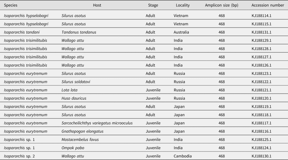
The phylogenetic trees were constructed using the maximum likelihood (ML) method applying the JC model for calculation by the MEGA-X program. Branch support was tested using bootstrap support with 10,000 replicates. The sequences of Isoparorchis spp. were compared with the nucleotide sequences reported in the GenBank database (table 1).
Summary data of species used to construct the phylogenetic trees of Isoparorchis spp. based on ITS2 including partial 5.8S and 28S rRNA sequences.

Results
Prevalence and mean intensity of infection
A total of 276 specimens of freshwater shrimp were found infected with the metacercarial stage of Isoparorchis sp. Most infections were found in the cephalothorax (encysted) and muscle in the abdomen of the freshwater shrimp. The overall prevalence and mean intensity of Isoparorchis sp. infections in freshwater shrimp were 92% and 1.89, respectively.
Morphological description
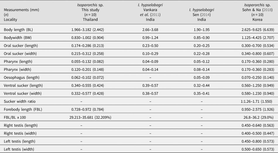
Encysted metacercariae of Isoparorchis sp. (n = 10) are spherical and brown, with a diameter ranging from 0.650 to 0.754 (0.697) mm. Cyst wall transparent and somewhat thin (one layer). Oral sucker sub-terminal and well developed. Ventral sucker muscular and larger than oral sucker. Bifurcated intestinal caeca extend posteriorly to near extremity of body (fig. 1b, c). Excysted metacercariae based on ten specimens measured and compared with Isoparorchis spp. in previous reports (table 2). Overall body brown and yellow, thick and elongated. Oral sucker sub-terminal and well developed. Pharynx cone-shaped and short. Pre-pharynx absent. Oesophagus very short and links to intestinal caeca. Intestinal ceca initial bifurcation dumbbell-shaped and extends to approximately 98% of body length from anterior extremity. Ventral sucker well developed and larger than oral sucker. Testes and ovaries not distinguishable. Excretory pore on posterior end of body (fig. 1d, e).
Measurements of Isoparorchis sp. (excysted metacercariae) from freshwater shrimp compared with previous studies (Forebody length: distance from the anterior extremity of the fluke to the upper edge of the ventral sucker, BL: distance from the anterior extremity of the fluke to the bottom edge of the body).

Pairwise genetic distances
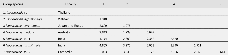
The nucleotide sequences of Isoparorchis sp. in this study were aligned with Isoparorchis taxa. There were no gaps in the alignment. The pairwise genetic distances of our sequences relative to I. hypselobagri from Vietnam showed very low intraspecific variability (1.948%) for ITS2, including partial 5.8S and 28S rRNA sequences, while there were nine different nucleotides between our sequences and I. hypselobagri from Vietnam (data not shown). On the other hand, the intraspecific variation in our sequences showed the highest variation relative to the sequences of Isoparorchis sp. 2 (5.083%), I. trisimilitubis (4.855%) and Isoparorchis sp. 1 (4.174%), respectively (table 3).
Pairwise genetic distances (%) between taxa of Isoparorchis sp. in Thailand and the sequences for Isoparorchis spp. of the concatenated nuclear DNA sequence of ITS2 including partial 5.8S and 28S rRNA sequences.

Phylogenetic analyses
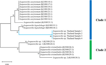
The BLAST results of Isoparorchis sp. specimens in this study demonstrated 98.08% identity with the ITS2 region, including partial 5.8S and 28S rRNA genes of I. hypselobagri from Vietnam (KJ188114.1). The phylogenetic trees were constructed with the nucleotide sequences of Isoparorchis taxa based on ITS2 sequences (468 bp after trimming), including I. hypselobagri, I. trisimilitubis, I. eurytremum, I. tandani, Isoparorchis sp. 1 and Isoparorchis sp. 2, as well as our four sequences. Based on the phylogenetic analyses, Isoparorchis taxa separate into two major clades: clade 1 with I. eurytremum (KJ188116.1–KJ188123.1) from Japan and Russia, I. tandani (KJ188131.1) from Australia, I. hypselobagri from Vietnam (KJ188114.1 and KJ188115.1) and our four sequences from Thailand and clade 2 with Isoparorchis sp. 1 (KJ188124.1 and KJ188125.1) from India, I. trisimilitubis (KJ188126.1–KJ188129.1) from India and Isoparorchis sp. 2 (KJ188130.1) from Cambodia. These results show a clear distinction among Isoparorchis species, and this topology was well supported by ML analyses. The Thai sequences cluster as a clade sister to I. hypselobagri from Vietnam, supported by high bootstrap values (fig. 2).
Phylogenetic relationships of Isoparorchis sp. and other related species analysed using the ML method based on ITS2, including partial 5.8S and 28S rRNA sequences (468 bp) inferred from the JC model with 10,000 bootstrap replicates (Blue branches shown the Isoparorchis sp. specimens in this study, scale bar shows the expected number of substitutions per site).

Discussion
The present study is the first report of the freshwater shrimp M. lanchesteri serving as a second intermediate host of Isoparorchis sp. We also provide the first data regarding the morphological descriptions of true metacercariae (encysted). The results show that a high overall prevalence may be because this area contains a number of biological and physical components, including the ecological conditions, for example, as this canal connects to major rivers (Pachee River and Kwai Noi River) and has potential a high diversity of intermediate and definitive hosts (snails, crustaceans, fishes, birds and mammals). Clearly, the definitive host must be present in this ecosystem, but it remains unidentified. Moreover, a variety of cercarial types of at least five trematode families have also been reported in this province (Anucherngchai et al., Reference Anucherngchai, Tejangkura and Chontananarth2017). For these reasons, it is an interesting place for this study.
The second intermediate hosts in this study are freshwater shrimps (M. lanchesteri and Caridina sp.), similar to the second intermediate hosts of I. tandani in eastern Australia (Cribb, Reference Cribb1988; Shimazu et al., Reference Shimazu, Cribb, Miller, Urabe, Van Ha, Binh and Shed'ko2014). Encysted metacercariae were found in the cephalothorax, whereas excysted metacercariae were found in the muscle, similar to those reported by Cribb (Reference Cribb1988). This contrasts with several reports of excysted metacercariae from parts of Asia – it is likely that those reports were based on fishes (Bhalerao, Reference Bhalerao1932, Reference Bhalerao1936; Yamashita & Nishida, Reference Yamashita and Nishida1955; Pandey, Reference Pandey1969; Sawada & Osako, Reference Sawada and Osako1969; Gibson, Reference Gibson, Gibson, Jones and Bray2002; Vankara et al., Reference Vankara, Mani and Vijayalakshmi2011; Nagasawa et al., Reference Nagasawa, Katahira and Nitta2013; Sen, Reference Sen2014; Shimazu et al., Reference Shimazu, Cribb, Miller, Urabe, Van Ha, Binh and Shed'ko2014; Sohn & Na, Reference Sohn and Na2018; Choudhary et al., Reference Choudhary, Ray and Agrawal2019). Typically, Isoparorchis (Asian species) can develop to adult stages through four hosts (mollusc, crustacean, fish and the fish definitive host). In these pathways, it can develop, respectively, from the first intermediate hosts to the final hosts and from foraging fishes to carnivorous fishes. As far as we know, only the life cycle of I. tandani (Australian species) uses three hosts (mollusc, crustacean and fish definitive host) to develop as the adult stage. Of note, the freshwater catfish (Tandanus tandanus) seemingly takes on the role of the third intermediate and definitive host of I. tandani since it does not eat fish that act as a fish third intermediate host (with the worms migrating in the tissues) and, thus, acts as the definitive host (Cribb, Reference Cribb1988; Shimazu et al., Reference Shimazu, Cribb, Miller, Urabe, Van Ha, Binh and Shed'ko2014); conversely, most if not all of the catfish species infected with Isoparorchis in Asia are indeed piscivorous (Cribb, Reference Cribb1988; Gibson, Reference Gibson, Gibson, Jones and Bray2002; Shimazu et al., Reference Shimazu, Cribb, Miller, Urabe, Van Ha, Binh and Shed'ko2014, Shimazu, Reference Shimazu2015). This information may indicate that the freshwater shrimps in this study are related to the second intermediate hosts of this genus and might be targeted by non-piscivorous catfish in these systems. Moreover, this pattern of the life cycles may further hint that the Isoparorchis sp. in this study is different from the other known Asian species.
Species identification based on the morphological appearance of the excysted metacercariae in this study is almost impossible because the larval stages do not have unique morphological characters that relate to the adult stages. We have attempted to survey the freshwater fishes in a more extensive area for a long time, especially around Suan Phueng district, Ratchaburi province, Thailand, but we have not found the adult stage to confirm the unique morphological characteristics for species identification, although the infection is common in this area. Based on pairwise genetic distances, we demonstrated that our sequences and those of I. hypselobagri from Vietnam (1.948%) are greater than between the other species of I. hypselobagri from Vietnam and I. eurytremum from Japan and Russia (1.076%). These results indicate that the Isoparorchis sp. in this study may be different from other Isoparorchis species previously described.
The phylogenetic analysis classified the Isoparorchis taxa into two major clades supported by 10,000 bootstrap replicates of the ML method, a classification in agreement with a previous study (Shimazu et al., Reference Shimazu, Cribb, Miller, Urabe, Van Ha, Binh and Shed'ko2014). Interestingly, our four samples were separated from the I. hypselobagri from Vietnam, with which they formed a clade well supported by high bootstrap values. Overall, our samples were also molecularly distinct from the other Isoparorchis based on the ITS2 sequences from the GenBank database. However, the pattern of geographical distinction in our samples is still ambiguous. In addition, other sequences based on 28S rDNA of Isoparorchis species have been presented (Choudhary et al., Reference Choudhary, Ray and Agrawal2019), including mt-cox1 (unpublished) from India. Unfortunately, those studies have few Isoparorchis sequences and, thus, they are insufficient to study species diversity and confirm species identities at the present time. However, we believe that ITS2 sequences are a good molecular marker to discriminate between Isoparorchis species and are suitable to study the phylogenetic relationship because of the completeness of Isoparorchis sequences in databases and the well-verified sources of specimens. This study used ITS2 sequences alone for the analysis, which are quite short for inference on intraspecific variation. Furthermore, the intraspecific levels of the ITS region are low and it may sometimes be difficult to distinctly differentiate between closely related digeneans (Morgan & Blair, Reference Morgan and Blair1995; Liu et al., Reference Liu, Gasser, Young, Song, Ai and Zhu2014). We suggest that the phylogenetic tree should use another genetic marker for additional analyses, especially in mitochondrial DNA, or use multiple genes in combination to achieve improved resolution.
Overall, although the morphological characteristics of the adult remain unknown, the molecular analysis of our samples shows a clear distinction from previously described Isoparorchis species; this likely represents a new species. However, we need additional information on the characteristics of adult worms to clarify and support this idea; this is the next challenging destination for us. For these reasons, we carefully consider that our Isoparorchis specimens are Isoparorchis sp. of Thailand until we can find the adult stages to further clarify specific characteristics. Finally, resolving the taxonomic status of Isoparorchis species in Thailand may help us understand the dynamics of parasite/disease dispersal and the mechanisms underlying parasite speciation, including the other factors involved in parasite epidemiology. Such an understanding may serve as the basis for developing a detection tool to prevent disease and monitor ongoing epidemics.
In conclusion, Isoparorchis sp. was detected in the cephalothorax (encysted) and the muscles of M. lanchesteri and Caridina sp. collected from the Suan Phueng district, Ratchaburi province, Thailand. This worm was identified as Isoparorchis sp. using the morphological characters together with molecular identification based on the ITS2 region. The molecular analysis demonstrated that the genetics of Isopaorchis sp. in Thailand differ from those of other previously reported Isoparorchis species. The results showing that Caridina sp. serves as the second intermediate host of Isoparorchis in this study were well supported.
Author contributions
All authors read and approved the final version of the manuscript and contributed equally.
Financial support
This work was supported by Srinakharinwirot University, which provided research funding and granted us the use of many of their facilities (grant numbers 574/2564 and 023/2564).
Conflicts of interest
None.
Ethical standards
All experimental methods related to animals were preceded by the National Research Council of Thailand and approved by the Committee for Animal Care and Use Committee Srinakharinwirot University (license number 024/2563).