I. INTRODUCTION
Multiferroic perovskite crystals are an important class of functional oxides that possess interesting ferroelectric, magnetoelectric, magnetoresistive, magnetoelastic, light-mechanical, and magneto-optical properties.Reference Dagotto1–Reference Eerenstein, Mathur and Scott5 As one of the few multiferroic materials which ferroelectricity and ferromagneticity can be operated above room temperature,Reference Wang, Neaton, Zheng, Nagarajan, Ogale, Liu, Viehland, Vaithyanathan, Schlom, Waghmare, Spaldin, Rabe, Wuttig and Ramesh6 bismuth ferrite BiFeO3 (BFO) has attracted a great attention. BFO has a rhombohedrally distorted perovskite structure with the unit cell parameters of a = b = c = 5.634 Å and α = 59.348°.Reference Smolenskii and Chupis7 The multifunctionality of BFO provides not only opportunities to fundamental physics study but also potential applications in information storage, spintronics, and sensors.Reference Wang, Neaton, Zheng, Nagarajan, Ogale, Liu, Viehland, Vaithyanathan, Schlom, Waghmare, Spaldin, Rabe, Wuttig and Ramesh6–Reference Leontsev and Eitel10
The morphology and microstructure of a material plays a crucial role in determining its physical properties. In this context, the controlled synthesis and self-organization of inorganic materials with regular geometrical shapes has been studied extensively.Reference Yang11–Reference Zhang, Lai, Zhao, Wang and Dai13 The morphology-dependent magnetic and optical properties were also observed in BFO nanostructures.Reference Park, Papaefthymiou, Viescas, Moodenbaugh and Wong14,Reference Selbach, Tybell, Einarsrud and Grande15 The hydrothermal method has been widely used to synthesize BFO with different morphologies by using bismuth nitrate and ferric nitrate as precursors in an alkaline solution.Reference Chen, Cheng, Yu, Che and Meng16–Reference Fei, Yuan, Hu, Wu, Wang and Wang19 It is found that the alkaline concentration has a strong effect on the morphologies of the final BFO product. For example, Joshi et al.Reference Joshi, Jang, Borse and Lee20 prepared BFO nanocubes in 11 M of KOH and 1 M of Na2CO3 solution with the microwave assisted hydrothermal method. Zhu et al.Reference Zhu, Hang, Xing, Yang, Zhu, Liu, Ming, Zhou, Song, Li, Yu and Zou21 followed Joshi’s procedure and got well dispersed nanocrystals by changing the molar ratio of KOH and Na2CO3 to 8:1. Li et al.Reference Li, Lin, Zhang, Wang and Nan18 reported the preparation of BFO microspheres in 8 M of KOH solution, while BFO microcubes were produced with the concentration of KOH increasing to 12 M. Fei et al.Reference Fei, Yuan, Hu, Wu, Wang and Wang19 also reported the formation of BFO pills in 1 M KOH solution, rods in 2 M KOH solution and cubes in 15 M KOH solution. In the case of the formation of Bi2Fe4O9 plate, the authors suggested that hydroxide ions preferentially got absorbed on one of the facets of the crystals and acted as a capping ligand, resulting in the enhanced growth along another facet.Reference Ruan and Zhang22,Reference Zhang, Lv, Bourgeois, Cui, Wu, Wang and Webley23 Hydroxide ions probably play a similar role in the morphology changes in the case of BFO. So an interesting question arises. How about other anions? Do they have the similar effect on the morphology changes of BFO?
For the hydrothermal preparation of BFO, it is also a big challenge to obtain pure phase perovskite BFO nanopowders due to its metastable phases and complex perovskite structures. As illustrated in the phase diagram,Reference Palai, Katiyar, Schmid, Tissot, Clark, Robertson, Redfern, Catalan and Scott8 two impurities phases of sillenite-type Bi25FeO39 and the mullite-type Bi2Fe4O9 could be formed along with BFO. Bernardo et al.Reference Bernardo, Jardiel, Peiteado, Caballero and Villegas24 confirmed that the secondary phases such as Bi25FeO39 and Bi2Fe4O9 are unavoidable during the single-phase BFO product formation. The reaction time,Reference Wang, Zhu, Xu, Sui, Peng and Tang25 rate of heating and cooling,Reference Chen, Qiu, Zhou, Zhu, Bian and Liu26 the pH value and temperatureReference Chen, Xing, Watson, Wang, Yu, Deng, Yan, Sun and Chen27 all contribute to the purity of BFO. To get pure BFO, a simple and effective postsynthesis purification process is highly desired. The ultrasonic method has been widely used in the sample dispersion and cleaning.Reference Cao, Choi, Konishi, Kou, Lakes and Li28,Reference Gonzalez-Avila, Prabowo, Kumar and Ohl29 Ultrasonic energy can produce acoustic cavitations, resulting in the formation, growth, and implosive collapse of bubbles in liquid.Reference Ashokkumar30,Reference Suslick, Choe, Cichowlas and Grinstaff31 The implosive collapse of bubbles produces strong strength to separate the agglomeration particles. Due to the different densities of BFO and impurities, it is possible to use the ultrasonic method to purify BFO.
Herein, we report a simple one-pot method to prepare well-defined BFO microcrystals with the assistance of microwave heating. A polyanion PMVEMA was used to investigate the morphology evolution of BFO microcrystals. An ultrasonic purification method was also developed to obtain pure BFO microcrystals from the crude products, which avoided the usage of any chemicals.
II. EXPERIMENTAL SECTION
A. Chemicals
Bismuth nitrate [Bi(NO3)3·5H2O], ferric nitrate [Fe(NO3)3·9H2O], PMVEMA were purchased from Aladdin Chemistry Co., Ltd. (Shanghai, China). Nitric acid and potassium hydroxide were purchased from Shanghai Lingfeng Chemical Reagent Co., Ltd. (Shanghai, China). All the chemicals were of analytical grade or better and used without further purification.
B. Preparation of PMVEMA modulated BFO microcrystals
1.2 M bismuth nitrate solution was prepared by adding nitric acid to dissolve the Bi precursors completely. Twenty milliliters of ferric nitrate solution (1.2 M) was slowly added to 20 mL of the prepared bismuth nitrate solution and stirred for 30 min, followed by the addition of PMVEMA and stirred for another 60 min. The obtained solution was dropped into 76 mL KOH and Na2CO3 solution (8 and 1 M) under magnetic stirring for 2 h. The obtained mixture was then transferred into a Teflon reactor and placed in a microwave oven (MDS-8, Shanghai Sineo Microwave Chemistry Technology Co., Ltd., Shanghai, China). The microwave reaction was carried out at 180 °C for 60 min.
For ultrasonic purification, the crude product solution in a centrifuge tube was ultrasonicated for 40 min at 30 °C by using a digital ultrasonic cleaner (KQ-250DE, Kun Shan Ultrasonic Instruments Co., Ltd., Jiangsu, China). After standing for 24 h, the crude products were divided into two layers distinguished by the color of black and brown from bottom to up in the centrifuge tube. The brown layer was collected and transferred to another centrifuge tube by using a tubularis. The process of ultrasonication and separation was repeated for 5 times. The color of the brown layer gradually changed from brown to yellow as the repetition of ultrasonication and separation was in progress. The final yellow and black products were thoroughly washed with deionized water and alcohol, and collected by centrifugation at 6000 r/min, respectively. At last all the obtained samples were dried at 65 °C for 48 h before further characterization.
The purified BFO sample is named “Px,” where “P” denotes “PMVEMA” and “x” is the molar ratio of PMVEMA to the amount of bismuth nitrate. The letters “c,” “b,” and “y” emerging in the sample name below denote “crude,” “black,” and “yellow,” respectively.
C. Characterizations
X-ray diffractometry of the samples was carried out using a Ni filtered Cu Kα (λ = 1.5418 Å) radiation source (D/Max-2550 V, Rigaku Co., Tokyo, Japan). The XRD scans were collected from 10 to 70° (2θ), with a step of 0.02° and a data collection time of 0.5 s. The JCPDS PDF database32 was utilized for the phase identification. The IR transmittance measurements were performed over the frequency range from 400 to 4000 cm−1 using a Fourier transform infrared spectrometer (FTIR, Bruker Vertex 80 V, Billerica, MA) with the KBr pellet technique.
The morphologies of these samples were investigated by using scanning electron microscopy (SEM, JSM7500SF, JEOL, Tokyo, Japan). Magnetic measurements were performed on a superconducting quantum interference device (SQUID, PPMS-9, Quantum Design Inc., San Diego, CA). The samples were filled in a small container made of polyvinyl chloride, whose diamagnetic moment was subtracted from the measured magnetization values.
III. RESULTS AND DISCUSSION
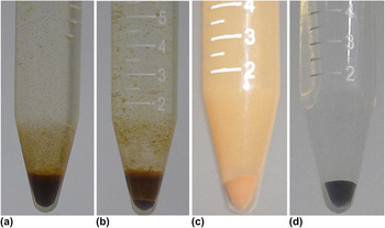
To illustrate the capability of the developed ultrasonic purification method (the sample P1.5 was taken as an example), photographs were taken to show the evolution of the as-prepared crude product before and after ultrasonication, as shown in Fig. 1. It can be seen that the as-prepared powder was obviously divided into two layers after ultrasonication for 40 min. The other two pictures [Figs. 1(c) and 1(d)] show the images of the obtained yellow and black samples.
Photographs of the samples obtained by microwave-assisted hydrothermal method: (a) P1.5_c, the crude products without ultrasonication; (b) as-prepared sample P1.5_c after ultrasonication for 40 min; (c) P1.5_y, the yellow part; (d) P1.5_b, the black part.

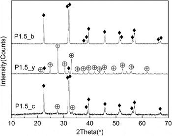
To identify the purity and crystallinity of the products prepared by ultrasonic purification method, the powder x-ray diffraction measurements (XRD) was carried out. As shown in Fig. 2, the XRD pattern of the sample P1.5_c is dominated by the diffraction peaks that correspond to the rhombohedral phase BFO (JCPDS No. 86-1518), and accompanied by small peaks corresponding to Bi25FeO39 (JCPDS No. 77-0865). For the black sample P1.5_b, the diffraction peaks can be readily indexed to the rhombohedral phase BFO (JCPDS No. 86-1518) and no other impurity phase was observed, demonstrating that the well-crystallized single phase BFO was obtained under current synthesis conditions and purification method. For the yellow sample P1.5_y, nearly all the diffraction peaks can be indexed to the cubic phase Bi25FeO39 (JCPDS No. 77-0865) except for relative weak peaks corresponding to BFO (JCPDS Card No. 86-1518). The above-discussed XRD results confirmed that the highly pure BFO crystals can be obtained by the developed ultrasonic purification process.
XRD diffraction patterns of the samples prepared under different conditions: P1.5_c, the as-prepared crude sample; P1.5_y, the yellow part; P1.5_b, the black part. (♦) BiFeO3, (⊕) Bi25FeO39.

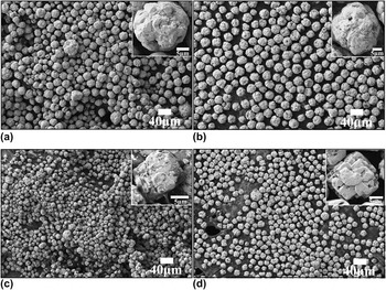
In this work, we chose PMVEMA which contains lots of –COOH groups as anions to avoid bringing other metal ions or changing the concentrations of metal ions such as Na+ and K+ in the reaction system. Because the metal ions also affect the morphologies of bismuth ferrites as illustrated by Wang et al.Reference Wang, Xu, Yang, Ren, Wei, Weng, Du, Shen and Han33 and Tsai et al.Reference Tsai, Yang, Liao and Chueh34 The effect of PMVEMA amount on the morphology and uniformity of BFO samples was investigated by using SEM. The results are represented in Fig. 3. In the absence of PMVEMA, the morphologies of the obtained BFO crystals were microspheres which were not uniform and the diameter was in a range of 15–30 μm, as shown in Fig. 3(a). From an enlarged view of the microcrystals [the inset of Fig. 3(a)], it can be seen that the microspheres exhibited a superstructure consisting of large amounts of small particles. After the addition of PMVEMA with a molar ratio of 0.5 to bismuth nitrate, the morphologies of the obtained BFO crystals were still microspheres [Fig. 3(b)], but they were much more uniform and the average diameter was about 25 μm. With a further increase of the molar ratio of PMVEMA to 1.5, most of the BFO microcrystals exhibited sphere-like morphology [Fig. 3(c)], while some of them were cubic-like shapes as shown in the inset of Fig. 3(c). The estimated edge length of this cubic BFO microcrystal was about 11 μm. When the molar ratio of PMVEMA to bismuth nitrate reached 2, a microcube superstructure of BFO crystal was formed with an edge length of 14 μm, as shown in Fig. 3(d). The microcubes consisted of BFO bricks which clamped each other [the inset of Fig. 3(d)]. The SEM results clearly illustrate that the morphology of BFO crystals can be modulated by PMVEMA from microsphere to microcube. Under our experimental conditions, the concentration of hydroxide ions actually decreased due to the reaction with carboxyl group. However, by increasing the concentration of PMVEMA, the morphology of BFO crystals was tuned from microsphere to microcube, similar to the results obtained by the increase of the hydroxide ion concentration. So it is reasonable to draw a conclusion that the polyanion PMVEMA can play a similar role in the formation of BFO microcube as the hydroxide ion.
SEM images of BFO microcrystals. (a) P0_b, BFO microcystals without PMVEMA; (b) P0.5_b, the molar-ratio of PMVEMA to the amount of bismuth nitrate is 0.5; (c) P1.5_b, the molar-ratio of PMVEMA to the amount of bismuth nitrate is 1.5; (d) P2.0_b, the molar-ratio of PMVEMA to the amount of bismuth nitrate is 2.0.

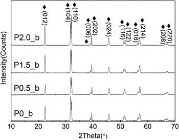
The BFO samples prepared under different conditions were further characterized by XRD. It can be seen from Fig. 4 that all the diffraction peaks in the patterns of these samples can be readily indexed to the rhombohedral phase BFO (JCPDS No. 86-518) and no peaks from other phases were observed, indicating the highly crystalline nature of the BFO microcrystals. It is worth noting that the full width at half maximum (FWHM) of diffraction peaks in the XRD patterns (Fig. 4) almost unchanges when the morphology of BFO changes from the microsphere to microcube. This result indicates that the sizes of small particles which make up the microsphere and microcube are almost the same.
XRD diffraction patterns of the BFO samples. Crystal orientation of the pure BFO was labeled corresponding to the JCPDS Card No. 86-1518.

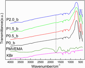
To investigate the contribution of PMVEMA to the morphologies of BFO crystals, the FTIR spectra measurements were performed with the KBr pellet technique. The obtained spectra are shown in Fig. 5. The IR spectrum of PMVEMA has a strong band at 1726 cm−1, which can be ascribed to –COO– asymmetrical stretching vibrations.Reference Busch and Bailar35 The band due to hydroxyl stretching is observed at 3250 cm−1, whereas the bands at 1176 and 1094 cm−1 are assigned to C–O stretching vibrations and the band located at 2945 cm−1 can be owed to C–H stretching vibrations.Reference Nakanishi36 The FTIR spectra of the BFO samples P0.5_b and P1.5_b are same as that of the sample P0_b prepared in the absence of PMVEMA without the observation of the characteristic bands of PMVEMA. This result illustrates that PMVEMA was completely removed from the products during the purification process. But for the sample P2.0_b, a very tiny peak at 1726 cm−1 ascribed to –COO– can be observed, indicating that a very small amount of PMVEMA was entrapped in the product. All BFO samples show two absorption peaks at 546 and 442 cm−1, which are attributed to the Fe–O stretching and bending vibrations of the octahedral FeO6 groups in the perovskite compounds.Reference Rao, Rao and Ferraro37,Reference Kaczmarek and Graja38
FTIR spectra of the BFO microcrystals with different molar ratios of PMVEMA to the amount of bismuth nitrate, plus polymer PMVEMA and the pellet BrK.

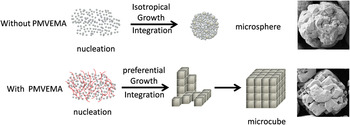
Grounded on the above polymer-dependent morphology evolution process, a possible mechanism for the formation of BFO microspheres and microcubes is suggested as illustrated in Scheme 1. First, hydroxyl bismuth and iron compounds are formed under alkaline condition in aqueous solution. Under high temperature and autogenous pressure in the reaction system, BFO amorphous colloids are formed by the reaction between solution Bi3+ and FeO3+ species which comes from the dissolution of the hydroxyl bismuth and iron compounds.Reference Fei, Yuan, Hu, Wu, Wang and Wang19 The aggregation and crystallization of primary particles result in the formation of BFO nuclei which grow bigger to form small crystals with further mutual reaction. SEM images (Fig. 3) clearly show that the microspheres and microcubes were composed of small particles. Furthermore, FWHM of diffraction peaks in the XRD patterns (Fig. 4) almost unchanges when the morphology of BFO changes from the microsphere to microcube. So it is reasonable to assume that these small crystals act as “building unit” for the formation of BFO microspheres and microcubes. In the absence of PMVEMA, these building units isotropically integrate into microspheres. While in the presence of PMVEMA, the polyanion which contains many –COO– groups preferentially absorbs on one of the facets of the building units through specific and/or noncovalent interactions, such as van der Waals interactions, electrostatic interactions and molecular surface forces. When the amount of absorbed PMVEMA reaches a certain level, it causes the preferential integration of the building units to form a cube-like architecture. The FTIR spectra support this explanation. In the BFO microspheres, PMVEMA was not detected or under the detection limit of the instrument. In other words, the absorbed PMVEMA was not enough to bring a preferential integration of the building units. While in the BFO microcubes, PMVEMA was detected.
Formation and shape evolution process of BFO microcrystals.

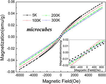
To characterize the magnetic properties of the BFO microcrystals, M-H hysteresis loops were measured by using a SQUID. All the M-H hysteresis loops obtained at 300 K show a nearly linear change with the magnetic field (not shown here), which is similar to that of bulk BFO, a typical behavior for the antiferromagnetic arrangement of the Fe3+ magnetic moments.Reference Lebeugle, Colson, Forget, Viret, Bonville, Marucco and Fusil39 This is mainly because the crystal size of our samples is larger than the superimposing wave length in the spiral spin arrangement of BFO.Reference Park, Papaefthymiou, Viescas, Moodenbaugh and Wong14 So the magnetic properties of the microcrystals with typical morphology of microcubes were chosen to be presented here. Fig. 6 gives the M-H loops of the microcube sample P2.0_b at different temperatures of 5, 100, 200, and 300 K. It can be seen that the magnetization at 6 kOe decreases as the temperature increases, in accordance with the results reported by Jaiswal et al.Reference Jaiswal, Das, Vivekanand, Abraham, Adyanthaya and Poddar40 The weak ferromagnetic hysteresis loop measured at 5 K might come from the existed paramagnetic Fe3+ which behaves the superparamagnetic (SPM) performance at low temperature.Reference Gao, Yuan, Wang, Chen, Chen and Liu41 In the enlarged view of the M-H hysteresis loops (the inset in the right corner of Fig. 6), it can be seen that the microcubes show no coercivity at room temperature and down until 100 K.
The magnetization hysteresis loops of BFO microcubes (P2.0_b) at 5, 100, 200, and 300 K. The inset in the right corner shows the partially enlarged curves, respectively.

IV. CONCLUSIONS
In this work, we successfully modulated the morphology of BFO from microsphere to microcube by using a polyanion PMVEMA in a microwave assisted hydrothermal route. An ultrasonic purification method has been developed to obtain pure phase BFO from the crude products without using any chemicals. The XRD results confirmed the capability of this method. The dependence of the BFO morphology on the amount of PMVEMA has been illustrated by using SEM. The experimental results show that the polyanion PMVEMA can play a similar role in the formation of BFO microcubes as the hydroxide ion. The polyanion preferentially absorbed on one of the facets of the BFO nuclei and brought the preferential integration of these building units to form a cube-like architecture. There is no obvious difference in the magnetic properties of the BFO microspheres and microcubes, because the crystal grain size of the samples is all larger than the superimposing wave length in the spiral spin arrangement of BFO.
ACKNOWLEDGMENT
This work was supported by the State Key Basic Research Program of China (Grant No. 61176011), National Basic Research Project (Grant No. 2013CB922301), KLIFMD-2011-06, Shanghai Pujiang Program (Grant No. 11PJ1403000), Innovation Program of Shanghai Municipal Education Commission (Grant No. 12ZZ041), NCET-11-0143 and PCSIRT.