Introduction
Primary pulmonary vein stenosis with normal pulmonary venous connections is a rare congenital cardiac condition (0.4%) and carries a poor prognosis. Reference J.E.1 Reye et al. first reported an 8-year-old patient who died of severe stenosis of three pulmonary veins and atresia of the fourth pulmonary vein. Reference R.D.2 There are often concomitant lesions, such as patent ductus arteriosus, atrial septal defect, and ventricular septal defects, which affect 30%–80% of patients with primary pulmonary vein stenosis. Reference Latson and Prieto3 These associated conditions elevate pulmonary blood flow and atrial load, which in turn promote pulmonary vein stenosis. Reference Endo, Yamaki, Ohmi and Tabayashi4 The reported estimates of overall mortality range from 30 to 86%. Reference Holt, Moller, Larson and Johnson5– Reference Feng, Mao and Ma8 In general, overall outcomes remain disappointing. Approximately half of patients have unilateral pulmonary vein stenosis, 30%–35% have four pulmonary vein stenosis, and 10%–15% have a single pulmonary vein stenosis. The clinical presentation of patients with pulmonary vein stenosis depends on the number of involved pulmonary veins and the severity of the stenosis. Especially when there is bilateral stenosis, symptoms are more likely to appear early. The mortality rate of patients with lesions involving all four pulmonary veins is 80%–100%. Reference Holt, Moller, Larson and Johnson5,Reference Breinholt, Hawkins and Minich9 Patients with only one or two pulmonary veins involved have a more benign course. Reference Sadr, Tan, Kieran and Jenkins10 Stenosis of the pulmonary veins may be manifested as relatively localised narrow ridges or rings, tubular hypoplasia at the junction of the pulmonary veins and the left atrium, or diffuse hypoplasia of the pulmonary veins. Reference Fong, Anderson, Park and Zuberbuhler11 From the histopathologic perspective, neointimal proliferation and medial hypertrophy have been described as main causative pathogenetic processes leading to occlusion of the luminal contour; proliferation of myofibroblast-like cells accounts for this progression. Reference Sadr, Tan, Kieran and Jenkins10,Reference Riedlinger, Juraszek and Jenkins12,Reference Abe, Deguchi and Matsumoto13 This undoubtedly explains why the results of stent implantation using cardiac catheterisation intervention have been disappointing. The use of covered stents and coated stents has also been tried, but only the short-term effect is acceptable because restenosis still occurs at both ends of the stent. Reference Muller, Krause, Paul and Schneider14 Chemotherapy with vinblastine and methotrexate has also been tried in the hope of inhibiting myofibroblast proliferation and reducing recurrent stenosis. However, initial results have been disappointing. Reference Rehman, Jenkins and Juraszek15 Recently, an interesting study of an animal model led by Masaki’s group presented a new strategy of applying rapamycin-eluting films to prevent pulmonary vein anastomotic stenosis, but with limited effects. Reference Masaki, Adachi and Katahira16 Ryan Callahan et al. Reference Callahan, Kieran and Baird17 found that survival to 72 weeks was 77% in a referral population with multivessel intraluminal pulmonary vein restenosis undergoing multimodal treatment, including antiproliferative tyrosine kinase blockade. In a single-centre study, Jay D. Patel et al. Reference Patel, Briones and Mandhani18 found a survival benefit associated with systemic sirolimus therapy in infants and children with moderate-to severe pulmonary vein restenosis. This survival benefit persisted after adjusting the analysis for survival bias. There were two mild adverse events associated with systemic sirolimus therapy during the study period; both patients were able to resume therapy without recurrence. As a result, more research is therefore warranted to verify the effectiveness of medical therapy to prevent the recurrence of pulmonary vein stenosis.
The main surgical techniques include endovenectomy, patch venoplasty, and sutureless technique. Reference Feng, Mao and Ma8 Most of the published literature reported the application of sutureless technique in pulmonary vein restenosis after total anomalous pulmonary venous connection repair and achieved encouraging achievements. Reference Devaney, Chang, Ohye and Bove19,Reference Azakie, Lavrsen, Johnson and Sapru20 However, there are few reports on the efficacy of sutureless technique in patients with primary pulmonary vein stenosis, most of which are case series, and the results are still controversial. Moreover, the risk factors for restenosis and reintervention after primary pulmonary vein stenosis repair are still unclear due to small sample size. This study aimed to compare the surgical effects of different surgical techniques, identify the risk factors for pulmonary vein restenosis and pulmonary vein restenosis-related reintervention, and provide a theoretical basis for the selection of the optimal surgical strategy for primary pulmonary vein stenosis patients by reviewing the surgical results of 64 patients with primary pulmonary vein stenosis in a single centre.
Materials and methods
Study design
This study was approved by our Institutional Ethics Review Management Committee, and because it was a retrospective study, patients’ informed consent was waived. The clinical data of 64 patients who underwent surgical repair of primary pulmonary vein stenosis in Beijing Children’s Hospital from December 2008 to December 2023 were retrospectively analysed. Patients with primary pulmonary vein stenosis were included if they underwent repair before the age of 18 years. Patients with acquired and secondary pulmonary vein stenosis were excluded. Preoperative echocardiography was performed to obtain the initial diagnosis that included the location and degree of stenosis and associated cardiac lesions. CT angiography was used to further define the level of stenosis and characteristics of each pulmonary vein at the left atrial junction. Final confirmation would be determined by intraoperative exploration. Sixty-four patients were divided into non-sutureless group (n= 42) and sutureless group (n= 22) according to different surgical techniques. Perioperative clinical data and postoperative follow-up data of all children were collected from the ward medical record system, outpatient medical record system, and telephone follow-up in our hospital. Patients underwent outpatient review at 1, 3, 6, and 12 months in the first postoperative year and annually thereafter. Echocardiography chest X-ray and electrocardiogram were routinely performed during follow-up.
Definition
Early mortality was defined as death occurring less than or equal to 30 days after the initial repair or during the same hospitalisation. In contrast, late mortality was considered as death occurring greater than 30 days after the initial repair or after discharge. Prematurity is defined as gestational age that was born before 37 weeks. Primary pulmonary vein stenosis was defined as anatomically normal pulmonary venous connections without previous surgery. Echocardiography was performed on all patients, and vessels were considered stenotic when the colour Doppler technique revealed a turbulent, continuous flow with an abnormally high peak velocity (>1.6 m/s). Reference Shi, Zhu, Chen, Zhang, Zheng and Liu21 Postoperative pulmonary vein restenosis was defined as an abnormally high peak flow velocity of greater than 1.6 m/s in the pulmonary vein ostia or the anastomosis between pulmonary venous confuence and left atrium on postoperative echocardiography and further confirmed by CT angiography. Pulmonary vein restenosis-related reintervention was suggested if a severe postoperative pulmonary venous obstruction (≥1.8 m/s) in pulmonary vein ostia or the anastomosis was diagnosed. The primary endpoints of follow-up were death and postoperative pulmonary vein restenosis, and the secondary endpoint was pulmonary vein restenosis-related reintervention.
Primary pulmonary vein stenosis severity score
A specific primary pulmonary vein stenosis severity score was described by Kalfa and colleagues Reference Kalfa, Belli and Bacha22 in their multi-institutional study. In this series, we used this severity score to evaluate the status of each pulmonary vein, which was based on the comprehensive assessment of preoperative echocardiography, CT angiography, and auxiliary angiography, if necessary. The stenosis was described as “focal” when affecting a very short length of the pulmonary vein and associated with upstream dilatation of the pulmonary vein. The stenosis was described as “diffuse” when affecting a significant length of the pulmonary vein with no upstream dilatation of the pulmonary vein. The scores assigned to each vein were 0 (no stenosis), 1.0 (mild focal), 1.5 (mild diffuse), 2.0 (moderate focal), 2.5 (moderate diffuse), 3.0 (severe focal), 3.5 (severe diffuse), or 4.0 (atresia). Finally, the severity score for each patient was the sum of individual scores from all four pulmonary veins, with an additional score of 2.0 added if there was bilateral involvement (score range, 0–18). The severity score was then confirmed by a single cardiologist, independent of the surgical outcome of different pulmonary vein procedures.
Surgical techniques
All patients underwent surgical repair with standard aortobicaval cardiopulmonary bypass. Surgical techniques were used based on the morphologic assessment of the involved pulmonary veins by CT angiography or catheterisation, including resection of endocardial or fibrous tissue (endovenectomy, Supplementary Figure A), pericardial patch augmentation of the stenotic segments (patch venoplasty, Supplementary Figure B), or the sutureless technique (Supplementary Figure C). For those patients with sutureless technique, the involved veins were opened widely from their orifices to the distal bifurcation, and autologous in situ pericardial flaps were used to create a “neoleft atrium” by sewing around the opening of the left atrium. Concomitant cardiac lesions were corrected.
Statistical analyses
The normality of the distribution was tested using the Shapiro–Wilk test. The normally distributed measurement data were expressed as mean±standard deviation, and the t-test was used for comparison between groups; the non-normally distributed measurement data were expressed as median (interquartile range), and the Wilcoxon–Mann–Whitney U-test was used for comparison between groups. Descriptive statistics for categorical data were reported as frequency/percentage and were compared using Pearson’s χ2 or Fisher’s exact test. Freedom from time-dependent endpoints was studied using the Kaplan–Meier and Log-rank test. Freedom from postoperative pulmonary vein restenosis, pulmonary vein restenosis-related reintervention, and survival were determined by Kaplan–Meier analysis with a 95% confidence interval. A univariate Cox proportional hazards regression mode was used to identify variables that were associated with postoperative pulmonary vein restenosis and pulmonary vein restenosis-related reintervention. Only variables statistically significant (P < 0.05) in univariate analysis were included in multivariate Cox regression analysis to verify the independent risk factors of the studied endpoints mentioned previously. Results from all models are reported as the hazard ratio with a 95% confidence interval. All P-values<0.05 were considered statistically significant. All statistical analyses were performed by IBM SPSS Statistics for Windows, version 27.0 (IBM Corp., Armonk, N.Y., USA).
Results
Patient characteristics
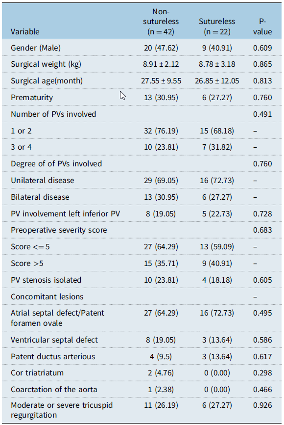
Sixty-four infants underwent surgical repair of primary pulmonary vein stenosis. Patient characteristics are presented in Table 1. All patients were divided into the non-sutureless group (n = 42, endovenectomy/patch venoplasty = 15/27) and the sutureless group (n= 22) according to different surgical techniques. Among them, 29 cases (45.31%) were male, and 19 cases (29.69%) were premature. One or two pulmonary veins were involved in 47 patients (73.44%), and bilateral pulmonary veins were involved in 19 patients (29.69%). No veins had atresia. The primary pulmonary vein stenosis severity score was greater than 5 in 37.5%. Among all patients, 14 infants had isolated pulmonary vein stenosis, and the remaining associated congenital cardiac lesions included atrial septal defect/patent foramen ovale in 43, moderate or severe tricuspid regurgitation in 17, ventricular septal defect in 11, patent ductus arteriosus in 7, cor triatriatum in 2, and coarctation of the aorta in 1. There was no significant difference in preoperative demographic baseline data between the non-sutureless group and the sutureless group.
Comparison of clinical data between the two groups [Mean ± SD/ case (%) /M (P25, P75)]

Early outcomes
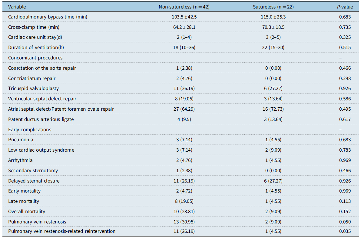
There were three early deaths, resulting in an early mortality rate of 4.69%. There were two early deaths in the non-sutureless group and one early death in the sutureless group, respectively. One patient died of refractory low cardiac output on the third day after patch venoplasty. One patient received extracorporeal membrane oxygenation support for 6 days after patch venoplasty due to low intraoperative cardiac output and died of circulatory failure 1 hour after weaning from extracorporeal membrane oxygenation. The other died of respiratory failure due to acute respiratory distress syndrome on the fifteenth day after sutureless repair. Bilateral pulmonary veins were involved in all three patients. Two patients had three pulmonary veins involved and one patient had four pulmonary veins involved. The preoperative severity score of three patients was greater than 5. There were no significant differences in cardiopulmonary bypass time, cross-clamp time, cardiac care unit stay, duration of ventilation, and early complications between the two groups. See Table 2, Figures 1, 2, 3, and 4.
(a) Overall survival. ( b) Survival in different surgical techniques.

(a) Freedom from pulmonary vein restenosis-related reintervention in the whole cohort. ( b) Freedom from pulmonary vein restenosis-related reintervention in different surgical techniques. (c) Freedom from pulmonary vein restenosis-related reintervention comparison between unilateral pulmonary vein involvement group and bilateral pulmonary vein involvement. ( d) Freedom from pulmonary vein restenosis-related reintervention comparison between preoperative severity score <= 5 group and preoperative severity score>5 group. ( e) Freedom from pulmonary vein restenosis-related reintervention comparison between one or two pulmonary veins (PVs) involvement group and three or four PVs involvement group. ( f) Freedom from pulmonary vein restenosis-related reintervention comparison between non-involvement of the left inferior pulmonary vein group and involvement of the left inferior pulmonary vein group.

(a) Freedom from pulmonary vein restenosis in the whole cohort. ( b) Freedom from pulmonary vein restenosis comparison in different surgical techniques. ( c) Freedom from pulmonary vein restenosis comparison between unilateral pulmonary vein involvement group and bilateral pulmonary vein involvement. ( d) Freedom from pulmonary vein restenosis comparison between preoperative severity score <= 5 group and preoperative severity score >5 group. ( e) Freedom from pulmonary vein restenosis comparison between one or two pulmonary vein (PVs) involvement group and three or four PVs involvement group. ( f) Freedom from pulmonary vein restenosis comparison between non-involvement of the left inferior pulmonary vein group and involvement of the left inferior pulmonary vein group.

Flow chart.

Comparison of perioperative and postoperative follow-up data between two groups [Mean±SD/ case (%) /M (P25, P75)]

Late outcomes
Survival
Follow-up was 100% complete, with a median period of 59(15,110) months. Late death occurred in nine patients (14.06%). Among those late deaths, one patient died of pulmonary hypertension crisis without additional pulmonary vein restenosis-related intervention in the non-sutureless group. The remaining eight patients underwent ineffective pulmonary vein restenosis-related reintervention and died, including seven patients in the non-sutureless group and one patient in the sutureless group. See Table 2 and Figure 4. The survival rates at 1, 5, and 15 years were 91.9%, 84.2%, and 69.8%, respectively, in the whole cohort. See Figure 1A. The survival rates at 1, 5, and 15 years in the sutureless group were 95.5%, 88.6%, and 88.6%, respectively, and the survival rates at 1, 5, and 15 years in the non-sutureless group were 90.2%, 82.3%, and 67.4%, respectively (log-rank, P = 0.484). Compared with the non-sutureless group, the sutureless group did not significantly improve the long-term survival of primary pulmonary vein stenosis patients.
Postoperative pulmonary vein restenosis and pulmonary vein restenosis-related reintervention
During follow-up, 15 patients (23.44%) met the criteria for postoperative pulmonary vein restenosis. Pulmonary vein restenosis occurred in 13 patients in the non-sutureless group and two patients in the sutureless group. Pulmonary vein restenosis-related reintervention occurred in 11 of 13 pulmonary vein restenosis in the non-sutureless group and 1 of 2 pulmonary vein restenosis in the sutureless group. See Table 2 and Figure 4. In the whole cohort, freedom from pulmonary vein restenosis-related reintervention at 1, 5, and 15 years were 81.3%, 79.1%, and 79.1%, respectively. See Figure 2A. Freedom from pulmonary vein restenosis-related reintervention at 1, 5, and 15 years in the non-sutureless group were 74.5%, 71.7%, and 71.7%, respectively, and freedom from pulmonary vein restenosis-related reintervention at 1, 5, and 15 years in the sutureless group were 95.0%, 95.0%, and 95.0%, respectively (log-rank, P = 0.049). See Figure 2B. In the whole cohort, freedom from pulmonary vein restenosis at 1, 5, and 15 years were 75.0%, 75.0%, and 75.0%, respectively. See Figure 3A. Freedom from pulmonary vein restenosis at 1, 5, and 15 years in the non-sutureless group were 67.5%, 67.5%, and 67.5%, respectively, and freedom from PVS at 1, 5, and 15 years in the sutureless group were 89.1%, 89.1%, and 89.1%, respectively (log-rank, P = 0.005). See Figure 3B. Using the sutureless technique was associated with less pulmonary vein restenosis and less PVS-related reintervention. Freedom from pulmonary vein restenosis-related reintervention at 1, 5, and 15 years in the unilateral pulmonary vein involvement group were 93.0%, 93.0%, and 93.0%, respectively, and freedom from PVS-related reintervention at 1, 5, and 15 years in the bilateral pulmonary vein involvement group were 50.0%, 43.8%, and 43.8%, respectively (log-rank, P < 0.001). See Figure 2C. Freedom from pulmonary vein restenosis at 1, 5, and 15 years in the unilateral pulmonary vein involvement group were 86.3%, 86.3%, and 86.3%, respectively, and freedom from pulmonary vein restenosis at 1, 5, and 15 years in the bilateral pulmonary vein involvement group were 43.8%, 43.8%, and 43.8%, respectively (log-rank, P < 0.05). P = 0.001). See Figure 3C. Bilateral pulmonary vein involvement was associated with higher pulmonary vein restenosis and higher pulmonary vein restenosis-related reintervention. Freedom from pulmonary vein restenosis-related reintervention at 1, 5, and 15 years in preoperative severity score <= 5 group were 92.4%, 92.4%, and 92.4%, respectively, and freedom from pulmonary vein restenosis-related reintervention at 1, 5, and 15 years in preoperative severity score>5 group were 60.5%, 60.5%, and 60.5%, respectively (log-rank, P < 0.001). See Figure 2D. Freedom from pulmonary vein restenosis at 1, 5, and 15 years in preoperative severity score <= 5 group were 87.5%, 87.5%, and 87.5%, respectively, and freedom from pulmonary vein restenosis at 1, 5, and 15 years in preoperative severity score>5 group were 50.8%, 50.8%, and 50.8%, respectively (log-rank, P = 0.003). See Figure 3D. Increased preoperative severity score was associated with higher pulmonary vein restenosis and higher pulmonary vein restenosis-related reintervention. Freedom from pulmonary vein restenosis-related reintervention at 1, 5, and 15 years in 1 or 2 pulmonary veins involvement group were 93.3%, 93.3%, and 93.3%, respectively, and freedom from pulmonary vein restenosis-related reintervention at 1, 5, and 15 years in three or four pulmonary veins involvement group were 41.7%, 33.3%, and 33.3%, respectively (log-rank, P < 0.001). See Figure 2E. Freedom from pulmonary vein restenosis at 1, 5, and 15 years in 1 or 2 pulmonary veins involvement group were 87.0%, 87.0%, and 87.0%, respectively, and freedom from PVS at 1, 5, and 15 years in three or four pulmonary veins involvement group were 35.7%, 35.7%, and 35.7%, respectively (log-rank, P < 0.001). See Figure 3E. Increased number of involved pulmonary veins was associated with higher pulmonary vein restenosis and higher pulmonary vein restenosis-related reintervention. Likewise, involvement of the left inferior pulmonary vein was associated with higher pulmonary vein restenosis and higher pulmonary vein restenosis-related reintervention. See Figure 2F and Figure 3F.
Factors associated with postoperative pulmonary vein restenosis and pulmonary vein restenosis-related reintervention
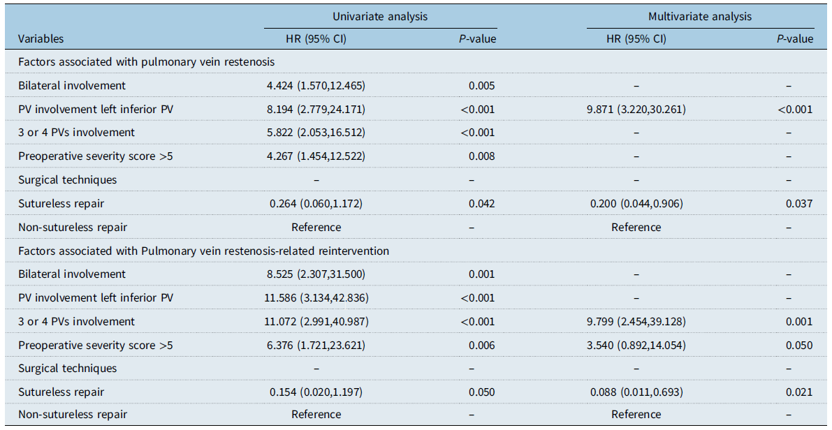
Involvement of the left inferior pulmonary vein (hazard ratio:9.871, 95% confidence interval: 3.220-30.261, P < 0.001) was associated with postoperative pulmonary vein restenosis, but sutureless repair (hazard ratio:0.200, 95% confidence interval: 0.044-0.906, P = 0.037) was protective against postoperative pulmonary vein restenosis by multivariate Cox regression. Multivariate analysis showed that three or four pulmonary veins involvement (hazard ratio:9.799, 95% confidence interval: 2.454-39.128, P = 0.001) and preoperative severity score exceeding 5 (hazard ratio:3.540, 95% confidence interval: 0.892-14.054, P = 0.050) were independent risk factors, but sutureless repair (hazard ratio:0.088, 95% confidence interval: 0.011-0.693, P = 0.021) was protective against pulmonary vein restenosis-related reintervention. These results are summarised in Table 3.
Risk factors for pulmonary vein restenosis (n = 15) and pulmonary vein restenosis-related reintervention (n = 12) by multivariate Cox proportional hazard model

Discussion
Primary pulmonary vein stenosis is a progressive and recurrent disease with death occurring secondary to right ventricular failure. It is difficult to ascertain the true survival of primary pulmonary vein stenosis since it is such a rare entity, but survival has been noted to be abysmal upon analysing early case studies of this disease. The reported estimates of overall mortality range from 30 to 86%. Reference Holt, Moller, Larson and Johnson5–Reference Feng, Mao and Ma8 In general, overall outcomes remain disappointing.
Surgical techniques have evolved since Kawashima and colleagues Reference Kawashima, Ueda, Naito, Morikawa and Manabe23 reported the first surgical repair of primary pulmonary vein stenosis. The conventional surgical intervention included endarterectomy and patch venoplasty; however, outcomes generally have been less than optimal. Reference Song, Bae and Jeong24,Reference Charlagorla, Becerra, Patel, Hoyer and Darragh25 The sutureless technique has now been commonplace for almost 2 decades and has been shown to decrease pulmonary vein restenosis in patients undergoing primary surgical repair of total anomalous pulmonary venous connection or pulmonary vein restenosis subsequent to total anomalous pulmonary venous connection surgery. Reference Harada, Nakano, Ando and Hashimoto26–Reference Wang, Ding and Yi28 The sutureless technique has become the preferred procedure for the initial repair of primary pulmonary vein stenosis in many institutions ever since its introduction in 1999. It minimises injury to the pulmonary veins and surgical trauma since trauma to the veins is found to stimulate regrowth of the obstructive tissue; however, a pericardial sac is made over the pulmonary vein and the left atrium adjacent to it, thus avoiding suturing the pulmonary vein. Reference Najm, Caldarone, Smallhorn and Coles29 This approach can thereby help prevent geometric distortion of the suture line and reduce the stimulus for regrowth of the obstructive tissue Reference Devaney, Chang, Ohye and Bove.19,Reference Shi, Zhu, Chen, Zhang, Zheng and Liu21 However, the results are inconclusive for this procedure as well. Some researchers Reference Devaney, Chang, Ohye and Bove19,Reference Azakie, Lavrsen, Johnson and Sapru20 believe that the prognosis of patients after sutureless repair is improved compared with standard endarterectomy and patch venoplasty. However, other researchers Reference Holt, Moller, Larson and Johnson5,Reference Song, Bae and Jeong24,Reference Viola, Alghamdi, Perrin, Wilson, Coles and Caldarone30 hold exactly the opposite view. Guocheng Shi et al. Reference Shi, Zhu, Chen, Zhang, Zheng and Liu21 believed that moderate or severe primary pulmonary vein stenosis is worse than mild primary pulmonary vein stenosis, and no differences were found in effectiveness among endarterectomy, pericardial patch venoplasty, and sutureless technique in treating primary pulmonary vein restenosis. Zicong Feng et al. Reference Feng, Mao and Ma8 found the sutureless technique was not significantly associated with improved results compared with the other two techniques. Joshua M. Rosenblum et al. Reference Rosenblum, Altin and Gillespie31 showed that sutureless technique was not associated with improved outcomes in their current series. Pradeepkumar Charlagorla et al. Reference Charlagorla, Becerra, Patel, Hoyer and Darragh25 also found that no statistically significant difference in mortality or reintervention rate was present among these different therapeutic modalities. However, in this study, we found that compared with non-sutureless repair, sutureless repair was significantly associated with decreased pulmonary vein restenosis and decreased pulmonary vein restenosis-related reintervention, respectively. This is likely due to the heterogeneous nature of this disease and the small number of patients who underwent sutureless repair at their institution. Considering the large sample size of the study in this cohort, the statistical errors were relatively small and the inferences drawn were more robust.
The postoperative outcome of primary pulmonary vein stenosis patients is very worrisome, and postoperative pulmonary vein restenosis has always been the main cause of long-term prognosis. Some published reports Reference Holt, Moller, Larson and Johnson5,Reference Devaney, Chang, Ohye and Bove19– Reference Shi, Zhu, Chen, Zhang, Zheng and Liu21,Reference Song, Bae and Jeong24,Reference Rosenblum, Altin and Gillespie31 have shown that the long-term restenosis rate of primary pulmonary vein stenosis patients varies from 20% to 65.5%. A retrospective study of 40 patients led by Zicong Feng et al. Reference Feng, Mao and Ma8 found that overall death, pulmonary vein reintervention, and restenosis occurred in 15%, 12.5%, and 25% of patients, respectively, with a median follow-up duration of 37.5 ± 31.5 months. A multicentre study led by Kalfa and colleagues Reference Kalfa, Belli and Bacha22 reported a survival rate of 70% and freedom from pulmonary vein reintervention of 62% at 8 years of follow-up. In this study, the restenosis rate was 23.44% and pulmonary vein restenosis-related reintervention rate was 17.19%. Our results are generally consistent with previously published results. In this study, in the non-sutureless group, 11 patients underwent reintervention due to pulmonary vein restenosis, of which three patients with sutureless repair all survived, and eight patients with patch venoplasty only one patient survived. Therefore, we advocate the use of sutureless techniques in patients who underwent pulmonary vein restenosis-related reintervention after primary repair of primary pulmonary vein stenosis.
Independent predictors for adverse outcomes remain uncertain due to the small number of patients in previous reports. Risk factors for adverse events have been reported in only a few small studies. It has been reported that risk factors for mortality and progressive stenosis are prematurity, chronic lung disease, associated congenital heart lesions, and bilateral disease. Reference Devaney, Chang, Ohye and Bove19,Reference Drossner, Kim, Maher and Mahle32 Joshua M. Rosenblum et al. Reference Rosenblum, Altin and Gillespie31 found that factors associated with mortality included bilateral disease and primary pulmonary vein stenosis severity score>6, additionally, bilateral disease and primary pulmonary vein stenosis severity score>5 were associated with reintervention. Zicong Feng et al. Reference Feng, Mao and Ma8 reported that bilateral pulmonary vein involvement was an independent risk factor for death or pulmonary vein reintervention and involvement of the left inferior pulmonary vein was an independent risk factor for postoperative restenosis of the left inferior pulmonary vein. Pradeepkumar Charlagorla et al. Reference Charlagorla, Becerra, Patel, Hoyer and Darragh25 reported that higher initial pre-and post-intervention gradients were significantly associated with decreased reintervention-free survival. In this study, multivariable analysis showed that involvement of the left inferior pulmonary vein was the only independent risk factor for pulmonary vein restenosis, while sutureless repair was a protective factor. Three or four pulmonary veins involvement and preoperative severity score exceeding 5 were found to be independent risk factors associated with pulmonary vein restenosis-related reintervention; however, sutureless repair was a protective factor. This demonstrates sutureless repair was significantly associated with decreased pulmonary vein restenosis and decreased pulmonary vein restenosis-related reintervention, respectively. Our series identified preoperative involvement of the left inferior pulmonary vein as an independent predictor for postoperative recurrence of left inferior pulmonary vein stenosis in multivariate analysis. Potentially, this might be the result of the combination of cardiomegaly and anterolateral displacement of the descending aorta. Reference Feng, Mao and Ma8 The possible mechanism might be that associated cardiac anomalies induce intracardiac shunting and subsequently cause volume overload, leading to cardiac enlargement and that the displacement of the aorta will ultimately increase the risk of anatomic compression of the pulmonary vein, especially the left inferior one. Similar findings were observed by Kotani Reference Kotani, Sano, Arai and Kasahara33 and Guocheng Shi. Reference Shi, Zhu, Chen, Zhang, Zheng and Liu21 This strengthened the major importance of creating a favourable anatomic relationship in the initial repair for patients with primary pulmonary vein stenosis. To summarise, care should be taken to avoid postoperative compression when addressing the stenotic pulmonary veins. Three or four pulmonary veins involvement and preoperative severity score exceeding 5 indicate that the degree of preoperative pulmonary vein stenosis is very severe, and its course is usually more malignant. So the incidence of pulmonary vein restenosis-related reintervention is undoubtedly higher after repair.
Limitations
The most important limitations of this study are its retrospective and single-institution nature. An additional limitation is the relatively small study size limiting the statistical
robustness of any inference that may be drawn. Selection bias is also unavoidable. Average age of patients is higher than other reported cohorts, potentially introducing a survival bias to our results. In the future, longer follow-up and larger sample size multicentre clinical studies are needed to further evaluate the long-term outcomes.
Conclusions
Management of infants with primary pulmonary vein stenosis is challenging. Surgical repair of primary pulmonary vein stenosis can be safely achieved using different techniques, with similar long-term mortality. Compared with non-sutureless repair, sutureless repair was significantly associated with decreased pulmonary vein restenosis and decreased pulmonary vein restenosis-related reintervention, respectively. Preoperative severity scores exceeding 5, 3, or 4 pulmonary vein involvement, and left inferior pulmonary vein involvement are independent risk factors for adverse outcomes.
Supplementary material
The supplementary material for this article can be found at https://doi.org/10.1017/S1047951124026180.
Data availability statement
All data are incorporated into the article, and the data underlying this article are available in the article.
Acknowledgements
We acknowledge the roles of our colleagues, perfusionists, nurses, and others involved in the care of the study participants.
Author contribution
Zhangwei Wang Conceptualisation; Data curation; Formal analysis; Investigation; Methodology; Software; Validation; Visualisation; Writing—original draft. Yang Yang Writing—review & editing.
Financial support
No funding was received for this work.
Competing interests
None declared.