INTRODUCTION
Trypanosoma cruzi, the causative agent of Chagas' disease, is a parasitic protozoan endemic in Central and South America. Despite the significant reduction in transmission observed in several countries in the last 20 years, this parasite nowadays affects 18 million people and, in approximately one third of the infected individuals, causes incurable heart or digestive dysfunction (WHO, 2002). This situation is worsened by the lack of a vaccine, and the undesirable side-effects of chemotherapeutic T. cruzi-specific drugs currently used.
Mice and humans infected with T. cruzi develop strong immune responses to trans-sialidase (TS)-derived antigens from the parasite. The TS superfamily proteins share a common structure consisting of 1–4 complete and/or degenerate copies of a bacterial sialidase motif and a subterminal FLY motif at their N-terminal domain, and a C-terminal domain, according to which they are classified into 4 families. Though only very few of the TS antigens have trans-sialidase activity, a large number of enzymatically inactive TS proteins are simultaneously expressed in the surface membrane of the parasite and shed to the milieu presenting a complex network of cross-reacting epitopes (Cross and Takle, Reference Cross and Takle1993; Frasch, Reference Frasch2000). Tc13 is a TS family protein bearing 5 amino-acid (EPKSA) repeats at its C-terminal region (Campetella et al. Reference Campetella, Sanchez, Cazzulo and Frasch1992). It has not been determined whether Tc13 antigens posses TS activity; however, it is known that the EPKSA portion is able to induce a high antibody response (Burns et al. Reference Burns, Shreffler, Rosman, Sleath, March and Reed1992).
It has been widely documented that a robust T cell and type 1 cytokine response is necessary to control T. cruzi infection. Furthermore, a recent study demonstrates that in T. cruzi-infected mice, as well as in humans, the CD8+T-cell response is highly focused on epitopes encoded by members of the TS superfamily (Martin et al. Reference Martin, Weatherly, Laucella, Cabinian, Crim, Sullivan, Heiges, Craven, Rosenberg, Collins, Sette, Postan and Tarleton2006). Many of these antigens, such as the catalytic domain of TS, TSA-1 and SA85-1, are able to elicit CD8 and CD4 T cells that produce IFN-γ, being the epitopes recognized on the N-terminal domain sequences of these proteins (Wizel et al. Reference Wizel, Nunes and Tarleton1997; Rodrigues et al. Reference Rodrigues, Ribeirao, Pereira-Chioccola, Renia and Costa1999; Millar et al. Reference Millar, Wleklinski-Lee and Kahn1999; Fujimura et al. Reference Fujimura, Kinoshita, Pereira-Chioccola and Rodrigues2001). Nevertheless, the epitope variations expressed during the infection may influence the protective responses allowing a partial escape from the host immune system (Wrightsman et al. Reference Wrightsman, Dawson, Fouts and Manning1994; Millar et al. Reference Millar, Wleklinski-Lee and Kahn1999; Martin et al. Reference Martin, Weatherly, Laucella, Cabinian, Crim, Sullivan, Heiges, Craven, Rosenberg, Collins, Sette, Postan and Tarleton2006). Otherwise, some TS superfamily proteins have been implicated in pathological processes developed during T. cruzi infection, such as induction of apoptosis in components of the immune system (Mucci et al. Reference Mucci, Risso, Leguizamon, Frasch and Campetella2006) or down-regulation of the immune response through interaction with host cellular receptors (Gorelik et al. Reference Gorelik, Cremaschi, Borda and Sterin-Borda1998). We had previously shown that the repetitive EPKSA C-terminal domain of Tc13Tul antigen interacts with β1-adrenergic receptor (García et al. Reference Garcia, Joensen, Bua, Ainciart, Perry and Ruiz2003) and displays an agonist-like activity in rat myocardium in vitro (Joensen et al. Reference Joensen, Borda, Kohout, Perry, Garcia and Sterin-Borda2003), suggesting that it may be involved in the pathogenesis of Chagas' disease. We have also recently demonstrated that Tc13Tul mainly induces immune responses associated with pathology rather than protection, since genetic immunization with the gene encoding this antigen produces harmful effects in tissues, does not elicit IFN-γ production and does not protect against the acute phase of the infection (García et al. Reference Garcia, Arnaiz, Laucella, Esteva, Ainciart, Riarte, Garavaglia, Fichera and Ruiz2006). As genetic immunization should mimic an intracellular infection (Whitton et al. Reference Whitton, Rodriguez, Zhang and Hassett1999), we may speculate that these results would represent the effect produced by the Tc13 antigens present in the amastigote surface in the outcome of a natural infection with T. cruzi.
In order to deepen the role of Tc13 antigens in the development of T. cruzi infection, in the present study we evaluated the humoral and cellular immune responses triggered by these antigens in BALB/c mice either infected with T. cruzi or immunized with soluble recombinant Tc13Tul. As a result of this research, we report B- and T-cell epitope localization in Tc13 molecules, describe a CD8+T-cell epitope capable of inducing IFN-γ secretion in chronically infected mice and provide evidence that soluble Tc13 antigens do not participate in protective immune responses.
MATERIALS AND METHODS
Mice and parasites
Male BALB/c mice were used in all the experiments. Parasites of the Tulahuén strain, stock Tul2 of T. cruzi were used in this study. Bloodstream trypomastigotes were maintained in vivo by serial passage of blood-form trypomastigotes in BALB/c mice. A T. cruzi lysate was obtained from trypomastigotes collected from the culture medium of infected Vero cell monolayers by freeze-thawing with liquid N2. The lysate was used for DTH and ELISA assays.
Animal handling and experimental design conformed to the ethical treatment of animals established by the Argentinean Animal Protection Society.
Recombinant proteins
The Tc13Tul protein is encoded by a 2391-bp fragment (GenBank Accession no. AF092099) which lacks the 5′ end but codifies for 2 of 3 SXDXGXTW motifs and 45 EPKSA repeats. Tc13Tul protein was expressed as a maltose-binding protein (MBP) fusion and purified by amylose resin (García et al. Reference Garcia, Joensen, Bua, Ainciart, Perry and Ruiz2003). The clone coding for EPKSA (GenBank Accession no. M92046) repeats fused to glutathione-S-transferase (GST) was kindly provided by Dr O. Campetella (Instituto de Investigaciones Biotecnológicas, Universidad de San Martín, Buenos Aires, Argentina). The GST-EPKSA recombinant protein was purified by glutathione-agarose resin.
Infection, immunization and challenge
To study the immune response raised against Tc13 antigens in the outcome of the infection, 9-week-old mice were infected by intraperitoneal injection of 500 bloodstream trypomastigotes of Tulauhén strain, stock Tul2/mouse. According to our experimental data, mice were considered acutely infected at 30 days post-infection (p.i.) and chronically infected after 4 months p.i.
To study the effect of Tc13Tul immunization, 6-week-old male BALB/c mice were injected with MBP-Tc13Tul or MBP. The immunization protocol consisted of 5 weekly subcutaneous (s.c.) doses of 50 μg of recombinant protein/mouse/dose emulsified with an equal volume of incomplete Freund's adjuvant (IFA). A non-immunized group injected with IFA alone served as control. In order to evaluate the protective effect of Tc13Tul immunization, mice were challenged by intraperitoneal injection of 750 bloodstream trypomastigotes of Tulauhén strain, stock Tul2 at 4 weeks after the last dose. Survival was recorded daily and parasitaemia was determined by counting in a haematocytometer the number of bloodstream trypomastigotes in fresh blood diluted 10 times in a red blood cell lysis solution. Experiments were performed 3 times, independently, using 6–7 mice per group.
Measurement of humoral immune response
The presence of anti-Tc13Tul and anti-EPKSA IgM, total IgG and IgG isotype antibodies in serum samples from T. cruzi-infected and Tc13Tul-immunized mice were evaluated by ELISA using as target antigens MBP-Tc13Tul, GST-EPKSA, MBP and GST at a concentration of 5 μg/ml and a trypomastigote lysate at 20 μg/ml. The OD490 for Tc13Tul antigen was calculated as follows: OD490 using MPB-Tc13Tul as antigen – OD490 using MBP as antigen and the OD490 for EPKSA antigen as: OD490 using GST-EPKSA as antigen – OD490 using GST as antigen. Sera from infected mice were collected in different stages of the infection and sera from immunized mice were obtained 21 days after the last immunization.
Briefly, flat-bottomed plates were coated overnight at 4°C with different antigens and afterwards were blocked with 5% skimmed milk in PBS. After being washed with PBS-0·05% Tween20 (PBS-T), plates were incubated for 1 h at RT with pooled sera from 5 mice for each group or individual sera diluted 1/100 or 1/500 for infected and immunized mice, respectively. When using MBP-Tc13Tul (or MBP) and GST-EPKSA (or GST) as target antigens sera were diluted in PBS-1% skimmed milk with 20 μg/ml of MBP and GST, respectively and pre-incubated for 15 min. After washing, plates were incubated for 1 h at RT with horseradish peroxidase-labelled goat anti-mouse IgG (Jackson) diluted 1:2000 or biotinylated anti-mouse IgM (Vector Laboratories) diluted 1:250 or biotinylated anti-mouse IgG isotypes (IgG1, IgG2a, IgG2b or IgG3) (PharMingen) diluted 1:200. After incubation with the biotin conjugates, a dilution of 1:1000 of streptavidin-horseradish peroxidase (Vector Laboratories) was applied and incubated for 1 h at RT. Colour was developed with o-phenylenediamine dihydrochloride (OPD) and optical density (OD) was read at 490 nm using an ELISA microplate reader (Dynatech).
To evaluate anti-Tc13 antibodies in immunized animals by Western blot, GST-EPKSA (5 μg/lane) and a trypomastigote lysate (1·5×107 trypomastigotes/lane) were run in 10% SDS-PAGE, electroblotted onto nitrocellulose membranes and incubated overnight at 4°C with a pool of 6 sera from non-immunized and MBP- or MBP-Tc13Tul- immunized mice, diluted 1:50 for the lysate and 1:2000 for GST-EPKSA antigen. Membranes were washed and then incubated for 1h at RT with biotinylated anti-mouse IgG (Jackson) diluted 1:2000. Afterwards, membranes were washed and incubated with streptavidin-horseradish peroxidase (Jackson) diluted 1:1000 during 30 min at RT. Detection was performed with chloro-1-naphthol as substrate.
Measurement of delayed-type hypersensitivity (DTH)
The Tc13-specific DTH response in BALB/c mice ranging over different stages of the infection was evaluated by footpad inoculation with 50 μg of MBP-Tc13Tul or GST-EPKSA. MBP or GST was also inoculated in the same concentration to serve as a control. The DTH response in immunized mice was evaluated 27 days after the last immunization by injection of 50 μg of a trypomastigote lysate. Footpad induration was measured with a micrometer (Mitutoyo) 24 and 48 h after the injection and the induration index was calculated as the rate between the induration at 24 h or 48 h with respect to the value obtained before the injection in the same footpad. Experiments were performed 3 times, independently, with 3–6 mice per group.
Peptides
H-2Kd-binding peptides and proteosomal cleavage sites from the Tc13Tul sequence were predicted by using the following programs: RANKPEP (http://bio.dfci.harvard.edu/Tools/rankpep.html), MAPPP (http://www.mpiib-berlin.mpg.de/MAPPP) and SYFPEITHI (http://syfpeithi.de/Scripts/MHCServer.dll/EpitopePrediction.htm) and the 2 peptides with the highest score were selected, GYGATSSSL (Tc13Tul p-1) and EYLGVNAVV (Tc13Tul p-2). Peptides were synthesized by SigmaGenosys with a final purity varying from 91 to 93%.
ELISPOT assay for the detection of Tc13-specific IFN-γ- and IL-4-producing cells
Enzyme-linked immunospot (ELISPOT) assay was used to quantify IFN-γ and IL-4 secreting cells in chronically infected (6 month post-infection) or immunized mice (20 days after the last dose) after stimulation with recombinant MBP-Tc13Tul (or MBP alone as control) and the peptides Tc13Tul p-1 and Tc13Tul p-2. Nitrocellulose bottomed 96-well plates (Millipore Corp.) were coated with 0·5 μg of anti-mouse IFN-γ or anti-mouse IL-4 antibodies (BD Biosciences) overnight at 4°C, washed with PBS and blocked with RPMI-10% Fetal Bovine Serum (FBS) for 2 h at RT. Spleen cells from individual animals were plated in triplicate at 1×10⁵ or 2×10⁵ cells/well in wells containing 100 μl of either RPMI-10% FBS, MBP-Tc13Tul 20 μg/ml, MBP 6·8 μg/ml, Tc13Tul p-1 20 μg/ml or Tc13Tul p-2 20 μg/ml and incubated overnight at 37°C, 5% CO2. As positive control, cells were plated with 5 ng/ml phorbol myristic acetate (PMA) and 500 ng/ml ionomycin. After washes, plates were incubated with 0·2 μg of biotinylated anti-mouse IFN-γ or biotinylated anti-mouse IL-4 (BD Biosciences) overnight at 4°C. Afterwards, plates were washed and incubated with peroxidase-labelled streptavidin (Kirkegaard & Perry Laboratories) diluted 1:500 during 1 h 30 min at RT. Individual IFN-γ or IL-4 producing cells were detected as dark spots, after 15 min of colour development with 3-amino-9 ethylcarbazol (Sigma). Spots were counted with a stereoscopic microscope (Carl Zeiss). The experimental values correspond to the mean number of spots/1×105 (or 2×10⁵) splenocytes from 3–4 animals.
Histopathological analysis
Mice immunized with MBP-Tc13Tul or MBP and a control group immunized with IFA were sacrificed 24 days after the last immunization. Challenged mice were killed in the acute (38 days post-infection) or in the chronic (4 months post-infection) phases of the infection. Mice were ether anaesthetized, bled by eyeball puncture for serological studies and killed by cervical dislocation. Complete autopsies and histopathological analyses were performed on immunized and/or infected and uninfected control mice. Heart, skeletal muscle, liver and spleen were removed and fixed in 10% buffered formalin, dehydrated in absolute ethanol, cleared in xylene and embedded in paraffin. Sections (5 μm) were stained with haematoxylin and eosin and evaluated by light microscopy. The extent of inflammation in the myocardium was scored according to: normal (−); focal (+); multifocal (++); diffuse with partial wall involvement (+++) and total wall involvement (++++) (Sun and Tarleton, Reference Sun and Tarleton1993). The scoring of skeletal muscle inflammation was based on a modification of a previously described method (Ben Younes-Chennoufi et al. Reference Ben Younes-Chennoufi, Said, Eisen, Durand and Hontebeyrie-Joskowicz1988) as normal (−); few cellular infiltrates (+); diffuse infiltrates (++); abundant infiltrates (+++) and granulomatous infiltrates (++++).
Statistical analysis
The Student's t-test was used to compare the possible differences in the mean values of induration of MBP-Tc13Tul and MBP inoculated footpads in DTH assays with infected mice and the number of spots obtained with or without stimulus in ELISPOT. For ELISA assays using Tc13Tul or EPKSA as target antigens (Fig. 1), one-way ANOVA followed by Tukey's test was used to compare the optical density at 490 nm at different time-points post-infection. Two-way ANOVA followed by Bonferroni's multiple comparison test was used to compare induration in DTH reactions with immunized animals. Parasitaemia observed in the different groups at each time-point were compared by one-way ANOVA and log-rank test was used to compare mouse survival times after challenge with T. cruzi. Differences were considered significant when the P value was <0·05.
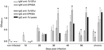
IgM and IgG responses against Tc13 antigens in Trypanosoma cruzi-infected BALB/c mice. IgM and IgG production specific for Tc13 in different stages of T. cruzi infection was evaluated by ELISA using recombinant Tc13Tul and the Tc13 C-terminal repetitive portion (EPKSA) as antigens. IgG reactivity against a trypomastigote lysate (Tc lysate) served as a positive control. Results are presented as the mean±s.e.m. of the optical density at 490 nm of 3 pools of sera. P values obtained by one-way ANOVA followed by Tukey test: (x) P<0·05 vs non-infected mice; (○) P<0·05 vs 10 days post-infection (p.i.); (●) P<0·05 vs 20 days p.i.; (*) P<0·05 vs 30 days p.i.

RESULTS
Humoral and cellular T cell responses specific for Tc13 antigens in the outcome of T. cruzi infection
With the aim to determine the role of Tc13 antigens in the outcome of T. cruzi infection, the humoral and cellular T cell responses against these antigens were investigated at different stages of the infection.
The levels of anti-Tc13 antibodies were evaluated by ELISA using the recombinant proteins EPKSA, derived from the C-terminal repeat portion, and Tc13Tul, a Tc13 antigen from the Tulahuén strain of T. cruzi, which also contains a large section of the N-terminal region. IgM antibodies specific for Tc13Tul were not detected until 10 days post-infection (p.i.), reaching at maximum levels at 20 days p.i. Thereafter, IgM anti-Tc13Tul remained relatively constant throughout the infection (Fig. 1). In contrast, the IgM response against EPKSA displayed delayed appearance, being detectable at 20 days p.i., peaking at 30–40 days p.i., and decreasing significantly thereafter (Fig. 1), with 50% of the mice showing anti-EPKSA negative IgM responses in the chronic phase of the infection (data not shown). Otherwise, for both antigens, IgG antibodies were not detected until 20 days p.i. The IgG response against Tc13Tul reached a maximum at 40 days p.i., declined in the post-acute phase and returned to the peak level in the chronic phase of the infection. IgG levels specific for EPKSA markedly increased between 30 and 90 days p.i., being 1·6- to 3-fold higher than those obtained with Tc13Tul, even exceeding the response detected using a trypomastigote lysate preparation as target antigen. In the chronic phase, anti-EPKSA IgG slightly decreased (Fig. 1). Altogether, these results suggest that while epitopes with the ability to induce IgM responses are mainly contained in the N-terminal domain of Tc13Tul, epitopes efficient in promoting IgG switching are mainly localized in the EPKSA repeat portion of the antigen.
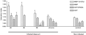
In order to determine whether Tc13 antigens elicit T cell responses, delayed-type hypersensitivity (DTH) was measured after injection of recombinant Tc13Tul (MBP-Tc13Tul) or EPKSA (GST-EPKSA) in the footpad of mice coursing different stages of T. cruzi infection. Mice infected with T. cruzi showed a positive cutaneous reaction against MBP-Tc13Tul up to 90 days p.i. Thereafter, induration produced by this antigen was not significantly different from that induced by MBP alone, indicating the lack of an effective memory T cell response against Tc13 antigens (Fig. 2). In contrast, GST-EPKSA-induced induration never exceeded GST induction, demonstrating that the EPKSA repeats portion of Tc13 antigens are not able to induce T cell responses as measured by DTH assay (Fig. 2).
Delayed-type hypersensitivity against Tc13 antigens in Trypanosoma cruzi BALB/c infected mice. Footpad swelling was measured after local inoculation of MBP-Tc13Tul (or MBP as control) and GST-EPKSA (or GST as control) in mice ranging over different stages of T. cruzi infection. Non-infected mice inoculated with these antigens served as negative controls. Values are presented as mean induration index±s.e.m. of 3–4 mice per group. The induration index was calculated as the rate between the induration at 24 h with respect to the value obtained before the injection in the same footpad. (*) P<0·05 vs MBP inoculation.

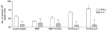
Taking into account that IFN-γ plays a main role in protective immunity against T. cruzi (Reis et al. Reference Reis, Higuchi Mde, Benvenuti, Aiello, Gutierrez, Bellotti and Pileggi1997) and that CD8+T cells are crucial to control the infection (Martin and Tarleton, Reference Martin and Tarleton2004) we evaluated whether Tc13 antigens were able to elicit IFN-γ secretion measured by ELISPOT assays. Spleen cells from chronically infected mice were stimulated with recombinant Tc13Tul and 2 peptides selected by the presence of putative sites for binding to the MHC class I H-2Kd, Tc13Tul p-1 and Tc13Tul p-2. The number of IFN-γ secreting T cells induced after stimulation with MBP-Tc13Tul did not differ from those elicited by non-stimulated or MBP-stimulated controls. However, we found that there was a significant IFN-γ secretion when spleen cells were stimulated with the putative H-2Kd binding peptide Tc13Tul p-2 (Fig. 3). Conversely, neither MBP-Tc13Tul nor the peptides induced IL-4 ELISPOT responses (Fig. 3).
Tc13 specific-IFN-γ and IL-4 release by spleen cells from Trypanosoma cruzi-infected mice. IFN-γ and IL-4 ELISPOT responses to MBP (3·4 μg/ml), MBP-Tc13Tul (10 μg/ml) and individual peptides from Tc13Tul sequence (Tc13Tul p-1 and Tc13Tul p-2) (10 μg/ml) were assessed in chronically T. cruzi-infected mice. Values representative of 2 independent experiments with 3 different mice each are presented as mean±s.e.m. Spleen cells from each animal were tested independently and in triplicate wells. (*) P<0·05 vs unstimulated cells.

Humoral and cellular immune responses induced by immunization with recombinant Tc13Tul
With the aim to determine the immune responses induced by soluble Tc13 antigens, BALB/c mice were immunized with recombinant Tc13Tul in the presence of incomplete Freund's adyuvant (IFA) to further characterize the antibody and T cell responses. As this antigen is fused to MBP, a control group was immunized with this protein plus the corresponding adjuvant.
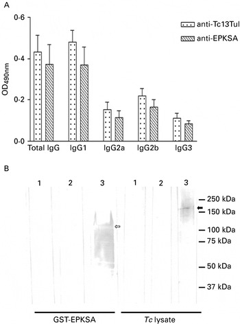
In order to study the humoral immune response elicited by immunization with recombinant Tc13Tul, the levels of specific IgG antibodies were determined by ELISA using MBP-Tc13Tul and GST-EPKSA as target antigens. Sera from MBP-Tc13Tul-immunized animals reacted with both antigens similarly (Fig. 4A). Sera from MBP-immunized mice, although they were competed with free MBP, lightly reacted with the MBP portion of MBP-Tc13Tul antigen but no reactivity was observed against GST or GST-EPKSA in this control group (data not shown). When the IgG isotype profile against Tc13Tul and EPKSA molecules was evaluated in Tc13Tul immunized animals, IgG1 was the predominant subclass presenting similar reactivity to both antigens (Fig. 4A). Western blot analysis showed that sera from MBP-Tc13Tul immunized mice recognized GST-EPKSA antigen and a protein of 200 kDa in a trypomastigote lysate, previously identified as a Tc13 protein (Ibañez et al. Reference Ibanez, Affranchino and Frasch1987; Burns et al. Reference Burns, Shreffler, Rosman, Sleath, March and Reed1992), while sera from MBP-immunized as well as sera from mice immunized with adjuvant alone did not show positive reactivity (Fig. 4B).
Humoral immune response of BALB/c mice immunized with recombinant Tc13Tul. (A) Determination of the total IgG and IgG isotype levels against Tc13Tul and its EPKSA portion in Tc13Tul-immunized mice. Sera from MBP-Tc13Tul-immunized mice were tested individually by ELISA using MBP-Tc13Tul or GST-EPKSA as target antigens. Sera were also simultaneously tested on MBP and GST and the OD490 nm specific for MBP or GST were subtracted (see Material and Methods for details). (B) Sera reactivity of Tc13Tul-immunized mice against EPKSA repeats and a Trypanosoma cruzi lysate. Pooled sera from non-immunized (lane 1), MBP- (lane 2) or MBP-Tc13Tul-immunized mice (lane 3) were tested by Western blot against the recombinant protein GST-EPKSA and a trypomastigote lysate (Tc lysate) separated in 10% SDS-PAGE. The white and black arrows show the GST-EPKSA antigen and Tc13 molecule on parasite lysate recognized by sera from MBP-Tc13Tul-immunized animals, respectively.

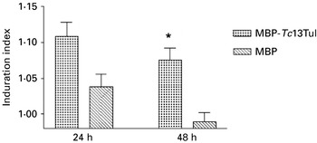
Memory T cell responses specific for Tc13Tul in immunized mice were evaluated by DTH measurement after footpad inoculation with a trypomastigote lysate. The cutaneous reaction observed in the MBP-Tc13Tul immunized group was significantly higher than in the MBP immunized group at 48 h post-inoculation (Fig. 5). To further characterize the T-cell responses elicited by Tc13Tul immunization, the secretion of IFN-γ by splenic cells from Tc13Tul immunized mice was assessed 3 weeks after the last dose by in vitro stimulation with MBP-Tc13Tul (or MBP as control) and the putative H-2Kd binding peptides Tc13Tul p-1 and Tc13Tul p-2. Immunization with the recombinant antigens, MBP-Tc13Tul or MBP, did not induce net IFN-γ secretion as evidenced by the negative ELISPOT responses after stimulation of splenic cells with the recombinant proteins or peptides (data not shown).
Delayed-type hypersensitivity against a Trypanosoma cruzi lysate of mice immunized with recombinant Tc13Tul. Footpad swelling in mice immunized with MBP-Tc13Tul or MBP after inoculation of trypomastigote lysate was measured. Values are presented as mean induration index±s.e.m. of 6 mice per group. (*) P<0·05 by Bonferroni vs MBP-immunized mice at 48 h post-inoculation.

Evaluation of the protective response against T. cruzi infection induced by immunization with recombinant Tc13Tul
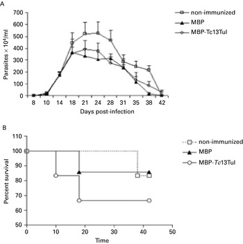
To evaluate whether immunization with recombinant Tc13Tul could provide protection against T. cruzi infection, Tc13Tul-immunized mice were challenged with a sublethal dose of 750 T. cruzi bloodstream trypomastigotes. No differences were observed in either parasitaemia or survival rates among the MBP-Tc13Tul immunized group and non-immunized or MBP-immunized control groups (Fig. 6A and B). Likewise, both parameters were not altered when using non-lethal and lethal doses of 500 and 2000 bloodstream trypomastigotes, respectively (data not shown).
Parasitaemia and survival of BALB/c mice immunized with recombinant Tc13Tul and challenged with Trypanosoma cruzi. MBP-Tc13Tul- and MBP-immunized mice and a control non-immunized group were challenged with 750 blood trypomastigotes 4 weeks after the last immunization. The data show a representative experiment out of 3 performed with 6 mice per group. (A) Course of infection estimated by the number of parasites/ml of blood. Values are presented as the mean±s.e.m. (B) Mortality rate of immunized mice infected with T. cruzi (0·43>P>0·61 by log-rank test).

Histological findings elicited by immunization with recombinant Tc13Tul
We have previously demonstrated that genetic immunization with Tc13Tul induces hepatotoxicity and reactive changes in heart, liver and spleen and modulates the inflammatory infiltrate generated by T. cruzi infection in heart and skeletal muscle (García et al. Reference Garcia, Arnaiz, Laucella, Esteva, Ainciart, Riarte, Garavaglia, Fichera and Ruiz2006). In order to evaluate if immunization with recombinant Tc13Tul induces any pathological manifestation in tissues, the histopatological analysis of heart, spleen, liver and skeletal muscle sections from Tc13Tul-immunized mice and non-immunized and MBP-immunized control groups was performed. Recombinant Tc13Tul immunization did not induce pathological effects in tissues, although some non-specific alterations ascribable to the adjuvant, like oedema and hyperplasia, were found (data not shown).
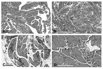
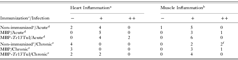
Histological analysis of heart and skeletal muscle sections from Tc13Tul-immunized mice challenged with T. cruzi was carried out with the aim to evaluate whether immunization with recombinant Tc13Tul is able to attenuate or exacerbate the tissue damage induced by T. cruzi infection in target organs. In the acute phase of the infection, heart samples from all animal groups mainly showed focal inflammatory infiltrates. However, multifocal inflammatory infiltrates was only observed in MBP-Tc13Tul-immunized mice. Interestingly, in the chronic phase of the infection inflammation was only evidenced in MBP-Tc13Tul-immunized group (Table 1 and Fig. 7A and B). In skeletal muscle tissues, inflammation was observed either in the acute or chronic phases of the infection in all the groups evaluated. Nevertheless, in the chronic phase of the infection, all of the animals immunized with MBP-Tc13Tul showed few cellular infiltrates while some of the animals from both control groups exhibited diffuse inflammatory infiltrates with foci of calcification (Table 1 and Fig. 7C and D). Although we found that immunization with MBP-Tc13Tul has no effect on tissues, these observations let us speculate that recombinant Tc13Tul immunization may somehow modulate the inflammation induced by T. cruzi infection in target organs.
Histopathology of heart and skeletal muscle of Tc13Tul-immunized mice challenged with Trypanosoma cruzi. (A) Serial heart paraffin section from non-immunized, acutely T. cruzi-infected mice showing an inflammatory focus (white arrow) and oedema (black arrow). (B) Heart sections from acutely T. cruzi-infected mice previously immunized with Tc13Tul displaying multifocal inflammatory infiltrates (white arrow). (C) Serial sections of skeletal muscle of non-immunized chronically T. cruzi-infected mice showing multiple foci of inflammation (white arrow) and important oedema in the interstitial space (black arrow). Insert shows areas of calcification accompanying the phlogosic process. (D) Serial sections of skeletal muscle of chronically T. cruzi-infected mice previously immunized with recombinant Tc13Tul displaying scattered lymphocytes and mild oedema (white arrow).

Histological analysis of heart and skeletal muscle of recombinant Tc13Tul-immunized mice challenged with Trypanosoma cruzi

a The scoring was based on Sun and Tarleton (Reference Sun and Tarleton1993). Normal (−), focal (+), multifocal (++).
b The scoring was based on Ben Younes-Chennoufi et al. (Reference Ben Younes-Chennoufi, Said, Eisen, Durand and Hontebeyrie-Joskowicz1988). Normal (−), few cellular infiltrates (+), diffuse cellular infiltrates (++).
c Immunized with adjuvant alone.
d Mice were challenged four weeks after the last dose with 750 bloodstream trypomastigotes and killed at 38 days post-infection.
e Mice were challenged four weeks after the last dose with 750 bloodstream trypomastigotes and killed 4 months post-infection.
f Both mice showed tissue calcifications.
DISCUSSION
Since the role of Tc13 antigens in the outcome of natural T. cruzi infection has not been fully investigated so far, we sought to characterize the immune responses induced by these antigens in an experimental murine model. Unlike previous reports in which only the Tc13 EPKSA domain was employed (Burns et al. Reference Burns, Shreffler, Rosman, Sleath, March and Reed1992; Peralta et al. Reference Peralta, Teixeira, Shreffler, Pereira, Burns, Sleath and Reed1994), the use of a Tc13 antigen containing part of the N-terminal portion allowed analysis of B- and T-cell epitope distribution along both Tc13 domains. The kinetics of the IgM responses specific for Tc13Tul and EPKSA throughout the infection suggest that epitopes with low ability to promote heavy chain isotype switching and capable of stimulating an early and sustained B lymphocyte activation may be mainly located at the N-terminal domain. According to previous publications (Ibañez et al. Reference Ibanez, Affranchino and Frasch1987; Burns et al. Reference Burns, Shreffler, Rosman, Sleath, March and Reed1992; Peralta et al. Reference Peralta, Teixeira, Shreffler, Pereira, Burns, Sleath and Reed1994), we found high levels of IgG specific for the Tc13 C-terminal domain during the acute phase of the infection with T. cruzi. This anti-EPKSA IgG response was even higher than those detected using a parasite lysate as source of antigen, indicating that EPKSA may be more sensitive than conventional diagnostic tests to detect recent infections. In contrast, animals immunized with recombinant Tc13Tul showed levels of IgG specific for Tc13Tul as high as those specific for EPKSA. These results suggest that Tc13 B-cell epitopes are differentially presented during infection and immunization. This is probably due to the adjuvant effect or, as it has been described for other TS antigens, the simultaneous expression of multiple Tc13 variants during the infection which may modulate the specific immune response (Wrightsman et al. Reference Wrightsman, Dawson, Fouts and Manning1994; Millar et al. Reference Millar, Wleklinski-Lee and Kahn1999; Martin et al. Reference Martin, Weatherly, Laucella, Cabinian, Crim, Sullivan, Heiges, Craven, Rosenberg, Collins, Sette, Postan and Tarleton2006).
A positive DTH reaction against Tc13Tul antigen but not EPKSA during T. cruzi infection shows that epitopes capable of activating T-cells may be mainly located at the N-terminal domain of Tc13 molecules. However, the negative DTH response in the chronic phase of the infection indicates that Tc13 antigens are not able to elicit a robust memory T-cell response in experimental T. cruzi infection (Black, Reference Black1999). Likewise, no Tc13-specific IFN-γ and IL-4 secretion was observed when spleen cells from chronically infected mice were stimulated with recombinant Tc13Tul. As it has been reported by others that cytokine secretion by spleen cells after in vitro re-stimulation with recombinant proteins is dependent on the activation of CD4+ T cells (Boscardin et al. Reference Boscardin, Kinoshita, Fujimura and Rodrigues2003; Vasconcelos et al. Reference Vasconcelos, Hiyane, Marinho, Claser, Machado, Gazzinelli, Bruna-Romero, Alvarez, Boscardin and Rodrigues2004), our results point to the absence of Tc13-specific CD4+ T-cells with ability to release IFN-γ and IL-4 in the chronic phase of the infection. As we previously showed with Tc13Tul DNA immunization (García et al. Reference Garcia, Arnaiz, Laucella, Esteva, Ainciart, Riarte, Garavaglia, Fichera and Ruiz2006), mice immunized with recombinant Tc13Tul developed a DTH response against a parasite lysate preparation but did not elicit positive IFN-γ ELISPOT responses, suggesting that neither methodologies induce Tc13Tul-specific CD4+ Th1 cells.
Taking into account that CD8+ T cells are crucial to the control of T. cruzi infection, which mainly act through IFN-γ secretion (Martin and Tarleton, Reference Martin and Tarleton2004), the Tc13Tul sequence was scanned for prediction of H-2Kd-binding regions and proteosomal cleavage sites. IFN-γ ELISPOT responses specific for 2 selected peptides were evaluated in chronically infected and recombinant Tc13Tul immunized mice. One of them, Tc13Tul p-2, induced IFN-γ release from splenocytes of T. cruzi chronically infected mice, evidencing that Tc13-specific CD8+ Tc1 cells are maintained in mice during the chronic phase of the infection. On the contrary, the lack of IFN-γ secretion by the Tc13Tul p-2 peptide in mice immunized with recombinant Tc13Tul agrees with the notion that immunization with recombinant proteins generates CD4+ rather than CD8+ T-cell responses. The analysis of predicted proteins database from TSK-TSC (El-Sayed et al. Reference El-Sayed, Myler, Bartholomeu, Nilsson, Aggarwal, Tran, Ghedin, Worthey, Delcher, Blandin, Westenberger, Caler, Cerqueira, Branche, Haas, Anupama, Arner, Aslund, Attipoe, Bontempi, Bringaud, Burton, Cadag, Campbell, Carrington, Crabtree, Darban, da Silveira, de Jong, Edwards, Englund, Fazelina, Feldblyum, Ferella, Frasch, Gull, Horn, Hou, Huang, Kindlund, Klingbeil, Kluge, Koo, Lacerda, Levin, Lorenzi, Louie, Machado, McCulloch, McKenna, Mizuno, Mottram, Nelson, Ochaya, Osoegawa, Pai, Parsons, Pentony, Pettersson, Pop, Ramirez, Rinta, Robertson, Salzberg, Sanchez, Seyler, Sharma, Shetty, Simpson, Sisk, Tammi, Tarleton, Teixeira, Van Aken, Vogt, Ward, Wickstead, Wortman, White, Fraser, Stuart and Andersson2005) demonstrated that the full and partial Tc13Tul p-2 epitope sequence is encoded by 27 and 71 TS genes, respectively, of the 734 recorded in the CL Brener strain genome. Further characterization of the frequency of Tc13Tul p-2-specific T cells in infected mice, the percentage of infected mice responsive to this peptide and the presence of antagonist epitopes will permit determination of whether IFN-γ release by Tc13Tul p-2-specific CD8+ T cells is indeed relevant in the infection.
As IFN-γ has been shown to be essential for controlling T. cruzi infection (Reis et al. Reference Reis, Higuchi Mde, Benvenuti, Aiello, Gutierrez, Bellotti and Pileggi1997; Martin and Tarleton, Reference Martin and Tarleton2004), the failure to secrete Tc13Tul-specific IFN-γ in either recombinant (this study) or DNA (García et al. Reference Garcia, Arnaiz, Laucella, Esteva, Ainciart, Riarte, Garavaglia, Fichera and Ruiz2006) Tc13Tul-immunized mice is in concordance with the lack of protection achieved after challenge with T. cruzi. Since we did not detect histological changes after immunization with recombinant Tc13Tul, we may speculate that over-exposure to the multiple Tc13 variants expressed during the infection may evidence the putative non-detectable effects that recombinant Tc13Tul induced on heart tissues. A similar synergistic phenomenon was observed in T. cruzi re-infections, where hosts show more severe myocardial lesions (Bustamante et al. Reference Bustamante, Rivarola, Fernandez, Enders, Ricardo, d'Oro Gloria, Palma and Paglini-Oliva2003; Gómez-García et al. Reference Gomez-Garcia, Alejandre-Aguilar, Aranda-Fraustro, Lopez and Monteon2005). Unlike recombinant immunization, we have previously described that genetic immunization with Tc13Tul decreased myocarditis and increased myositis induced during T. cruzi infection (García et al. Reference Garcia, Arnaiz, Laucella, Esteva, Ainciart, Riarte, Garavaglia, Fichera and Ruiz2006). Differences observed between DNA and recombinant immunization may be mainly attributed to the induction of dissimilar global and local immune responses as well as the pathway whereby the antigen reaches target organs.
We conclude that in the outcome of T. cruzi infection Tc13 antigens mainly induce non-protective immune responses. In conjunction with previous findings demonstrating that the Tc13Tul antigen has agonist β-adrenergic activity and induces harmful effects as a DNA vaccine, data presented herein allowed us to hypothesize that the Tc13 TS family protein is mainly involved in physiological mechanisms deleterious to the host, and this may favour parasite persistence.
This work was supported by grant PICT03 01-14188 from Agencia Nacional de Promoción Científica y Tecnológica (ANPCyT-FONCyT) and the Instituto Nacional de Parasitología “Dr. Mario Fatala Chaben” – A. N. L. I. S. “Dr. Carlos G. Malbrán”. AMR and SAL are members of the Research Career of CONICET. We specially thank Dr Lilian Joensen for critically reviewing the manuscript and Dr Brent Weatherly for his advice about peptide selection.