Introduction
The corticogeniculate circuit is an evolutionarily conserved feedback pathway that links the visual cortex to the visual portion of the thalamus in the mammalian visual system (Jones, Reference Jones1985; Sherman & Guillery, Reference Sherman and Guillery2006; Briggs & Usrey, Reference Briggs, Usrey and Squire2007a ). Corticogeniculate neurons are a subpopulation of corticothalamic neurons. The term “corticothalamic” refers to all cortical neurons that project axons to thalamic structures (e.g. pulvinar, superior colliculus, basal forebrain), while “corticogeniculate” refers only to cortical neurons that project axons to the lateral geniculate nucleus (LGN) of the thalamus. The cell bodies of corticogeniculate (CG) neurons are located exclusively in layer 6 of the primary and secondary visual cortex (Gilbert & Kelly, Reference Gilbert and Kelly1975; Lund et al., Reference Lund, Lund, Hendrickson, Bunt and Fuchs1975; Lin & Kaas, Reference Lin and Kaas1977; Swadlow & Weyand, Reference Swadlow and Weyand1981; Katz, Reference Katz1987; Conley & Raczkowski, Reference Conley and Raczkowski1990; Jiang et al., Reference Jiang, Johnson and Burkhalter1993; Fitzpatrick et al., Reference Fitzpatrick, Usrey, Schofield and Einstein1994; Usrey & Fitzpatrick, Reference Usrey and Fitzpatrick1996; Brumberg et al., Reference Brumberg, Hamzei-Sichani and Yuste2003; Ichida et al., Reference Ichida, Mavity-Hudson and Casagrande2014; Kim et al., Reference Kim, Matney, Blankenship, Hestrin and Brown2014; Briggs et al., Reference Briggs, Kiley, Callaway and Usrey2016). CG neurons often receive direct feedforward geniculocortical input (Hendrickson et al., Reference Hendrickson, Wilson and Ogren1978; Bullier & Henry, Reference Bullier and Henry1980; Blasdel & Lund, Reference Blasdel and Lund1983; Ferster & Lindstrom, Reference Ferster and Lindstrom1983; Briggs & Usrey, Reference Briggs and Usrey2007b ; Da Costa & Martin, Reference Da Costa and Martin2009) as well as local input from neurons spanning the cortical layers (Lund & Boothe, Reference Lund and Boothe1975; Katz, Reference Katz1987; Wiser & Callaway, Reference Wiser and Callaway1996; Briggs & Callaway, Reference Briggs and Callaway2001; Zarrinpar & Callaway, Reference Zarrinpar and Callaway2006; Kim et al., Reference Kim, Matney, Blankenship, Hestrin and Brown2014; Briggs et al., Reference Briggs, Kiley, Callaway and Usrey2016). CG neurons are all excitatory neurons, releasing glutamate as their primary neurotransmitter (Sherman & Guillery, Reference Sherman and Guillery2006). All CG neurons project axons to the LGN and the thalamic reticular nucleus (TRN), where they form modulating synapses (Sherman & Guillery, Reference Sherman and Guillery1998, Reference Sherman and Guillery2006). The corticogeniculate circuit serves a vital role in visual information processing by providing the visual cortex the opportunity to dynamically influence its own input. Experimental and theoretical evidence suggests that CG feedback increases the salience of relevant visual stimuli by modulating the gain of LGN neuronal responses, altering the mode, timing, and precision of LGN spiking activity, and sharpening LGN receptive fields (Briggs & Usrey, Reference Briggs and Usrey2008). The goal of this review is to examine similarities and differences in corticogeniculate anatomy, physiology, and function across commonly studied mammalian species: primates, carnivores, rabbits, and rodents.
Anatomy of CG neurons
Like most cortical areas, primary visual cortex (V1) is a laminar structure with 6 main cortical layers. The deepest layer of V1, layer 6, is relatively large and contains a morphologically diverse neuronal population in mammals (Tombol, Reference Tombol, Peters and Jones1984; Ferrer et al., Reference Ferrer, Fabregues and Condom1986a ,Reference Ferrer, Fabregues and Condom b ; Briggs, Reference Briggs2010). In primates and insectivores, layer 6 of V1 is further subdivided into tiers based on a separation of distinct corticothalamic neuronal subpopulations with cell bodies in the upper, middle, and lower portions of layer 6 (Conley & Raczkowski, Reference Conley and Raczkowski1990; Fitzpatrick et al., Reference Fitzpatrick, Usrey, Schofield and Einstein1994; Usrey & Fitzpatrick, Reference Usrey and Fitzpatrick1996). Morphologically distinct neuronal types in layer 6 receive different patterns of local cortical input (Briggs & Callaway, Reference Briggs and Callaway2001; Zarrinpar & Callaway, Reference Zarrinpar and Callaway2006). Additionally, nonoverlapping neuronal subpopulations within layer 6 project axons to different extrastriate cortical and subcortical targets (LeVay & Sherk, Reference LeVay and Sherk1981; Katz, Reference Katz1987; Conley & Raczkowski, Reference Conley and Raczkowski1990; Casagrande & Kaas, Reference Casagrande, Kaas, Peters and Rockland1994; Usrey & Fitzpatrick, Reference Usrey and Fitzpatrick1996; Sincich & Horton, Reference Sincich and Horton2005; Sherman & Guillery, Reference Sherman and Guillery2006; Nhan & Callaway, Reference Nhan and Callaway2012; Olsen et al., Reference Olsen, Bortone, Adesnik and Scanziani2012).
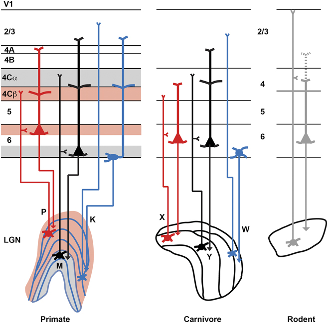
CG neurons make up a fraction of the total population of excitatory neurons in layer 6 of V1, on the order of ∼15% in primates and closer to ∼50% in carnivores (Gilbert & Kelly, Reference Gilbert and Kelly1975; Ferster & Lindstrom, Reference Ferster and Lindstrom1985; Fitzpatrick et al., Reference Fitzpatrick, Usrey, Schofield and Einstein1994). Within the CG neuronal population, there is also diversity in local dendritic morphologies, axonal projection patterns, and stratification of cell bodies within layer 6, described in detail below. Some organizational motifs appear to be conserved across mammals. Figure 1 illustrates schematic representations of CG subtypes in primates, carnivores, and rodents, including local dendritic morphologies, putative axonal projection patterns, and stratification of cell bodies within layer 6.
Schematic representations of corticogeniculate (CG) subtypes in primates, carnivores, and rodents. Parvocellular (P), magnocellular (M), and koniocellular (K) and homologous X, Y, and W stream neurons are illustrated in red, black, and blue, respectively. Axons are thinner and dendrites thicker. Layers within V1 are indicated to the left for primate V1 and between the laminar boundaries for carnivore and rodent V1. LGN relay axons project to layers 1 and 4 in rodents (thin gray lines). Dashed gray dendrite to layer 2/3 illustrates possible, but yet unconfirmed, arborization of rodent CG neurons.

Morphology and local circuit connections
CG neurons fall into three broad morphological categories: short, tall, and stellate or unusual morphology (Briggs, Reference Briggs2010). Short CG neurons are the most abundant morphological type with cell bodies in the upper tier of layer 6 and apical dendrites targeting layer 4. Tall CG neurons have cell bodies in either the top or bottom tier of layer 6 with apical dendrites extending into layer 2/3, sometimes including apical dendritic tufts. Less abundant and more unusual CG neuronal types include spiny stellate neurons that lack apical dendrites and tilted, sideways, or upside-down pyramidal-like neurons that have been observed in upper and lower tiers of layer 6 and also in the white matter below the cortical layers.
A recent anatomical study performed in macaque monkeys (Briggs et al., Reference Briggs, Kiley, Callaway and Usrey2016) has expanded our understanding of the morphological diversity of CG neurons, including the presence of rare and previously unobserved CG types. In the macaque, CG cell bodies are restricted to the top and bottom tiers of layer 6 (Fitzpatrick et al., Reference Fitzpatrick, Usrey, Schofield and Einstein1994). CG neurons with cell bodies in the upper tier include Iβ neurons (Wiser & Callaway, Reference Wiser and Callaway1996), neurons with large cell bodies, and spiny stellate neurons. Following the general pattern for short CG neurons, Iβ neurons have apical dendrites that terminate in layer 4, where they preferentially target parvocellular stream input layer 4Cβ; and the basal dendrites of Iβ neurons are restricted to the upper tier of layer 6, which is the parvocellular stream input zone within layer 6 (Hubel & Wiesel, Reference Hubel and Wiesel1972; Hendrickson et al., Reference Hendrickson, Wilson and Ogren1978). Large neurons have tall apical dendrites with apical tufts near the pia in layer 1. Their basal dendritic arbor is more extensive than that of Iβ neurons, with basal dendrites targeting all three tiers of layer 6. Stellate CG neurons have dendrites restricted to the upper tier of layer 6.
Briggs et al. (Reference Briggs, Kiley, Callaway and Usrey2016) identified two distinct CG morphological subtypes with cell bodies in the bottom tier of layer 6: IC neurons (Wiser & Callaway, Reference Wiser and Callaway1996) and tilted neurons. IC neurons follow the general pattern for tall CG neurons. Interestingly, the apical dendrites of IC neurons show no preference for the parvocellular or magnocellular stream input sublamina of layer 4C, however the basal dendrites of IC neurons are restricted to the magnocellular stream input zone at the bottom of layer 6 (Hubel & Wiesel, Reference Hubel and Wiesel1972; Hendrickson et al., Reference Hendrickson, Wilson and Ogren1978). Tilted neurons had not been observed previously in monkey V1, however neurons with similar morphologies have been observed in rodent and cat visual cortex (Tombol, Reference Tombol, Peters and Jones1984; Miller, Reference Miller1988). Tilted CG neurons are observed less frequently and have unique morphology. First, their oval-shaped cell bodies are restricted to a narrow band in the deepest part of layer 6, often encroaching into the white matter. Second, the apical dendrites of tilted neurons exit at the side of the cell body (rather than the top of the cell body, as for pyramidal neurons) and turn to continue up through the cortical layers toward the pia. The dendritic and axonal analyses performed by Briggs et al. (Reference Briggs, Kiley, Callaway and Usrey2016) suggest that CG neurons form local connections with neurons in the same tier within layer 6 and receive segregated parvocellular or magnocellular stream input within those same tiers.
While the majority of CG neurons are in V1, CG neurons have also been identified in V2 of primates (Lin & Kaas, Reference Lin and Kaas1977; Briggs et al., Reference Briggs, Kiley, Callaway and Usrey2016) and the homologous area 18 in cats (Gilbert & Kelly, Reference Gilbert and Kelly1975; Updyke, Reference Updyke1975; Murphy et al., Reference Murphy, Duckett and Sillito2000). V2 CG neurons probably made up a smaller fraction of excitatory neurons in layer 6 compared to the fraction of CG neurons in V1. V2 CG neurons also include three morphologically distinct subtypes with similar morphological features as V1 CGs. Short V2 CGs are located in the upper tier of layer 6 while tall and tilted V2 CGs are located in the lower tier (Briggs et al., Reference Briggs, Kiley, Callaway and Usrey2016).
The results of most retrograde labeling studies in primates and insectivores support the notion that distinct CG subtypes are segregated into upper and lower tiers of layer 6 (Conley & Raczkowski, Reference Conley and Raczkowski1990; Fitzpatrick et al., Reference Fitzpatrick, Usrey, Schofield and Einstein1994; Usrey & Fitzpatrick, Reference Usrey and Fitzpatrick1996). However detailed data on CG dendritic morphology is only available from a limited number of primate studies. Ichida et al. (Reference Ichida, Mavity-Hudson and Casagrande2014) performed a morphological analysis of retrogradely labeled CG neurons in galagos and observed only short dendritic morphology patterns, however technical limitations may have led to an under-sampling of superficial arbors. Still, it is possible that some species have more homogeneous CG neuronal populations than others.
CG neurons in cats follow many of the same organizational principals observed in primates. Cat CG neurons are pyramidal neurons with cell bodies restricted to layer 6 and the CG population does not overlap with other corticothalamic projecting neuronal populations (Gilbert & Kelly, Reference Gilbert and Kelly1975; LeVay & Sherk, Reference LeVay and Sherk1981; Katz, Reference Katz1987). Katz (Reference Katz1987) observed both short and tall CG neurons (called type II and type I, respectively) in cat V1. Interestingly, the cell bodies of short and tall CGs in cats do not appear to segregate into different tiers of layer 6 (Katz, Reference Katz1987), suggesting that sub-laminar organization of CG subtypes may be a primate specialization.
Compared to the wealth of anatomical data available about CG neurons in primates and carnivores, relatively little is known about CG morphology in rodents. Similar to other mammals, rat and mouse CG neurons have cell bodies restricted to layer 6 in V1, however CG dendritic morphology may be more homogeneous in rodents (Jiang et al., Reference Jiang, Johnson and Burkhalter1993; Brumberg et al., Reference Brumberg, Hamzei-Sichani and Yuste2003). Following the recent introduction of the Ntsr1-cre mouse line (Gong et al., Reference Gong, Doughty, Harbaugh, Cummins, Hatten, Heintz and Gerfen2007), which enables genetic targeting of corticothalamic neurons, more focus has been placed on the CG circuit in the mouse. Olsen et al. (Reference Olsen, Bortone, Adesnik and Scanziani2012) characterized the morphology of Ntsr1 neurons in mouse V1, observing both short and tall pyramidal neurons. It is important to note that while the Ntsr1 population includes CG neurons (Kim et al., Reference Kim, Matney, Blankenship, Hestrin and Brown2014; Denman & Contreras, Reference Denman and Contreras2015), it also includes additional corticothalamic populations both in layer 5 and layer 6 of the cortex (Olsen et al., Reference Olsen, Bortone, Adesnik and Scanziani2012). One notable difference between CG neurons in mice versus primates and carnivores involves their local connectivity. While CG neurons in primates and cats are strongly reciprocally connected with neurons in layer 4, Ntsr1 neurons in mice are more strongly connected to layer 5 and mediate inhibition across the cortical layers (Katz, Reference Katz1987; Briggs & Callaway, Reference Briggs and Callaway2001; Olsen et al., Reference Olsen, Bortone, Adesnik and Scanziani2012; Bortone et al., Reference Bortone, Olsen and Scanziani2014; Kim et al., Reference Kim, Matney, Blankenship, Hestrin and Brown2014; Briggs et al., Reference Briggs, Kiley, Callaway and Usrey2016). Further characterization of CG neurons in rodents is necessary before detailed comparisons can be made between rodent and primate or carnivore CG neurons. It is important to note that CG neurons in both V1 and V2 project local axons throughout the cortical depth, including the superficial layers and layer 5, both of which contain cortico-cortical projecting neurons. CG neurons are therefore positioned to influence not only visual signals relayed back to the LGN but also feedforward signals transmitted to extrastriate visual cortex.
Axon projections
By definition, CG neurons target the LGN with their axonal projections. Importantly, CG synapses onto LGN relay neurons far outnumber retinogeniculate synapses even though their influence appears to be more modulatory than driving (Guillery, Reference Guillery1969; Erisir et al., Reference Erisir, Van Horn, Bickford and Sherman1997a ; Erisir et al., Reference Erisir, Van Horn and Sherman1997b ; Sherman & Guillery, Reference Sherman and Guillery2006). Given the diversity of morphologically distinct CG subtypes across a variety of mammalian species, it seems reasonable to predict diverse CG axonal projection patterns within the LGN. While CG axonal projection data are largely consistent with the notion of diverse CG subtypes, it is technically challenging to confirm the actual specificity of CG-to-LGN connectivity. Interestingly, in primates, cats, and ferrets, CG axons target the LGN and TRN (Robson, Reference Robson1983; Claps & Casagrande, Reference Claps and Casagrande1990; Murphy & Sillito, Reference Murphy and Sillito1996; Ichida & Casagrande, Reference Ichida and Casagrande2002; Ichida et al., Reference Ichida, Mavity-Hudson and Casagrande2014), while in rodents, some CG neurons have axons in the LGN, TRN, and/or the lateral posterior nucleus of the thalamus (Bourassa & Deschenes, Reference Bourassa and Deschenes1995; Olsen et al., Reference Olsen, Bortone, Adesnik and Scanziani2012), suggesting that CG projections in rodents may be less selective or specific than those in more visual mammals. In ferrets and cats, CG axons tend to target multiple LGN layers even though boutons are often clustered (Robson, Reference Robson1983; Claps & Casagrande, Reference Claps and Casagrande1990). In primates, CG axons are mainly restricted to individual LGN layers or to functionally-matched layers (e.g., pairs parvocellular layers or pairs of magnocellular layers) although CG axonal arbors often encroach into neighboring koniocellular layers (Ichida & Casagrande, Reference Ichida and Casagrande2002; Ichida et al., Reference Ichida, Mavity-Hudson and Casagrande2014). Thus at least in visual mammals such as primates and carnivores, CG axon termination patterns are consistent with distinct CG subtypes targeting select populations of LGN neurons. Whether or not CG axons make functionally stream-specific connections with LGN neurons has not yet been established.
The structure and physiological properties of CG synapses onto LGN relay neurons, LGN interneurons, and TRN neurons have been studied in a number of species, including primates, carnivores, and rodents. For thorough reviews of the ultrastructure and physiology of CG synapses across species, please refer to (Bickford, Reference Bickford2016) and to (Sherman & Guillery, Reference Sherman and Guillery2006), chapters 3 and 5. The general characteristics of CG synapses appear to be largely conserved across species. While CG synapses outnumber retinal synapses, they display the characteristics of “modulating” rather than “driving” synapses. CG synapses are smaller, contain less synaptic vesicles, and are located further out along distal dendrites of LGN relay neurons compared to retinal inputs. Postsynaptic terminals of CG synapses include both ionotropic and metabotropic glutamate receptors, while postsynaptic terminals of retinal synapses contain only ionotropic glutamate receptors. The fact that these general structural characteristics are conserved across species suggests that the relative contributions of retinal and cortical inputs to LGN relay neurons are also evolutionarily conserved.
Physiology of CG neurons
In visual mammals such as primates and carnivores, there are precise relationships between CG axon conduction latency and visual physiology. Here visual physiology is used to describe the receptive field properties of neurons. One of the most common visual physiological metrics is the classification of V1 neurons as Simple or Complex. Simple cells in V1 have separate receptive field subregions that respond to opposite luminance polarity, while Complex cells respond equally well to stimuli of opposite luminance polarity within their receptive fields. The distinct CG subtypes defined by axon conduction latencies and visual response properties can be directly related to the feedforward parallel processing streams that are a hallmark of visual systems in highly visual mammals. Similar information is not available in rodents, however CG physiology has been well characterized in rabbits so comparisons between primates, carnivores, and rabbits are described below.
Axon conduction latencies
Measurements of CG axon conduction latencies provide some of the strongest, and oldest, evidence in favor of distinct CG subtypes. Antidromic stimulation studies, where CG axons in the LGN are electrically stimulated and resultant action potentials are recorded at cell bodies in layer 6 of V1, provide valuable physiological information about CG axon conduction speed. In visual mammals such as ferrets, cats, and monkeys, axon conduction latencies correlate with Simple/Complex visual response properties whereby fast-conducting CG neurons are Complex, medium-conducting CG neurons are Simple, and slowly-conducting CG neurons are Complex or nonresponsive to visual stimuli (Harvey, Reference Harvey1978; Tsumoto & Suda, Reference Tsumoto and Suda1980; Grieve & Sillito, Reference Grieve and Sillito1995; Briggs & Usrey, Reference Briggs and Usrey2005, Reference Briggs and Usrey2007b ). In rabbits, CG axon conduction latencies are generally slower, but similar relationships exist between axon conduction speed and visual response properties (Swadlow & Weyand, Reference Swadlow and Weyand1987). In all of these species studied to date, it is clear that CG neurons with different axon conduction latencies form distinct and nonoverlapping cell types. For example, in the monkey, CG subtypes defined based on axon conduction latency are clear-cut: fast/Complex CGs always have conduction latencies <7 ms; medium/Simple CGs always have conduction latencies between 7–15 ms; and slow/Complex CGs always have conduction latencies >15 ms (Briggs & Usrey, Reference Briggs and Usrey2007b , Reference Briggs and Usrey2009). Given the diversity in CG conduction latencies that is strictly defined and conserved across many species, it is likely that CG subtypes with different axon conduction speeds contribute to different aspects of visual processing. Along these lines, only fast-conducting CG neurons in primates also receive suprathreshold feedforward input from the LGN, creating a fast, reciprocal loop for transferring visual information between LGN and V1 (Briggs & Usrey, Reference Briggs and Usrey2007b ). Accordingly, these fast-conducting CG neurons may contribute primarily to motion perception. The diversity in axon conduction speeds also suggests that CG circuits operate on a variety of timescales, perhaps to accommodate a variety of integration windows for accumulating visual information. As we learn more about the functional contributions of CG circuits to vision, it will be important to consider the variety of timescales over which CG circuits operate.
Receptive field properties
In carnivores, primates, and rabbits, the visual response properties of CG neurons strongly correlate with axon conduction latencies, as described above. For example, CG neurons with fast and medium conduction latencies are often tuned for orientation and direction in cats and rabbits (Tsumoto & Suda, Reference Tsumoto and Suda1980; Swadlow & Weyand, Reference Swadlow and Weyand1987; Grieve & Sillito, Reference Grieve and Sillito1995). A more systematic study of the visual response properties of CG neurons in alert monkeys revealed that CG subtypes defined based on axon conduction latencies display unique receptive field properties that align with the feedforward magnocellular, parvocellular, and koniocellular processing streams (Briggs & Usrey, Reference Briggs and Usrey2009). Fast-conducting Complex CGs are sensitive to low contrast and high temporal frequency stimuli, and thus optimized to process motion signals conveyed by the magnocellular stream. Medium-conducting Simple CGs are less sensitive to contrast, but are orientation tuned and responsive to red and green color signals, in line with signals conveyed by the parvocellular stream. Slow-conducting Complex CGs are poorly tuned for orientation and direction but modulated by blue color signals, aligning with signals conveyed by the koniocellular stream. These data suggest that inputs from the magnocellular, parvocellular, and koniocellular parallel processing streams are segregated through the local circuitry in V1 such that CG feedback maintains stream-specificity.
There is ongoing debate about whether some of the slowest-conducting CG neurons are visually responsive. In ferrets, cats, and rabbits, some slow-conducting CG neurons are not responsive to visual stimuli (Tsumoto & Suda, Reference Tsumoto and Suda1980; Swadlow & Weyand, Reference Swadlow and Weyand1987; Briggs & Usrey, Reference Briggs and Usrey2005). Lack of responsiveness cannot necessarily be attributed to anesthesia as visually nonresponsive CG neurons are observed in awake rabbits (Swadlow & Weyand, Reference Swadlow and Weyand1987). Interestingly, in alert monkeys, all CG neurons, including slow-conducting CG neurons, are visually responsive (Briggs & Usrey, Reference Briggs and Usrey2009). The utility of the slowest-conducting CG neurons and whether or not they are responsive to visual stimuli in a variety of species remain unknown.
Stream specificity
A major hallmark of retino-geniculo-cortical circuits in visual mammals is the separation of feedforward signals into parallel processing streams made up of morphologically and physiologically distinct neuronal classes. In primates, the parvocellular, magnocellular, and koniocellular parallel processing streams carry information about form/acuity, motion, and blue color, respectively (Kaplan, Reference Kaplan, Chalupa and Werner2004). Homologous X, Y, and W streams are present in carnivores (Sherman & Guillery, Reference Sherman and Guillery2006). Evidence for parallel streams in the visual systems of rodents is less clear (Krahe et al., Reference Krahe, El-Danaf, Dilger, Henderson and Guido2011). Mounting evidence supports the notion that in visual mammals such as primates and carnivores, CG circuits are also organized into parallel streams that align with the feedforward parallel streams. The dendritic arborization and axon projection patterns of primate and carnivore CG neurons, described in detail above, support distinct CG subtypes. Similarly, physiological measurements including axon conduction latency and visual response properties also support distinct CG subtypes with physiological properties aligned with the properties of neurons in the feedforward streams. Together, these data provide strong support for the idea that separate channels of CG feedback preserve the segregation of visual information into distinct feedforward processing streams in highly visual mammals.
In the primate, the alignment of feedforward and CG feedback circuits is precise. The following predictions arise from combinations of morphological and physiological observations in monkeys. CG neurons with fast axon conduction latencies and Complex visual physiology are the presumptive magnocellular-projecting CG neurons. Magnocellular feedforward retino-geniculo-cortical and fast Complex CG neurons have the fastest conducting axons, the greatest contrast and temporal frequency sensitivity, the most surround suppression, the shortest visual response latencies, and the highest firing rates (Benardete et al., Reference Benardete, Kaplan and Knight1932; Schiller & Malpeli, Reference Schiller and Malpeli1978; Kaplan & Shapley, Reference Kaplan and Shapley1982, Reference Kaplan and Shapley1986; Maunsell et al., Reference Maunsell, Ghose, Assad, McAdams, Boudreau and Noerager1999; Usrey & Reid, Reference Usrey and Reid2000; White et al., Reference White, Solomon and Martin2001; Solomon et al., Reference Solomon, White and Martin2002; Briggs & Usrey, Reference Briggs and Usrey2007b , Reference Briggs and Usrey2009, Reference Briggs and Usrey2011b ; Alitto & Usrey, Reference Alitto and Usrey2008). Additionally, fast Complex CG neurons have sharper orientation tuning and greater direction selectivity, reminiscent of neurons in layer 4Cα that receive direct magnocellular input from the LGN (Hawken et al., Reference Hawken, Parker and Lund1988; Merigan & Maunsell, Reference Merigan and Maunsell1993; Gur et al., Reference Gur, Kagan and Snodderly2005; Briggs & Usrey, Reference Briggs and Usrey2009). The presumptive magnocellular-projecting fast Complex CG neurons are also the only CG neurons to receive direct, feedforward suprathreshold LGN input (Briggs & Usrey, Reference Briggs and Usrey2007b ). It is likely that the type IC CG neurons, located in the bottom tier of layer 6, are the magnocellular-projecting, fast Complex CG neurons because magnocellular LGN layers receive CG feedback from the bottom tier of layer 6 (Fitzpatrick et al., Reference Fitzpatrick, Usrey, Schofield and Einstein1994).
CG neurons with medium axon conduction latencies and Simple visual physiology are the presumptive parvocellular-projecting CG neurons. Parvocellular feedforward retino-geniculo-cortical and medium Simple CG neurons have intermediary axon conduction speeds, low contrast and temporal frequency sensitivity, less surround suppression, slower visual response latencies, intermediary firing rates, and are sensitive to inputs from the L- and M-cones in the retina (Schiller & Malpeli, Reference Schiller and Malpeli1978; Bullier & Henry, Reference Bullier and Henry1980; Maunsell et al., Reference Maunsell, Ghose, Assad, McAdams, Boudreau and Noerager1999; Field & Chichilnisky, Reference Field and Chichilnisky2007; Briggs & Usrey, Reference Briggs and Usrey2009). The type Iβ CG neurons, located in the top tier of layer 6 are likely the parvocellular-projecting, medium Simple CG neurons because parvocellular LGN layers receive CG feedback from the top tier of layer 6 (Fitzpatrick et al., Reference Fitzpatrick, Usrey, Schofield and Einstein1994).
CG neurons with the slowest conducting axons and Complex visual physiology likely project to the koniocellular LGN layers. Like the feedforward koniocellular retino-geniculo-cortical neurons, slow Complex CG neurons have slower axon conduction speeds, intermediate contrast, and temporal frequency sensitivity, some surround suppression, and sensitivity to inputs from the S-cones in the retina (Irvin et al., Reference Irvin, Norton, Sesma and Casagrande1986; White et al., Reference White, Wilder, Goodchild, Sefton and Martin1998; Solomon et al., Reference Solomon, White and Martin1999; Hendry & Reid, Reference Hendry and Reid2000; White et al., Reference White, Solomon and Martin2001; Solomon & Lennie, Reference Solomon and Lennie2007; Briggs & Usrey, Reference Briggs and Usrey2009, Reference Briggs and Usrey2011b ). Interestingly, slow Complex CGs are not orientation or direction tuned, unlike the other CG subtypes (Briggs & Usrey, Reference Briggs and Usrey2009). It is likely that a number of morphologically distinct CG subtypes, including stellate, large, and tilted CG neurons, project to the various koniocellular layers in the LGN. However, the tilted CG neurons are presumably the slow Complex, koniocellular-projecting neurons encountered physiologically because these neurons were always located in the deepest part of layer 6 (Briggs & Usrey, Reference Briggs and Usrey2009).
In primates, and possibly also in carnivores, CG neurons retain physiological signatures of the feedforward parallel processing streams in spite of the fact that V1 contains numerous local circuits that could mix information from the three input streams (Merigan & Maunsell, Reference Merigan and Maunsell1993; Douglas & Martin, Reference Douglas and Martin2004; Sincich & Horton, Reference Sincich and Horton2005; Callaway, Reference Callaway, Werner and Chalupa2014). Accordingly, a clear precedent is set to ensure that CG circuits preserve the specificity of information relayed by feedforward streams. Preservation of segregated information channels through both feedforward and feedback circuits is evident in other species and sensory systems including the whisker/barrel system of rodents and the auditory system of bats (Ghazanfar et al., Reference Ghazanfar, Krupa and Nicolelis2001; Suga & Ma, Reference Suga and Ma2003; Bokor et al., Reference Bokor, Acsady and Deschenes2008). Stream-specific corticothalamic feedback may therefore be evolutionarily conserved for neuronal pathways that serve primary senses across a variety of species. Because stream-specificity is likely to be a critical organizational feature of CG feedback, CG function must be considered in the context of parallel processing streams.
Function of CG neurons
In the past 10 years, we have gained significant insight about the structure, organization, and visual physiology of CG neurons, especially in the primate visual system. However the functional role of CG feedback in vision has remained a particularly stubborn puzzle. It is obvious from LGN recordings that the influence of CG feedback on the visual responses of LGN neurons is subtle. The receptive fields of LGN neurons mimic their retinal inputs and not their cortical inputs. For example, individual LGN neurons are not orientation tuned even though many CG neurons are tuned for orientation. Although CG inputs onto LGN neurons are anatomically robust in terms of numbers of synapses, the smaller size and placement of CG synapses on distal dendrites of LGN relay neurons means CG influence is modulatory rather than driving (Sherman & Guillery, Reference Sherman and Guillery1998, Reference Sherman and Guillery2006). Until recently, it has also been technically challenging to manipulate CG neurons in a selective and reversible manner. With the advent of optogenetics (Huber et al., Reference Huber, Petreanu, Ghitani, Ranade, Hromadka, Mainen and Svoboda2008; Gradinaru et al., Reference Gradinaru, Mogri, Thompson, Henderson and Deisseroth2009), it is now possible to perform a more causal test of CG function by performing reversible manipulations selectively on CG neurons in intact animals. Results of experimental examinations of CG function, varying in invasiveness and/or selectivity, converge on three general candidate functions for CG feedback: LGN gain modulation, sharpening or shifting of LGN receptive fields, and changes in LGN spiking mode, timing, and precision (Briggs & Usrey, Reference Briggs and Usrey2008, Reference Briggs and Usrey2011a ).
Gain modulation
As the first feedback circuit in the visual system, CG neurons could function as filters that increase the salience of relevant visual signals and suppress the activity of non-relevant visual information. Accordingly, CG feedback could operate by increasing or decreasing the gain of LGN neuronal responses. Changes in both the magnitude and gain of LGN responses have been observed when CG feedback is manipulated (Molotchnikoff et al., Reference Molotchnikoff, Tremblay and Lepore1984; Gulyas et al., Reference Gulyas, Lagae, Eysel and Orban1990; Marrocco et al., Reference Marrocco, McClurkin and Alkire1996; Cudiero et al., Reference Cudiero, Rivadulla and Grieve2000; Przybyszewski et al., Reference Przybyszewski, Gaska, Foote and Pollen2000; Worgotter et al., Reference Worgotter, Eyding, Macklis and Funke2002; Wolfart et al., Reference Wolfart, Debay, Le Masson, Destexhe and Bal2005; Andolina et al., Reference Andolina, Jones, Wang and Sillito2007; Li et al., Reference Li, Ye, Song, Yang and Zhou2011; Olsen et al., Reference Olsen, Bortone, Adesnik and Scanziani2012; Denman & Contreras, Reference Denman and Contreras2015). Specifically, aspirating or cooling V1 in anesthetized cats and primates led to decreased LGN responses to moving visual stimuli without changing LGN tuning preferences (Gulyas et al., Reference Gulyas, Lagae, Eysel and Orban1990; Marrocco et al., Reference Marrocco, McClurkin and Alkire1996). Additionally, cooling V1 in anesthetized monkeys led to a reduction in contrast gain that was specific to parvocellular LGN neurons (Przybyszewski et al., Reference Przybyszewski, Gaska, Foote and Pollen2000). Similarly, lesioning V1 in anesthetized cats led to decreased contrast adaptation with greater effects on Y than X cells in the LGN (Li et al., Reference Li, Ye, Song, Yang and Zhou2011). These latter findings support the notion of stream-specific functions for CG subtypes. Interestingly, in the mouse, optogenetic suppression of corticothalamic feedback led to a variety of effects on LGN responses including increases, decreases, and no change in response magnitude and gain (Denman & Contreras, Reference Denman and Contreras2015).
A related function for CG feedback could be to convey cognitive signals about attention or arousal state from the cortex to the LGN. Corticothalamic circuits are strongly modulated by arousal state, demonstrating different spiking, and oscillatory modes during sleep and arousal (Steriade, Reference Steriade2001, Reference Steriade2003). Additionally, LGN neurons are modulated by visual attention (Vanduffel et al., Reference Vanduffel, Tootell and Orban2000; O’Connor et al., Reference O’Connor, Fukui, Pinsk and Kastner2002; McAlonan et al., Reference McAlonan, Cavanaugh and Wurtz2006, Reference McAlonan, Cavanaugh and Wurtz2008; Ling et al., Reference Ling, Pratte and Tong2015). The precise role of CG feedback in delivering arousal and/or attention signals to the LGN is not known. However, as the only path by which cortical information reaches the LGN, CG feedback is a good candidate for conveying information about cognitive state.
Receptive field changes
LGN neurons derive their receptive field properties from their retinal inputs, however it is possible that CG feedback sharpens and/or shifts the tuning properties of individual or ensembles of LGN neurons. There is evidence that the spatial receptive field of LGN neurons, with the antagonistic center/surround of the classical receptive field and the extra-classical suppressive surround, may be sharpened or modulated by CG feedback (Murphy & Sillito, Reference Murphy and Sillito1987; Sillito & Jones, Reference Sillito and Jones2002; Webb et al., Reference Webb, Tinsley, Barraclough, Easton, Parker and Derrington2002; Jones et al., Reference Jones, Andolina, Ahmed, Shipp, Clements, Grieve, Cudiero, Salt and Sillito2012; Andolina et al., Reference Andolina, Jones and Sillito2013). CG feedback could sharpen the border between the classical and extra-classical fields (Murphy & Sillito, Reference Murphy and Sillito1987; Jones et al., Reference Jones, Andolina, Oakely, Murphy and Sillito2000; Rivadulla et al., Reference Rivadulla, Martinez, Varela and Cudeiro2002; Webb et al., Reference Webb, Tinsley, Barraclough, Easton, Parker and Derrington2002; Jones et al., Reference Jones, Andolina, Ahmed, Shipp, Clements, Grieve, Cudiero, Salt and Sillito2012). Alternatively, CG feedback could reduce the spatial extent of the classical receptive field and enhance surround suppression as pharmacological inactivation of V1 in anesthetized cats led to an increase in LGN receptive size and a reduction in surround suppression (Andolina et al., Reference Andolina, Jones and Sillito2013). In the mouse, corticothalamic feedback does not modulate LGN receptive field size or surround suppression (Denman & Contreras, Reference Denman and Contreras2015).
While the influence of CG feedback on the receptive fields of individual LGN neurons may be subtle, it is possible that CG feedback coordinates the activity of ensembles of LGN neurons to produce “tuning” among small groups of LGN neurons. Experimental evidence suggests that CG feedback enhances the activity of ensembles of LGN neurons with receptive fields clustered along the orientation axis of their CG inputs (Sillito et al., Reference Sillito, Cudeiro and Murphy1993; Wang et al., Reference Wang, Jones, Andolina, Salt and Sillito2006; Andolina et al., Reference Andolina, Jones, Wang and Sillito2007). Theoretical evidence also supports a role for CG feedback in enhancing the activity of ensembles of LGN neurons with retinotopically aligned receptive fields and suppressing neighboring neurons (Bickle et al., Reference Bickle, Bernstein, Heatley, Worley and Stiehl1999). Together, these findings highlight the importance of future studies involving multielectrode recordings so that the broader impact of CG feedback on populations of LGN neurons can be examined.
A final argument in favor of a role for CG feedback in sharpening or shifting the receptive fields of LGN neurons is that similar functions have been attributed to corticothalamic circuits in other sensory systems. The term “egocentric selection” describes the enhancement and shifting of thalamic responses to match the tuning preferences of activated corticothalamic neurons and suppression of activity among thalamic neurons with nonmatched tuning (Suga & Ma, Reference Suga and Ma2003). Egocentric selection by corticothalamic feedback has been described in the auditory and somatosensory systems (Krupa et al., Reference Krupa, Ghazanfar and Nicolelis1999; Ghazanfar et al., Reference Ghazanfar, Krupa and Nicolelis2001; Suga & Ma, Reference Suga and Ma2003; Li & Ebner, Reference Li and Ebner2007; Wu & Yan, Reference Wu and Yan2007; Temereanca et al., Reference Temereanca, Brown and Simons2008; Zhang & Yan, Reference Zhang and Yan2008).
Modulation of LGN spiking mode, timing, and precision
In addition to modulating the gain of LGN response or sharpening LGN receptive fields, CG feedback could influence the feedforward transmission of visual signals by modulating the spike timing of LGN relay neurons. By coordinating the spike latencies and/or increasing the spike timing precision for an ensemble of LGN neurons, CG feedback could enhance the efficacy of visual information conveyed by the coordinated ensemble to V1. One way in which CG feedback could coordinate the activity of LGN neurons is to shift a select group of LGN neurons between burst and tonic modes of firing (Sherman, Reference Sherman1996). In support of this notion is the finding that selective destruction of CG neurons led to a broadening of the inter-spike-interval distribution of tonic spikes recorded during synchronized EEG states (Eyding et al., Reference Eyding, Macklis, Neubacher, Funke and Worgotter2003). However, in the mouse, selectively manipulating corticothalamic feedback had no net impact on tonic or burst spiking (Denman & Contreras, Reference Denman and Contreras2015).
Alternatively, CG feedback could coordinate slow timescale fluctuations in membrane potential, leading to an increase in coordinated spiking patterns among LGN ensembles (Destexhe et al., Reference Destexhe, Contreras and Steriade1999; Bal et al., Reference Bal, Debay and Destexhe2000; Blumenfeld & McCormick, Reference Blumenfeld and McCormick2000; Destexhe, Reference Destexhe2000; Rigas & Castro-Alamancos, Reference Rigas and Castro-Alamancos2007). Both experimental and theoretical studies support the notion that CG feedback alters the timing of LGN responses in order to improve the transfer of information from the LGN to V1 (Funke et al., Reference Funke, Nelle, Li and Worgotter1996; Bal et al., Reference Bal, Debay and Destexhe2000; Destexhe, Reference Destexhe2000; Worgotter et al., Reference Worgotter, Eyding, Macklis and Funke2002; Wolfart et al., Reference Wolfart, Debay, Le Masson, Destexhe and Bal2005; Andolina et al., Reference Andolina, Jones, Wang and Sillito2007; Yousif & Denham, Reference Yousif and Denham2007). Specifically, Andolina and colleagues (Andolina et al., Reference Andolina, Jones, Wang and Sillito2007) observed decreased stimulus modulation and increased response variability in LGN neurons when V1 was pharmacologically inactivated in anesthetized cats. In spite of these advances, exactly how CG feedback alters the timing and/or precision of LGN neurons to improve information transmission remains unclear.
There is some experimental evidence that CG feedback synchronizes the activity of LGN ensembles (Sillito et al., Reference Sillito, Jones, Gerstein and West1994), however the synchrony measurements in this study were confounded (Brody, Reference Brody1999) and the manipulations of CG feedback were nonselective with limited data. Nevertheless, the hypothesis proposed in this study, that CG feedback selectively synchronizes the activity of LGN ensembles carrying feature-specific information, must be tested using modern techniques. Interestingly, somewhat parallel analyses performed in the mouse, in which pairwise correlations were measured among LGN neurons when corticothalamic feedback was manipulated, yielded no influence of corticothalamic feedback on correlated activity among LGN neurons (Denman & Contreras, Reference Denman and Contreras2015).
Finally, CG feedback could play a role in the arrangement of synapses onto LGN relay neurons during development—a function that could have important consequences for LGN spiking synchrony and precision throughout life. A number of recent studies examining the development of visual thalamic circuits in the mouse have provided significant new insight into the timing and influence of CG activity during development. CG axonal innervation of the LGN is delayed relative to innervation of the LGN by retinal inputs (Jacobs et al., Reference Jacobs, Campagnoni, Kampf, Reyes, Kalra, Handley, Xie, Hong-Hu, Spreur, Fisher and Campagnoni2007; Seabrook et al., Reference Seabrook, El-Danaf, Krahe, Fox and Guido2013). Additionally, CG inputs become stronger during later developmental stages relative to retinal inputs (Jurgens et al., Reference Jurgens, Bell, McQuiston and Guido2012). However, CG inputs are required for correct retinogeniculate innervation of the LGN as retinogeniculate afferents fail to correctly innervate the LGN in mice lacking layer 6 projection neurons (Shanks et al., Reference Shanks, Ito, Schaevitz, Yamada, Chen, Litke and Feldheim2016). Furthermore, disruptions of CG feedback during development result in over-abundant retinogeniculate innervation of LGN relay neurons, similar to that observed during dark rearing (Thompson et al., Reference Thompson, Picard, Min, Fagiolini and Chen2016). Together these findings suggest that CG activity during specific developmental stages is required for correct retinal and cortical synaptic innervation of LGN neurons, perhaps setting the stage for LGN neuronal synchronization that is critical for visual function throughout life.
Concluding remarks
While a large body of work is now available describing the structure, organization, and physiology of CG neurons, the functional role of these enigmatic neurons in vision remains unresolved. A number of other important questions also remain, including the functional connectivity of CG axons with LGN relay neurons and whether or not CG feedback is functionally stream specific. Any examination of CG function must take into account the fact that CG subtypes operate on very different timescales and are specialized to convey stream-specific signals from V1 to the LGN. Additionally, it will be important to record from ensembles of neurons in the LGN to determine whether CG feedback alters the timing or population tuning properties of select groups of LGN neurons. Finally, as technical approaches improve, studies of CG circuits in alert and behaving animals will provide important new insight into the contributions of CG feedback to contextual and cognitive processes.
There are clear similarities in CG morphology and physiology across highly visual mammals such as primates and carnivores. These similarities suggest that evolution maintains a precedent for preserving the separation of visual information into parallel processing streams throughout the early visual pathways. However, this precedent is not apparent for nonvisual mammals such as rodents, as CG morphology and function appears more homogeneous and less selective. Precise and parallel organization is present in the rodent somatosensory system, however. Further studies of corticothalamic feedback in a variety of sensory modalities and species may provide significant insight into the senses that are must useful for each species and the general strategies employed by corticothalamic circuits to enhance sensory perception.