Significant outcomes
Prenatal stressed male and female mice in the adulthood display significant recognition memory impairments;
Prenatal stressed female mice show recognition memory deficits dependent on estrous cycle;
Male prenatal stressed displayed increased locomotion in an unfamiliar environment.
Limitations
The impact of female estrous cycle on recognition memory would be properly investigated analysing the animal behaviour in all estrous phase;
Evaluation of changes in mouse hippocampal neurons was not performed in this study.
Introduction
Maternal stress during pregnancy entrains long-term behavioural and neurobiological deficits in the offspring. The impact of the prenatal stress (PNS) seems more likely as a result of the release of several stress-related hormones and neurotransmitters which act as signals to “program” the development of the future infant depending on his genetic background and sex (Schroeder et al., Reference Schroeder, Sultany and Weller2013).
In particular, several epidemiological studies have documented that maternal psychological stress – due to bereavement, unwanted pregnancy, death of relatives or other serious life events – during the first or second trimester of pregnancy increases the risk in the offspring of developing severe mental disorders (for a review see: Van den Bergh et al., Reference Van den Bergh, van den Heuvel, Lahti, Braeken, de Rooij, Entringer, Hoyer, Roseboom, Räikkönen, King and Schwab2017), such as schizophrenia (Pugliese et al., Reference Pugliese, Bruni, Carbone, Calabrò, Cerminara, Sampogna, Luciano, Steardo, Fiorillo, Garcia and De Fazio2019), affective disorders (Brannigan et al., Reference Brannigan, Cannon, Tanskanen, Huttunen, Leacy and Clarke2019), bipolar disorder (Pugliese et al., Reference Pugliese, Bruni, Carbone, Calabrò, Cerminara, Sampogna, Luciano, Steardo, Fiorillo, Garcia and De Fazio2019), autism (Varcin et al. Reference Varcin, Alvares, Uljarević and Whitehouse2017) and attention-deficit/hyperactivity disorder (ADHD) (Rosenqvist et al., Reference Rosenqvist, Sjölander, Ystrom, Larsson and Reichborn-Kjennerud2018). A growing number of clinical evidence supports also the role of maternal stress during late gestation, as a risk factor for mental illness in the offspring (Li et al., Reference Li, Yang, Guldin, Vedsted and Vestergaard2015).
To determine the mechanisms underlying the susceptibility to mental disorders in the offspring born from stressed mothers, a myriad of behavioural, biochemical, morphological and genetic studies about the impact of PNS in rodents have been performed. These studies have shown that late PNS induces impairments in cognitive, executive, volition and social functions as well as deficits in brain plasticity and variations in hormonal availability during different neurodevelopmental phases (for review see: Maccari & Morley-Fletcher, Reference Maccari and Morley-Fletcher2007; Bock et al., Reference Bock, Wainstock, Braun and Segal2015; Miranda & Souza, Reference Miranda and Sousa2018). Additionally, altered hypothalamic–pituitary–adrenal (HPA) axis function and secretion of glucocorticoid hormones are found in the offspring of prenatal stressed dams (Owen et al., Reference Owen, Andrews and Matthews2005). PNS influences also the fetal transcriptome through epigenetic mechanisms which include the regulation of microRNAs involved in the altered gene expression patterns linked to the pathophysiology of schizophrenia and bipolar disorder (Hollins & Cairns, Reference Hollins and Cairns2016). The understanding of the underlying mechanisms of vulnerability/resilience to psychopathologies due to maternal PNS is of special concern because it might pave the way for the discovery of important targets for developing biomarkers and innovative therapies for the treatment of psychiatric disorders (Boersma & Tamashiro, Reference Boersma and Tamashiro2014).
Although the effects of PNS have been largely examined (Pallarés & Antonelli, Reference Pallarés and Antonelli2017), few studies have documented how the sex of the offspring might affect the behavioural outcome of maternal stress during pregnancy in an outbred strain of mice. Therefore, this study aimed to investigate the impact of prenatal restraint stress in cognition and exploration to an unfamiliar environment in mouse adult offspring of both sexes.
Material and methods
Ethical issues and animals
This project was developed in accordance with the provisions of Law N° 11 794/2008, and with the regulations issued by the National Council for the Control of Animal Experimentation (CONCEA), and it has been previously approved by the Ethic Committees for Animal Use of Federal University of Rio Grande do Norte (License N° 067/2016). This study has been reported according to the ARRIVE guidelines (Kilkenny et al., Reference Kilkenny, Browne, Cuthill, Emerson and Altman2010).
Nulliparous female and male Swiss mice (Department of Biophysics and Pharmacology of the Federal University of Rio Grande do Norte) of 90 days old were kept in a 12-h light-dark cycle (lights on at 06:00) at constant room temperature (22 ± 2 °C) were used as progenitors for this study. The animals were housed in polypropylene boxes (20 × 34 × 13 cm) with access to feed and water ad libitum. Female mice were kept in boxes with a male for one night, at the ratio of two females for one male. The day after copulation was considered as day 0 of gestation. Pregnant female mice were randomly selected for submission to the PNS protocol (n = 17). Control pregnant females (n = 12) were left undisturbed in their home cages.
Male and female offspring were weaned 21 days after birth. In this study, we used male and female offspring from litters containing an average of 7–10 pups with a comparable number of males and females were used for the experiments. A maximum of two male or female pups were taken from each litter. After weaning, male and female mice from each experimental group (control and prenatal stress) were housed in groups of 5 to 8 (polypropylene boxes 41 × 34 × 16 cm) and maintained under similar environmental conditions until the experiments were started (60 days of age). Pups were briefly removed from the home cage on day 1 after birth (in the morning) and weighed. A total number of 38 male and 36 female 60-day-old mice born from mothers exposed to PNS or control conditions were used.
Prenatal stress
The maternal stress procedure was carried out using the methodology described in previous studies (Dong et al., Reference Dong, Dzitoyeva, Matrisciano, Tueting, Grayson and Guidotti2015). In brief, from day 14 of pregnancy until delivery, pregnant females were subjected to three daily stress sessions of 45 min each during which they were placed in transparent plastic cylinders (diameter = 7 cm; length = 19 cm) and exposed to bright light. The sessions took place during the light cycle at 09:00, 12:00 and 17:00 h. Control pregnant females were left undisturbed in their home cages. Male and female offspring were weaned 21 days after birth, and a maximum of two male and female pups were taken from each litter. After weaning, male and female mice from each experimental group (control and prenatal stress) were housed in groups of five and maintained under similar environmental conditions until experiments were started (60 days of age).
Estrous cycle determination
In order to investigate the influence of the estrous cycle on the female behavioural responses, the estrous cycle phase was determined from vaginal smears taken after each behavioural test. The phases of the estrous cycle were identified using the following criteria: (1) proestrus: predominantly nucleated epithelial cells; (2) estrus: predominantly cornified epithelial cells; (3) metestrus: cornified epithelialcells and leukocytes; (4) diestrus: predominantly leukocytes, some nucleated epithelial cells and mucus (Singletary et al., Reference Singletary, Kirsch, Watson, Karim, Huso, Hurn and Murphy2005). According to their estrous cycle, females were then subdivided into two groups: a first group was composed of female mice in proestrus and estrus (pro/est). These two phases are characterised by high levels of estradiol and progesterone. A second group consisted of female mice in metestrus and diestrus (met/die) phases which are characterised by low levels of estradiol and progesterone. Similar subdivisions have been described previously in a study in which the effects of estrous cycle on memory were investigated (Van Goethem et al., Reference Van Goethem, Rutten, van der Staay, Jans, Akkerman, Steinbusch, Blokland, van’tKlooster and Prickaerts2012). Figure S1 illustrates the percentage of females in each estrous cycle phase observed in the test day of the object recognition task and open field test.
Behavioural tests
Behavioural tests were performed in adult male and female offspring mice in the following sequence: day 1 – open field test (30 min; habituation session of the object recognition task) and day 2 – object recognition task (training and test sessions – 6 h of intertrial interval). In order to avoid pheromone interferences, experiments with male and female mice were conducted in distinct days.
Open field test
The open field test was performed in the same arena (40 × 40 × 40 cm) in which the object recognition task was performed. The analysis took into account the total distance moved (measured in metres) and the time spent by the animal (in seconds) in the central (10 × 10 cm) and peripheral zones of the arena, in order to evaluate anxiety-related behaviours and spontaneous locomotion, respectively. This test was performed under dark condition. Each mouse was placed in the centre of the open field, and the time spent exploring the apparatus during 30 min was video recorded and further analysed with the Anymaze® videotracking software (Stoelting Co., Wood Dale, IL, USA). The following parameters were recorded: total distance moved in and time spent in the central zone (during the first 10 min of exposition to the open field), and distance moved in the arena during 30 min of test. The percentage of central distance moved was calculated by using the following equation: (central distance moved/total distance) × 100. Increase or decrease in the percentage of exploration in the central zone indicates an anxiolytic or anxiogenic-related behaviour, respectively (Seibenhener & Wooten, Reference Seibenhener and Wooten2015).
Object recognition task
The object recognition task was aimed to evaluate the impact of maternal stress on learning and memory of the offspring. The test was conducted in a square acrylic arena, with a black base and black walls (40 × 40 × 40 cm), following the experimental protocol described by Leger et al. (Leger et al., Reference Leger, Quiedeville, Bouet, Haelewyn, Boulouard, Schumann-Bard and Freret2013). All animals were submitted to a habituation session in the arena. During this session, free exploration of the apparatus was registered for 30 min (spontaneous locomotion). Twenty-four hours after habituation, the training session was performed. For this, animals were placed in the centre of the arena that contained two identical objects in shape, size, colour and texture (A1 and A2) positioned at two adjacent vertices and at 10 cm from the walls. The animals explored the open field in the presence of these two objects during 10 min and the time spent exploring each object was manually registered. Six hours later, the animals were introduced again into the arena. However, one of the previous objects was replaced by a new one, in order to expose the animal to two different objects: one familiar (the same as the previous one; A1) and a new one (different in shape and material, but with a similar size; B). Objects to be discriminated were made of plastic (i.e. lego piece) and plastic/glass (i.e. nail polish glass). Exposition to objects (A and B) was counterbalanced during training and test sessions, aiming to avoid inherent object bias for either the familiar or novel object, independent of cognitive performance.
The time spent exploring the objects (in seconds) was manually evaluated when the following behaviours were performed: sniffing or touching the object with the muzzle and/or the front paws. The capacity to discriminate the novel from the familiar object (recognition index, RI) was determined by the following formula: RI = [(time exploring the novel object)/(time exploring both objects)]. RI equal to 0.5 indicates no preference for object A and B. However, in the test session, RI significantly higher than the training session indicates a preference for the novel object. The task was performed under dark condition. An infrared video camera was used for recording the animals’ behaviour. After each mouse, the apparatus was cleaned with 5% ethanol.
Statistical analysis
Data are expressed as mean ± standard error of the mean (SEM) as detailed in the legends. Data were analysed for normality and unpaired Student’s t-test or Mann–Whitney test were used to compare PNS and control groups depending on the normal or not normal distribution of the data. Comparison of the recognition index between training and test sessions was performed using paired t-test. Differences were considered statistically significant when p < 0.05. All statistical analyses were performed using Prism version 6.0 (Graphpad Software Inc, San Diego, USA).
Results
Diary observation of gross maternal behaviour during breast feeding did not reveal any significant alteration (i.e. pups and maternal deaths, cannibalization, and neglected pups in the cage) which could contribute to postnatal effects observed in this study. Additionally, prenatal stressed offspring has significantly lower birth weights on day 1 than control (control mice: 1.755 ± 0.029 g, prenatal stressed mice: 1.681 ± 0.022 g; t(131) = 2.009, p = 0.047; Student’s t-test).
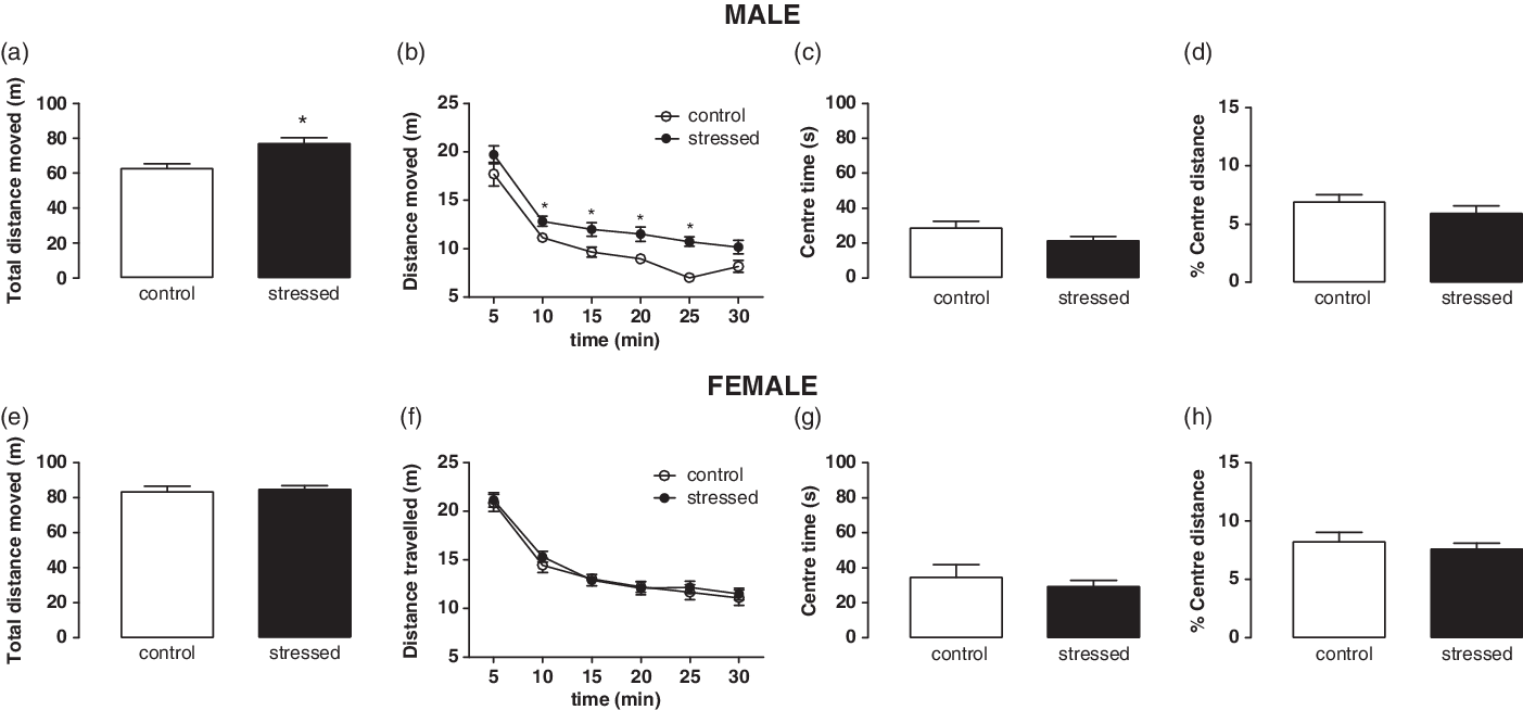
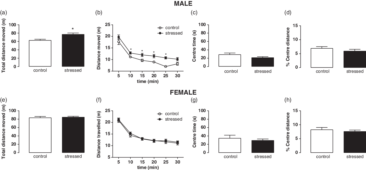
Figure 1 illustrates the effects of PNS on spontaneous locomotion in an unfamiliar environment. Male prenatal stressed mice moved more than their control counterparts during 30 min of observation in the open field (Fig. 1a, t(36) = 2.214, p = 0.032). After analysing the spontaneous locomotion in blocks of 5 min, a significant increase of distance moved was detected in prenatal stressed male mice compared to control (Fig. 1b, control: p > 0.05 for all; stressed: 0–5 min and 25–30 min: p > 0.05; 5–10, 10–15, 15–20, 20–25 min: p < 0.05). However, no differences between groups in the time spent in or the percentage of distance moved in the central area of the arena were observed (Fig. 1c, t(36) = 1.755, p = 0.088; Fig. 1d, t(36) = 1.088, p = 0.283). No significant differences were observed either in spontaneous locomotor activity or in the time spent exploring the central area between control and stressed female mice in any phase of the estrous cycle (Fig. 1e–h; p > 0.05 for all parameters). Females in estrus/proestrus and metestrus/diestrus phases also did not significantly differ in the spontaneous locomotion (Fig. 2S; p > 0.05).
Open field test in adult male and female prenatal stressed mice. (a, e) Total distance moved in the arena during 30-min test session; (b, f) Distance moved in blocks of 5 min (Student t-test, *p < 0.05); (c, g) Time spent exploring the centre zone of the arena during the first 10 min of test; (d, h) Percentage of distance travelled in the centre zone of the arena during the first 10 min of test. Data are expressed as mean ± SEM of male (16 and 22) and female (15 and 21) in control and prenatal stressed groups, respectively.
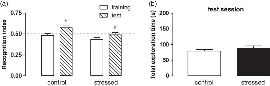
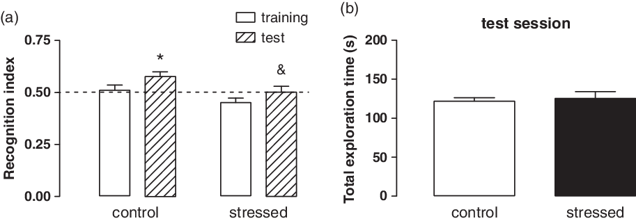
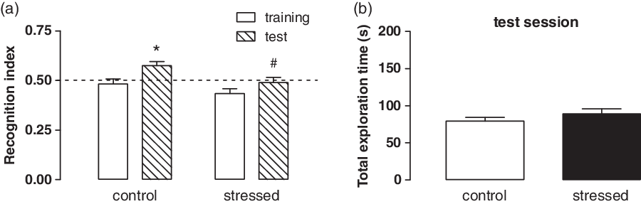
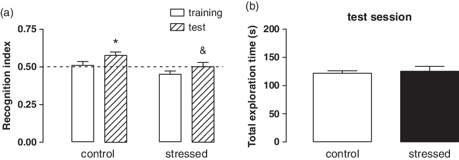
Figures 2 and 3 show the impact on cognitive performance of adult male and female mice exposed to prenatal stress. Control male and female mice exhibited preference for novel versus familiar object during the test session as demonstrated by Fig. 2a and 3a. In fact, a significant increase in the recognition index in the test session (when compared with training session) was observed in control group but not to prenatal stressed male and female mice (Fig. 2a, control: t(15) = 2.750, p = 0.015, stressed: t(21) = 1.495, p = 0.150; Fig. 3a, control: t(14) = 3.177, p = 0.007, stressed: t(20) = 1.276, p = 0.217). Additionally, a comparison in the test session between control and prenatal stressed mice detected a significant difference for male mice (Fig. 2a, t(36) = 2.455, p = 0.019) and a tendency to reach the significance for females (Fig. 3a, t(34) = 1.975, p = 0.056). In the test session, neither males nor females exibited differences between control and stressed groups in the total time spent exploring the two objects (Fig. 2b, t(36) = 1.076, p = 0.289; Fig. 3b, t(34) = 0.410, p = 0.684).
Impact of PNS on recognition memory in adult male prenatal stressed mice. (a) Recognition index estimated during the training and test sessions (Paired t-test, *p < 0.05 vs. training; #p < 0.05 vs. control); (b) Cumulative time spent exploring both familiar and novel objects in the test session. Dotted line represents no preference for any object. Data are expressed as mean ± SEM of 16 and 22 in control and stressed groups, respectively.
Impact of PNS on recognition memory in adult female prenatal stressed mice with no separation of estrous cycle. (a) Recognition index estimated during the training and test sessions (Paired t-test, *p < 0.05 vs. training; Student’s t-test, &p = 0.056 vs. control); (b) Cumulative time spent exploring both familiar and novel objects in the test session. Dotted line represents no preference for any object. Data are expressed as mean ± SEM of 15 and 21 mice in control and stressed groups, respectively.
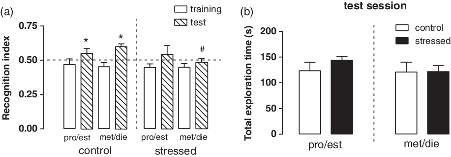
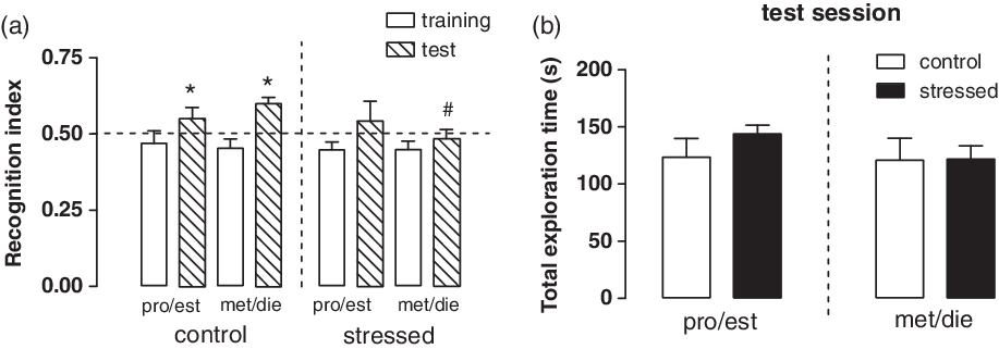
When female mice were categorised according to the phase of their estrous cycle, during the test session, control mice in proestrus/estrus and metestrus/diestrus preferred the novel object versus the familiar one. Of note, control mice in metestrus/diestrus displayed significant recognition index in the test session when compared to the training session. Importantly, control females in proestrus/estrus displayed a tendency to reach the statistical significance (p = 0.052); possibly an increase in animal number would reveal a statistical significance for this experimental group (Fig. 4a; pro/est control: t(6) = 2.421, p = 0.052, pro/est stressed: t(5) = 1.155, p = 0.301; met/die control: t(7) = 3.340, p = 0.012, met/die stressed: t(13) = 0.713, p = 0.488). Additionally, significant difference between control and prenatal stressed groups was observed for female mice in metestrus/diestrus (Fig. 4a, t(21) = 2.613, p = 0.016), while no differences were detected when comparing control and prenatal stressed female mice in proestrus/estrus (Fig. 4a, p > 0.05). No significant differences were observed in the time spent exploring the novel and familiar objects in the test session between control and stressed mice (Fig. 4b, pro/est: t(11) = 1.057, p = 0.313, met/die: U = 52, p = 0.628).
Impact of PNS on recognition memory in adult female prenatal stressed mice in distinct phases of the estrous cycle. (a) Recognition index estimated during the training and test sessions in female mice in proestrus/estrus and metestrus/diestrus (Paired t-test, *p ≤ 0.05 vs. training; Student’s t-test, #p < 0.05 vs. control); (b) Cumulative time spent exploring both familiar and novel objects in the test session. Dotted line represents no preference for any object. Data are expressed as mean ± SEM of 7 and 6 pro/est, and 8 and 15 met/die in control and stressed groups, respectively.
Discussion
The results of the present study showed that the memory deficits induced by maternal stress is observed in male and female offspring. Additionally, in females, the prenatal stress-induced memory deficits were dependent on their estrous cycle. Of note, control female mice in proestrus/estrus and metestrus/diestrus recognised the unfamiliar object during the test session. However, stressed females displayed cognitive impairments only in the metestrus/diestrus phases. In proestrus/estrus phases, female mice are under the effects of higher circulating levels of estradiol and progesterone and display beneficial effects on cognition (Contreras et al., Reference Contreras, Molina, Saavedra and Martínez-Mota2000; Van Goethem et al., Reference Van Goethem, Rutten, van der Staay, Jans, Akkerman, Steinbusch, Blokland, van’tKlooster and Prickaerts2012). The positive effects of estradiol and progesterone hormones on cognition may be the result of hormone-induced increases in hippocampal connectivity, while higher levels of estradiol correlate with increased hippocampal synaptic density (Woolley & McEwen, Reference Woolley and McEwen1992). Our findings showed that the natural increase of estradiol and progesterone in females during estrous cycle was able to counteract prenatal stress-induced cognitive impairments in female mice. This finding underscores the importance of assessing the effects of PNS in different animal species and sexes in order to determine the mechanisms of the developmental programming of psychiatric disorders.
The cognitive deficits induced by PNS have been largely documented in rats (for a review see: Maccari & Morley-Fletcher, Reference Maccari and Morley-Fletcher2007; Bock et al., Reference Bock, Wainstock, Braun and Segal2015). Only a small number of studies have investigated the effects of maternal stress during pregnancy on the cognitive capacities of the offspring using mice as animal model (Son et al., Reference Son, Geum, Chung, Kim, Jo, Kim, Lee, Kim, Choi, Kim, Lee and Kim2006; Behan et al., Reference Behan, van den Hove, Mueller, Jetten, Steinbusch, Cotter and Prickaerts2011; Abdul Aziz et al., Reference Abdul Aziz, Kendall and Pardon2012; Benoit et al., Reference Benoit, Rakic and Frick2015; Kiryanova et al., Reference Kiryanova, Meunier, Vecchiarelli, Hill and Dyck2016; Lee et al., Reference Lee, Kim and Goto2016; Gur et al., Reference Gur, Shay, Palkar, Fisher, Varaljay, Dowd and Bailey2017; Clarke et al., Reference Clarke, Sarkissian, Todd, Suraev, Bahceci, Brzozowska and Arnold2019). Among these, only two studies investigated the effects of PNS in outbred mouse strains (i.e. CD-1/ICR mice) (Lee et al., Reference Lee, Kim and Goto2016; Gur et al., Reference Gur, Shay, Palkar, Fisher, Varaljay, Dowd and Bailey2017). Similarly, there are a limited number of studies in which mice of both sexes were used to investigate the cognitive deficits of PNS (Abdul Aziz et al., Reference Abdul Aziz, Kendall and Pardon2012; Benoit et al., Reference Benoit, Rakic and Frick2015; Gur et al., Reference Gur, Shay, Palkar, Fisher, Varaljay, Dowd and Bailey2017). The results of the present study adds to the existing literature that for the first time, the cognitive deficits induced by PNS in female mice of an outbred strain (Swiss mice) are linked to the estrous cycle.
In this study, we adopted a 6-h interval between the training and the test sessions of the object recognition task as reported by Leger et al. (Reference Leger, Quiedeville, Bouet, Haelewyn, Boulouard, Schumann-Bard and Freret2013). In contrast, other authors have assessed the effects of PNS in mice on the object recognition task using an inter-trial interval of 3 to 4 h (Abdul Aziz et al., Reference Abdul Aziz, Kendall and Pardon2012; Gur et al., Reference Gur, Shay, Palkar, Fisher, Varaljay, Dowd and Bailey2017). As summarised by Cohen & Stackman (Reference Cohen and Stackman2015), the hippocampus is necessary for the retention of object recognition memory when an inter-trial interval greater than 10 min is used. If this interval is lesser than 10 min, then the perirhinal cortex seems to be mediating this process (Cohen & Stackman, Reference Cohen and Stackman2015). This indicates that under our experimental conditions, the prenatal stress-induced memory deficits are mainly related to an impairment of the hippocampal functions.
Novelty-induced hyperactivity was detected in male but not female offspring exposed to prenatal stress. This behavioural alteration has already been reported both in mice (Son et al., Reference Son, Chung, Geum, Kang, Choi, Kim and Choi2007) and rats (Kippin et al., Reference Kippin, Szumlinski, Kapasova, Rezner and See2008). Interestingly, an alteration of the midbrain dopaminergic system suggestive of a functional hyperdopaminergic state was reported for Son et al. (Reference Son, Chung, Geum, Kang, Choi, Kim and Choi2007) as a putative mechanism underlying this behavioural alteration. This alteration could contribute to the aetiology of some psychiatric disorders, such as ADHD, schizophrenia and bipolar mania. Another explanation that should be taken into account for novelty-induced hyperactivity in prenatal stressed male mice is impaired habituation to unfamiliar areas, which is also related to memory deficits. This explanation reinforces the view that prenatal stressed male mice display memory deficits. Further additional studies aimed to evaluate memory deficits of prenatal stressed male and female offspring could confirm this hypothesis.
Under our experimental conditions, we did not detect any significant difference in behavioural parameters related to the exploration of unprotected areas, such as the percentage of time spent in the central area of the arena or the distance travelled within the central area. This observation indicates that control and prenatal stressed mice exhibited similar fear-related behaviours and contrasts with previous studies showing enhanced anxiety-like behaviours in rodents exposed to PNS (Van den Hove et al., Reference Van den Hove, Blanco, Aendekerk, Desbonnet, Bruschettini, Steinbusch, Prickaerts and Steinbusch2005; Hiroi et al., Reference Hiroi, Carbone, Zuloaga, Bimonte-Nelson and Handa2016; Nakhjiri et al., Reference Nakhjiri, Saboory, Roshan-Milani, Rasmi and Khalafkhani2017; Zhang et al., Reference Zhang, Wang, Chen and Wang2019). We believe that the absence of effect on anxiety for prenatal stressed mice can be explained by a floor effect because lower exploration of unprotected areas, which is expected in an anxiety-like phenotype, could not be detected under our experimental conditions.
In conclusion, we demonstrated that late PNS induces significant memory recognition impairment in adult male and female mice. The impact on memory cognition observed in female offspring is observed in mice under lower progesterone and estradiol circulating levels (metestrus/diestrus phases). These results reinforce the view that the PNS is an aetiological component of the most common forms of psychiatric disorders associated with cognitive deficits.
Acknowledgements
This work was supported by funds from the Brazilian National Council Research (CNPq grant no. 302302/2015-8 to ECG) and CAPES, which supported research fellowships to CAM, FCC, RHL, VADH.
Conflict of interest
None.
Supplementary material
To view supplementary material for this article, please visit https://doi.org/10.1017/neu.2020.3