INTRODUCTION
The knowledge about the nemerteans inhabiting beaches is scarce due to their low abundance in these habitats and they are slightly documented in books about beach ecology (e.g. McLachlan & Brown, Reference McLachlan and Brown2006). Recent beach studies involving an intensive sampling (e.g. Junoy et al., Reference Junoy, Castellanos, Viéitez, De la Huz and Lastra2005, Reference Junoy, Castellanos, Viéitez and Riera2013) have discovered very unusual species such as Lineus acutifrons Southern, 1913 or Psammamphiporus elongatus (Stephenson, 1911) (Herrera-Bachiller et al., Reference Herrera-Bachiller, García-Corrales, Roldán and Junoy2008; Puerta et al., Reference Puerta, Andrade and Junoy2010).
Interstitial nemerteans are more elusive to collect in benthic samples due to their size (like many other invertebrates the nemerteans also have their smallest species in the interstitial fauna) and there are fixation problems that make their identification difficult.
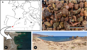
Sampling of beaches from Cabo de Gata Natural Park (south-east Spain) (Figure 1) revealed the presence of the nemertean Ototyphlonemertes duplex Bürger, Reference Bürger1895. The aim of this paper is to document this record and provide morphological data for the species.
Location of sampled specimens of Ototyphlonemertes duplex: (A) distribution of O. duplex in European waters, data from Bürger (Reference Bürger1895), Corrêa (Reference Corrêa1953) Kirsteuer (Reference Kirsteuer and Hulings1971) and Envall (Reference Envall1996), new record (red star); (B) volcanic coarse sand of the Embarcadero beach; (C) aerial photograph of the Embarcadero beach; (D) panoramic photograph of the Embarcadero beach. Scale bar: B, 1 cm.

MATERIALS AND METHODS
Sedimentary samples of volcanic coarse sand from the Embarcadero beach (36°47.603′N 2°3.695′W) (Figure 1) were collected and examined following the method described in Corrêa (Reference Corrêa1949). Nine specimens (five ♂, four ♀) of O. duplex were obtained on 21 June 2014. The specimens were examined alive both before and after anaesthetization in 7.5% MgCl2. The nine specimens were live squeezed to observe the stylet region. Sediment samples also contained specimens of the heteronemertean Ramphogordius lacteus Rathke, 1843.
RESULTS
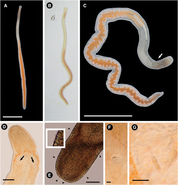
The body is extremely slender. Colour is orange in the intestine region, the anterior part is whitish and it is reddish around the cerebral ganglia (Figure 2A, C). Specimens measure up to 16 mm in length but no more than 1 mm in width. Head is bluntly rounded in shape (Figure 2D, E), not demarcated from the trunk, but in motion sometimes slightly oval; lacks eyes. A pair of statocysts is found dorsal to the cerebral ganglia; each one contains a statolith with two granules (Figure 2F). One cephalic furrow posterior to statocysts is observed reaching around body, dorsally forming an angle pointing caudally and ventrally pointing anteriorly (Figure 2C). Tactile cirri are present in the cephalic region; their number and position are variable (Figure 2E). The back end is slightly pointed with adhesive plate; caudal cirri are present. The proboscis occupies only the anterior third part of the body (Figure 2C). The stylet is smooth; all specimens have two accessory stylet pouches containing up to 3 stylets each (Figure 2G); the direction of the accessory stylets is both forward and backward in six specimens and forward in three specimens. The middle chamber is bulbous. Measurements of the stylet apparatus of the specimens are shown in Table 1. Specimens were sexually mature in June (Figure 2C). They move quickly in the trays with sand and survive in captivity in small containers without any special care for several weeks.
Ototyphlonemertes duplex: (A) photograph of a complete specimen (dorsal view); (B) original illustration, reproduced from Bürger (Reference Bürger1895: plate 2, figure 6); (C) photograph of a complete live squeezed female specimen showing the gonads. Cephalic furrow (arrow); (D) microphotograph of the head of a live squeezed specimen showing the position of the statocysts (arrows); (E) microphotograph of a live squeezed specimen showing the head and the position of the tactile cirri (arrowheads). Inset shows a detail of the cirri; (F) microphotograph of the statocyst; (G) microphotograph of a live squeezed specimen showing the central stylet and its basis and the two pouches of accessory stylets. Scale bars: A, 2 mm; C, 3 mm; D–E, 0.5 mm; F, 10 µm; G, 50 µm.

Measurements recorded for the stylet apparatus of nine specimens of Ototyphlonemertes duplex.

DISCUSSION
Ototyphlonemertes duplex has been recorded in Mediterranean waters of Naples (Italy), Villefranche-sur-Mer (France) and the south of England (Bürger, Reference Bürger1895; Corrêa, Reference Corrêa1953; Kirsteuer, Reference Kirsteuer and Hulings1971; Envall, Reference Envall1996). The specimens from Cabo de Gata Natural Park constitute the first record of the species in Spain. There were only two records of the genus Ototyphlonemertes from the Iberian Peninsula. Envall & Norenburg (Reference Envall and Norenburg2001) collected a single specimen of O. brunnea from Barcelona beaches and Norenburg (Reference Norenburg2008) listed O. macintoshi from Mindelo (Portugal).
The morphology of the Spanish specimens is consistent with the descriptions of O. duplex provided by Bürger (Reference Bürger1895), Corrêa (Reference Corrêa1953) and Envall (Reference Envall1996). They have the same colour pattern as the one represented in the original description by Bürger (Reference Bürger1895: plate 2, figure 6) (Figure 2B). Kirsteuer (Reference Kirsteuer1977) suggested an intraspecific constancy in number and distribution of cirri and Norenburg (Reference Norenburg1988) proposed a system to account for it. A basic distributional pattern has not been detected in the Spanish specimens; only some specimens showed a regular pattern similar to O. duplex represented by Envall (Reference Envall1996: figure 1b). The validity of the cirri as a specific character has been discussed by Envall & Norenburg (Reference Envall and Norenburg2001).
Spanish specimens were collected together with the heteronemetean Ramphogordius lacteus (= Lineus lacteus). The common occurrence of both species in the same area has been also reported by Bürger (Reference Bürger1895) and Corrêa (Reference Corrêa1953); the ecological significance of these data remain still unknown.
ACKNOWLEDGEMENT
We are extremely grateful to two anonymous referees for their constructive comments about the original manuscript.