Congenitally corrected transposition is a cardiac anomaly characterized by the combination of discordant connections at both the atrioventricular and ventriculoarterial junctions. It has a highly variable clinical presentation.
Case report
A 79 year old female patient was admitted to our centre with increasing signs of cardiac failure.
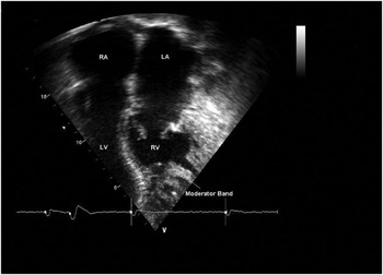
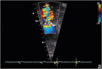
She had been followed up annually in our clinic for adults with congenital heart disease, being known to have congenitally corrected transposition associated with significant morphologically tricuspid regurgitation and controlled chronic atrial fibrillation. She had been a non-smoker for 5 years, but had subsequently developed hypertension, diabetes type II, and chronic obstructive disease of the airways. An arteriogram performed 2 years previously had shown normal coronary arteries, and a transthoracic echocardiogram had revealed a peak gradient of 51 mmHg across the aortic valve. Our investigation was performed in view of potential surgical replacement of her morphologically tricuspid valve. In the 2 weeks prior to admission, she started complaining of a productive cough and fever, with a deterioration of her shortness of breath, increased peripheral oedema, and progressive loss of weight. On examination, her pulse was 85 beats per minute; blood pressure was 110/70 mmHg, and saturation of oxygen whilst breathing air was 84%. A holosystolic murmur was audible at the apex, which radiated to the carotid arteries. Crackles were heard bilaterally at the bases of both lungs, more prominent on the right, and her jugular venous pressure was 6 cm, with bilateral oedema to the level of the knees, and signs of hepatomegaly. An electrocardiogram demonstrated controlled atrial fibrillation at 90 beats/minute, right axis deviation, and no ischaemic changes. The transthoracic echocardiogram confirmed the discordant atrioventricular and ventriculoarterial connections (Fig. 1) with moderate dilation and impairment of the right ventricular systolic function, the ejection fraction being calculated at 41%. The leaflets of the morphologically tricuspid valve were mildly thickened, with a moderate to severe jet of tricuspid regurgitation. This jet was broad and central, with a large vena contracta of 0.8 cm which extended to the roof of the morphologically left atrium. Both atriums were dilated. The aortic valve was severely calcified, with restricted movements of the leaflets indicating severe aortic stenosis. Colour Doppler interrogation of the aortic valve showed aliasing as sign of high velocity. This finding was subsequently confirmed by use of the continuous Doppler modality, which revealed velocity of 4.2 m/sec, and a calculated peak gradient of 70 mmHg, the mean gradient being 40 mmHg (Fig. 2). The right-sided morphologically left ventricle showed good systolic function, with an unobstructed outflow tract, and normal flow through the pulmonary valve. There was mild to moderate mitral valvar regurgitation, indicating moderate pulmonary hypertension, the pulmonary arterial pressure being estimated at 55 mmHg. Chest X-ray demonstrated a left-sided cardiac apex, with an upper left cardiac border bulging due to the left-sided location of the ascending aorta. The cardiothoracic ratio was increased as assessed on the posteroanterior view, and there were signs of pulmonary venous congestion and infection at the right basis. The patient was successfully treated with high doses of diuretics, antibiotics, and oxygen. She was discharged after a few days.
The four-chamber echocardiographic section shows discordant atrioventricular connections. The systemic morphologically right ventricle is characterized by heavy trabeculations and a prominent moderator band. The ventricular septum is intact. Note that the hinge of the septal leaflet of the tricuspid valve is displaced further towards the apex than the leaflets of the mitral valve. RV: right ventricle; LV: left ventricle; RA: right atrium; LA: left atrium.

Colour Doppler interrogation showing the pattern of flow through the aortic valve.

Discussion
Congenitally corrected transposition is a rare cardiac anomaly, with an incidence of approximately 0.5%, and a slight male preponderance.Reference Freedom, Dyck, Emmanouilides, Riemenschneider, Allen and Gutgesell1 The clinical presentation is highly variable. In the context of an intact ventricular septum and a normal outflow tract from the right-sided morphologically left ventricle, adults may present with heart block or signs of progressive tricuspid valvar regurgitation, which causes congestive cardiac failure. These patients with associated lesions can be in a reasonably good functional state, and in particular three-fifths report no symptoms, with another one-quarter describing only mild symptoms.Reference Graham, Bernard and Mellen2 In patients with no associated cardiac lesions, the diagnosis is often missed. These patients typically lead normal lives, presenting later with congestive cardiac failure related to severe tricuspid regurgitation or heart block. Our patient was admitted for a severe and rapid deterioration of her chronic heart failure due to a transient pulmonary infection. In her case, the regurgitation across the tricuspid valve positioned in the systemic circulation was exacerbated by progressive, degenerative aortic stenosis. We believe that this combination is extremely rare. To our knowledge, this is the first case to be reported in the medical literature.