Introduction
Bipolar disorder (BD) is clinically defined by elevated and depressed mood states, indicating altered emotion processing. It may additionally be characterized by altered reward processing: during hypomanic and manic episodes, BD patients show increased responsiveness to cues signalling future rewards, evidenced by exaggerated reward-seeking behaviour, increased sociability and hypersexual behaviour (Gray, Reference Gray, Van Goozen, Van De Poll and Sergeant1994; Kaplan & Sadock, Reference Kaplan and Sadock1997; American Psychiatric Association, 2000). Simultaneously, manic episodes are distinguished by decreased responsiveness to aversive cues and therefore underestimating risks, clinically indicated by, for example, loss of social inhibitions, unrestrained buying sprees or reckless driving (Bermpohl et al. Reference Bermpohl, Kahnt, Dalanay, Hägele, Sajonz, Wegner, Stoy, Adli, Krüger, Wrase, Ströhle, Bauer and Heinz2010). During depressive episodes, BD patients show symptoms of anhedonia and loss of energy, which might be explained by blunted responsiveness to reward stimuli (Satterthwaite et al. Reference Satterthwaite, Kable, Vandekar, Katchmar, Bassett, Baldassano, Ruparel, Elliott, Sheline, Gur, Gur, Davatzikos, Leibenluft, Thase and Wolf2015). While these clinical observations suggest altered reward processing in BD, only a few neuroimaging studies on reward processing have focused to date on euthymic BD (Nusslock et al. Reference Nusslock, Almeida, Forbes, Versace, Frank, Labarbara, Klein and Phillips2012; Caseras et al. Reference Caseras, Lawrence, Murphy, Wise and Phillips2013; Yip et al. Reference Yip, Worhunsky, Rogers and Goodwin2015). While studies in acutely depressed or manic patients may serve to identify state markers, investigations of euthymic patients may reveal trait markers of the disorder.
Neuroimaging findings in BD
Previous functional magnetic resonance imaging (fMRI) studies have suggested reward hypersensitivity during manic episodes (Abler et al. Reference Abler, Greenhouse, Ongur, Walter and Heckers2008; Bermpohl et al. Reference Bermpohl, Kahnt, Dalanay, Hägele, Sajonz, Wegner, Stoy, Adli, Krüger, Wrase, Ströhle, Bauer and Heinz2010), and blunted reward responsiveness during depressive states (Chase et al. Reference Chase, Nusslock, Almeida, Forbes, Labarbara and Phillips2013; Satterthwaite et al. Reference Satterthwaite, Kable, Vandekar, Katchmar, Bassett, Baldassano, Ruparel, Elliott, Sheline, Gur, Gur, Davatzikos, Leibenluft, Thase and Wolf2015). Studies on reward processing in euthymic BD are hitherto sparse. These studies have, in parts, used paradigms similar to the one employed here. Overall, reward findings in BD have not been consistent so far: some findings suggest that euthymic BD may be characterized by ‘reward hypersensitivity’, which may be a trait marker underlying emotional lability and mood dysregulation (Urošević et al. Reference Urošević, Abramson, Harmon-Jones and Alloy2008; Nusslock et al. Reference Nusslock, Almeida, Forbes, Versace, Frank, Labarbara, Klein and Phillips2012; Caseras et al. Reference Caseras, Lawrence, Murphy, Wise and Phillips2013). Specifically, increased ventral striatal (VS) activity was found in a card-guessing game, involving also a choice-making period (Delgado et al. Reference Delgado, Nystrom, Fissell, Noll and Fiez2000), during reward anticipation in medicated euthymic BD I (Nusslock et al. Reference Nusslock, Almeida, Forbes, Versace, Frank, Labarbara, Klein and Phillips2012; see, however, Caseras et al. Reference Caseras, Lawrence, Murphy, Wise and Phillips2013) and BD II (Caseras et al. Reference Caseras, Lawrence, Murphy, Wise and Phillips2013) subjects.
While these findings are compatible with ‘reward hypersensitivity’ in BD, more recent neuroimaging evidence rather suggest ‘blunted reward processing’ in euthymic BD. Yip et al. (Reference Yip, Worhunsky, Rogers and Goodwin2015) found decreased dorsal striatal (DS) activity during reward anticipation and decreased VS and DS activity during loss anticipation in unmedicated euthymic BD II patients. This study, contrary to previous findings of VS hyperactivation (see Nusslock et al. Reference Nusslock, Almeida, Forbes, Versace, Frank, Labarbara, Klein and Phillips2012; Caseras et al. Reference Caseras, Lawrence, Murphy, Wise and Phillips2013), involved young unmedicated, antipsychotic-naïve BD adults, who completed a simple reward-processing task (‘monetary incentive delay task’), which did not involve additional components of decision-making, and compared gain and loss anticipation with a neutral control condition. Similarly, Trost et al. (Reference Trost, Diekhof, Zvonik, Lewandowski, Usher, Keil, Zilles, Falkai, Dechent and Gruber2014) showed that euthymic to mildly depressed BD patients exhibited reduced neural responses in the VS when answering to previously conditioned reward stimuli and Linke et al. (Reference Linke, King, Rietschel, Strohmaier, Hennerici, Gass and Meyer-lindenberg2012) found an attenuated prediction error signal in a probabilistic reversal learning task in euthymic BD patients (Linke et al. Reference Linke, King, Rietschel, Strohmaier, Hennerici, Gass and Meyer-lindenberg2012). Taken together, these latter findings contradict a simple reward hypersensitivity model and rather suggest blunted reward processing in BD (Yip et al. Reference Yip, Worhunsky, Rogers and Goodwin2015).
It is yet unclear whether these discrepant findings may be due to differences in paradigms (i.e. decision-making component v. simple reward processing paradigm), BD subgroup (i.e. very young BD II v. older BD I and II patients), or medication status (i.e. medication-naïve BD patients v. medicated patients). We, therefore, used a reward paradigm that does not involve a decision-making component, to investigate medicated, middle-aged BD I and II patients in a sample size comparable with prior studies.
Functional connectivity
Besides such activation studies, connectivity studies may provide further insight into the neural mechanisms of reward processing underlying BD. While neuroanatomical studies showed white-matter abnormalities (indicating decreased structural connectivity) within the prefrontal–striatal–thalamic circuitry in BD (Strakowski et al. Reference Strakowski, Adler, Almeida, Altshuler, Blumberg, Chang, DelBello, Frangou, McIntosh, Phillips, Sussman and Townsend2012), functional connectivity studies of the reward network have emerged only recently. One connectivity study using a desire–reason dilemma paradigm in healthy subjects reported a functional connectivity of the bilateral antero-prefrontal cortex (aPFC, Brodmann area 10) with the nucleus accumbens (NAcc) and the ventral tegmental area (VTA), which was increased whenever subjects successfully inhibited impulses toward pursuing a small immediate reward for the sake of a greater future monetary goal (Diekhof & Gruber, Reference Diekhof and Gruber2010). Studying the same paradigm in euthymic and mildly depressed BD patients, Trost et al. (Reference Trost, Diekhof, Zvonik, Lewandowski, Usher, Keil, Zilles, Falkai, Dechent and Gruber2014) found altered functional connectivity between the aPFC (Brodmann area 10) and the NAcc. The question arises whether similar alterations of functional connectivity may be present also during more basic reward processes, i.e. independent of desire–reason conflicts. Studies investigating functional connectivity in euthymic BD patients undergoing a reward-processing paradigm free of a desire–reason component are still missing.
Aim of the current study
We here used a well-established monetary incentive delay paradigm similar to a task previously studied in BD patients (Kirsch et al. Reference Kirsch, Schienle, Stark, Sammer, Blecker, Walter, Ott, Burkart and Vaitl2003) to investigate reward anticipation in euthymic BD patients. Our main aim was to study (1) functional activation in the VS, and (2) functional connectivity of the fronto-striatal network during a basic reward task that does not involve a decision-making component. Distinguishing different types of reward (gain/loss v. neutral control condition) during anticipation of rewards allowed us to address the question whether these aspects influence hyper- and hypoactivation of reward areas. Using a straightforward reward task enabled us to investigate whether functional connectivity may be altered during reward processes independent of a desire–reason conflict.
Method
Subjects
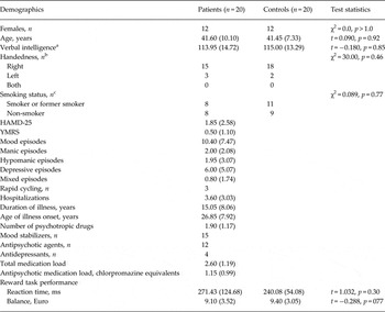
A total of 20 BD I and II patients (mean age = 41.6 years, s.d. = 10.1 years; eight males/12 females; 15 BD I/five BD II) were recruited via an out-patient facility at the Department of Psychiatry and Psychotherapy of the Charité – Universitätsmedizin Berlin. Euthymic mood state was confirmed by having a 25-item Hamilton Depression Scale (HAMD; Hamilton, Reference Hamilton1960) score <10 and a Young Mania Rating Scale (YMRS; Young et al. Reference Young, Biggs, Ziegler and Meyer1978) score <12. Patients had been in the euthymic state at the time of data collection for at least 5 months. Participants had been in remission for an average of 2.99 years. They were diagnosed by an experienced clinician using the German version of the Structured Clinical Interview for DSM-IV (SCID-I; First et al. Reference First, Spitzer, Gibbon and Williams1997; Wittchen et al. Reference Wittchen, Wunderlich, Gruschwitz and Zaudig1997). Exclusion criteria were current psychotic symptoms, diagnosis of dementia or organic brain disorder, diagnosis of substance dependency (except nicotine and caffeine), Axis II antisocial personality disorder, significant general medical illness (including neurological disorders and head trauma), and age below 18 or above 70 years. Patients did not suffer from any other Axis I disorder. Patients were medicated with mood stabilizers, antipsychotics and/or antidepressants, but not with benzodiazepines. Verbal intelligence of all subjects was approximated using the Multiple Choice Vocabulary Test (MWT-B; Lehrl, Reference Lehrl2005). Handedness was measured based on the Edinburgh Handedness Inventory (Oldfield, Reference Oldfield1971). Demographic sample characteristics are summarized in Table 1. BD patients and healthy controls (HC) did not differ significantly in age, verbal intelligence and gender. HC were recruited via media advertisements and mailing lists. HC had a negative lifetime history of psychiatric illness (including alcohol and drug dependence) and a negative family history (first-degree relatives) for Axis I disorders. They were free of current or past psychotropic pharmacological treatment and had no significant general medical illness, including neurological disorders and head trauma. All participants gave written informed consent before participation. The study was approved by the local ethics committee of the Charité – Universitätsmedizin Berlin.
Demographic sample characteristics and reward task performance

Data given as mean (standard deviation) unless otherwise indicated.
HAMD-25, Hamilton Rating Scale for Depression (25-item); YMRS, Young Mania Rating Scale.
a Measured using the Multiple Choice Vocabulary Test.
b Data missing from two participants.
c Data missing from four participants.
Functional imaging task
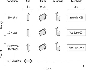
We used an event-related fMRI paradigm as previously described to examine neural mechanisms of anticipation and receipt of monetary reward and loss (Kirsch et al. Reference Kirsch, Schienle, Stark, Sammer, Blecker, Walter, Ott, Burkart and Vaitl2003; Grimm et al. Reference Grimm, Heinz, Walter, Kirsch, Erk, Haddad, Plichta, Romanczuk-Seiferth, Pöhland, Mohnke, Mühleisen, Mattheisen, Witt, Schäfer, Cichon, Nöthen, Rietschel, Tost and Meyer-Lindenberg2014) (Fig. 1). All subjects were instructed about the task and different conditions used in the experiment before scanning. The paradigm included four different conditions: a win condition, a loss avoidance condition, a verbal feedback condition, and a passive control condition. Each condition consisted of 10 trials, which appeared in a pseudorandom order. In each trial, participants were instructed to respond as fast as possible to a target stimulus (flashing of the screen, 0.1 s) by pressing a button with their right thumb. Before each target stimulus, a cue (arrow) was presented for 6 s, indicating the respective condition. After the target stimulus, feedback was presented for 2 s. In the win condition, a vertically oriented arrow pointing upward indicated a potential win (in the case of a fast enough response); in the loss avoidance condition an arrow pointing downward indicated a potential loss (in the case of a too slow response). A vertically oriented double-sided arrow conveyed no possible win or loss, but the occurrence of verbal feedback following the trial, and a horizontally oriented double-sided arrow indicated a passive control condition without a target, button press or win/loss feedback following the trial. The threshold for a fast response was adaptive for each subject. The adaptive algorithm was an increase of 5% of the threshold after a slow response and a 10% decrease after a fast response. This way, the outcome probabilities were fixed over the course of the paradigm with participants not being aware of this adaptive algorithm. Participants received the money they won after the experiment.
Monetary incentive delay paradigm. Depicted are the four conditions of our paradigm and the trial sequence.

Medication
To assess effects of psychotropic medication, we used the following parameters suggested by Almeida et al. (Reference Almeida, Versace, Hassel, Kupfer and Phillips2010) and Nusslock et al. (Reference Nusslock, Almeida, Forbes, Versace, Frank, Labarbara, Klein and Phillips2012): a general medication load reflecting the number and dose of different medications (Sackeim, Reference Sackeim2001), antipsychotic medication load (in chlorpromazine equivalents), the total number of psychotropic medication, and the particular medication group (taking v. not taking mood stabilizers, antipsychotics, antidepressants). No patient received dopaminergic antidepressants, such as bupropion, or benzodiazepines, so that we did not include those factors in the analysis.
Behavioural data analyses
Demographic and task performance measures were analysed using IBM SPSS Statistics version 22.0 (IBM Corp., 2013). Group differences in categorical variables were examined using χ2 tests and group effects on continuous variables were analysed employing analyses of variance as well as t tests for independent samples. The significance threshold for all analyses was set to p < 0.05.
Imaging parameters
Blood oxygen level-dependent fMRI was performed on a Siemens Trio 3 T MR scanner using an echoplanar imaging sequence (with 240 volumes comprising 28 slices of 4 mm + 25% gap, repetition time 2 s, echo time 30 ms, flip angle 80%, field of view 192 mm, descending slice order). Quality-control measurements were conducted on every day of data collection according to a multicentre quality assurance protocol (Friedman & Glover, Reference Friedman and Glover2006), revealing stable signals over time.
Functional imaging processing (pre-processing and first level)
Image processing and statistical analyses were conducted using statistical parametric mapping methods as implemented in SPM8 (http://www.fil.ion.ucl.ac.uk/spm/software/spm8/). Briefly, images were corrected for acquisition delay, realigned to a mean image (movement parameters were confined to <3 mm translation and <3° rotation between volumes), spatially normalized to a standard echoplanar imaging template (created by the Montreal Neurological Institute; MNI) with volume units (voxels) of 3 × 3 × 3 mm, smoothed with a 9 mm FWHM (full width at half maximum) Gaussian filter and ratio normalized to the whole-brain global mean. Six task regressors (gain anticipation, loss anticipation, verbal control, passive control, outcome, no outcome) as well as six regressors modelling head motion were included in the first-level analysis for each participant.
Statistical analyses (second level)
Group differences in brain activation during reward processing were analysed using two-sample t tests. We used the individual contrast images computed on the first level for gain anticipation > verbal control condition, loss anticipation > verbal control for group differences in activation. For group differences in functional connectivity we used the individual (gain/loss anticipation × seed) > (verbal control ( seed) contrasts from the psychophysiological interaction (PPI) first-level analysis. fMRI analyses were restricted to four a priori-defined functional regions of interest (ROIs) created as a 6 mm box around coordinates [using the same analysis approach by Yip et al. (Reference Yip, Worhunsky, Rogers and Goodwin2015) to allow direct comparisons of the results] reported by recent meta-analyses on reward processing (Knutson & Greer, Reference Knutson and Greer2008; Valentin & O'Doherty, Reference Valentin and O'Doherty2009). These include the left VS (−12, 10, −2), right VS (10, 8, 2). Small volume corrections (SVCs) were applied with a familywise error (FWE)-corrected threshold of p < 0.05.
Functional connectivity analyses
Functional connectivity was analysed with the generalized form of PPI analysis, which proved to have superior model fit compared with standard PPI analyses (McLaren et al. Reference McLaren, Ries, Xu and Johnson2012). Seeds were defined based on significant group differences in brain activity in the ROIs during reward anticipation observed in a t test directly comparing BD and HC (see Results). Accordingly, individual time series were extracted using first eigenvariates from voxels within the ROI regions centred on the left VS (−12, 10, −2) and right VS (10, 8, 2). Time courses were deconvolved from the haemodynamic response function (physiological variable) and added to additional individual first-level models. These models also included the aforementioned task regressors (psychological variables), the interaction of each task regressor with the respective seed time series (PPI regressors) as well as movement regressors. Studies on reward-processing comparable with our study are hitherto altogether missing. We thus defined ROIs using the aPFC region (±30, 51, 3; Brodmann area 10; 12 mm box) from Trost et al. (Reference Trost, Diekhof, Zvonik, Lewandowski, Usher, Keil, Zilles, Falkai, Dechent and Gruber2014), which is based on repeatedly reported aPFC–striatal connectivity in fMRI studies of reward–desire reasoning (Diekhof & Gruber, Reference Diekhof and Gruber2010; Diekhof et al. Reference Diekhof, Keil, Obst, Henseler, Dechent, Falkai and Gruber2012a , Reference Diekhof, Nerenberg, Falkai, Dechent, Baudewig and Gruber b ) and used a SVC p < 0.05, for multiple comparisons (FWE-correction).
Analyses on relationship of disorder-related variables with neural activity
We tested whether activation differences in the VS or functional connectivity between VS and the aPFC were influenced by variables regarding (1) residual, subthreshold depressive and manic symptoms as well as impulsivity traits, (2) severity of illness, or (3) medication effects. For residual symptoms we used values from the YMRS and HAMD. For impulsivity we used the Barratt Impulsiveness Scale (BIS). We tested for the effects of severity of illness by examining duration of illness, age of onset, number of depressive and manic episodes, total number of episodes and hospitalizations. We controlled for medication effects using total medication load, antipsychotic medication load (chlorpromazine equivalent), total number of psychotropic medication and taking v. not taking mood stabilizers, antipsychotic medication and antidepressants. In separate analyses, we correlated these variables with the extracted parameter estimates obtained from whole ROIs in significant contrasts (Spearman's ρ/point-biserial correlation, p < 0.05, uncorrected).
Results
Task performance
No significant differences in final monetary gains and reaction times were observed between patients with BD and HC participants (monetary gains: t 38 = −0.28, p = 0.77; reaction time: t 38 = 1.03, p = 0.30).
Activation patterns
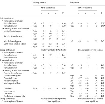
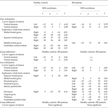
During reward and loss anticipation (gain/loss anticipation v. verbal control condition), both BD patients and HC showed reward-related brain activations in the bilateral VS (Table 2), but not in the DS. Exploratory whole-brain analysis revealed additional activations in a bilateral frontal and cerebellar network in the HC group during gain anticipation and additional activations of parietal regions in the BD group during loss anticipation (Table 2).
Brain regions activated during gain and loss anticipation
BD, Bipolar disorder; MNI, Montreal Neurological Institute.
* Significance level was set to p < 0.05, familywise error-corrected for a priori regions of interest (ventral striatum). For exploratory whole-brain analysis, the threshold criterion of p < 0.001, uncorrected, k = 25 voxels, was used.
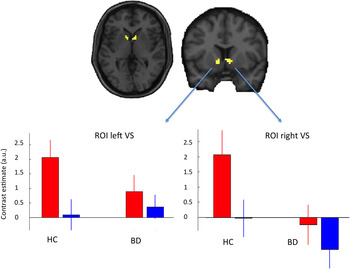
In the between-group comparisons, BD patients showed significantly weaker activation in the left (−9, 14, −5; SVC p FWE = 0.024) and right VS (6, 11, −2, SVC p FWE = 0.034, Table 2, Fig. 2) during reward anticipation (reward anticipation v. verbal control condition). We additionally applied a Bonferroni correction (p corr = 0.025) to account for the number of comparisons and found that the effect in the left VS was significant and the effect in the right VS showed a strong trend towards significance. There were no significant differences observed during loss anticipation. Exploratory whole-brain analyses did not reveal any additional group differences (HC > BD and BD > HC) during gain and loss anticipation (p < 0.05, FWE-corrected). We did not even find a group difference when the whole-brain threshold was tentatively lowered to p < 0.001, uncorrected.
Decreased bilateral ventral striatal (VS) activations during gain anticipation in bipolar disorder (BD) patients in a euthymic state (n = 20) compared with healthy controls (HC; n = 20). The top panel displays the anatomical location of the significant group difference during reward anticipation (two-sample t test: HC > BD patients, small volume correction p < 0.05, familywise error-corrected) in the left and right ventral striatal region of interest (ROI) mask. The bottom panel displays the mean bilateral VS activity for each condition and group during gain anticipation (red online) and verbal control (blue online). The y axis values reflect the mean parameter estimates in arbitrary units (a.u.); the error bars represent the standard errors of the mean. For the colour figure, see the online version of the paper.

Functional connectivity
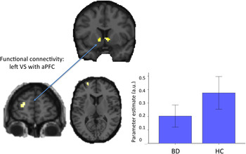
The comparison of BD patients v. HC yielded significantly reduced functional connectivity of the left VS seed region with the aPFC (−24, 59, 10, SVC p FWE = 0.043) during gain anticipation (Fig. 3). Exploratory whole-brain between-group analyses did not reveal any additional regions with altered functional coupling during gain or loss anticipation (p < 0.001, uncorrected).
Reduced functional connectivity of the left ventral striatum (VS) with the anterior prefrontal cortex (aPFC) during gain anticipation in bipolar disorder (BD) patients in a euthymic state (n = 20) compared with healthy controls (HC; n = 20). Top and bottom: anatomical location of reduced functional coupling between the left VS seed region and the left aPFC region of interest (ROI) (two-sample t test: HC > BD patients, small volume correction p < 0.05, familywise error-corrected). The histogram depicts decreased mean parameter estimates [connectivity values in arbitrary units (a.u.), reflecting the strength of connectivity] extracted from the left aPFC ROI during gain anticipation; the error bars represent the standard errors of the mean. For the colour figure, see the online version of the paper.

Relationship of disorder-related variables and neural activity
To assess the potential impact of residual symptomatology, illness severity and medication on the above reported neuroimaging findings, correlation analyses were carried out. Specifically, parameter estimates obtained in the main contrasts from the left and right VS ROIs were correlated with measures for residual symptoms, severity of illness and medication (for classification of medication effects see the Medication section in the Method). An analogous correlation was carried out for parameter estimates obtained in the connectivity analysis in the aPFC. As reported in detail in online Supplementary Table S1, none of these correlational analyses reached significance (at a threshold of p < 0.05, uncorrected).
Discussion
The present study investigated the neural circuitry underlying anticipation of monetary gain and loss in euthymic BD patients. We found decreased activation in the left and right VS during gain, but not during loss anticipation in middle-aged, medicated BD I and II patients undergoing a monetary incentive delay task compared with HC. Furthermore, this is the first study investigating functional connectivity in a monetary incentive delay task in BD patients. We found decreased functional connectivity between the left VS and the aPFC in BD patients during gain anticipation. Our study thus shows diminished responses of a reward circuitry in euthymic BD patients and suggests that blunted reward anticipation may be an underlying trait marker for BD.
Neural activation results
During reward and loss anticipation, both BD patients and HC showed activations in the bilateral VS. This general finding is consistent with previous fMRI studies associating the VS with reward prediction and anticipation of future reward and loss (Knutson et al. Reference Knutson, Fong, Adams, Varner and Hommer2001; McClure et al. Reference McClure, York and Montague2004; O'Doherty, Reference O'Doherty2004). The present study showed that decreased striatal activation during reward anticipation is present also in euthymic BD patients. Our study complements previous neuroimaging of reward processing, which suggests reward hypersensitivity during manic episodes, and blunted reward responsiveness during depressive states: Elevated reward system responses have been reported in manic BD patients in VS regions as well as in the orbitofrontal cortex during the anticipation of monetary reward (Abler et al. Reference Abler, Greenhouse, Ongur, Walter and Heckers2008; Bermpohl et al. Reference Bermpohl, Kahnt, Dalanay, Hägele, Sajonz, Wegner, Stoy, Adli, Krüger, Wrase, Ströhle, Bauer and Heinz2010). Additionally, increased valence ratings and amygdala responses to positive affective stimuli suggest a positive processing bias for mania (Bermpohl et al. Reference Bermpohl, Dalanay, Kahnt, Sajonz, Heimann, Ricken, Stoy, Hägele, Schlagenhauf, Adli, Wrase, Ströhle, Heinz and Bauer2009). Decreased functional activation in depressive BD patients undergoing a reward task (card-guessing paradigm) in the anterior cingulate cortex (ACC) as well as a correlation between depressive symptom severity and reduced activation in the bilateral VS, ACC, posterior cingulate cortex and right anterior insula have been reported recently (Chase et al. Reference Chase, Nusslock, Almeida, Forbes, Labarbara and Phillips2013; Satterthwaite et al. Reference Satterthwaite, Kable, Vandekar, Katchmar, Bassett, Baldassano, Ruparel, Elliott, Sheline, Gur, Gur, Davatzikos, Leibenluft, Thase and Wolf2015). Furthermore a study in a large and diverse sample of subjects including unipolar depression, BD (manic state), attention-deficit/hyperactivity disorder, alcoholism and schizophrenia undergoing a reward paradigm demonstrated that depressive symptoms are associated with a decreased VS activation during anticipation of reward (Hägele et al. Reference Hägele, Schlagenhauf, Rapp, Sterzer, Beck, Bermpohl, Stoy, Ströhle, Wittchen, Dolan and Heinz2015). Additionally, decreased VS activation was shown in relatives of patients with schizophrenia (Grimm et al. Reference Grimm, Heinz, Walter, Kirsch, Erk, Haddad, Plichta, Romanczuk-Seiferth, Pöhland, Mohnke, Mühleisen, Mattheisen, Witt, Schäfer, Cichon, Nöthen, Rietschel, Tost and Meyer-Lindenberg2014).
Because patients in our study were medicated and showed a prolonged course of illness, it is important to note that additional analyses revealed that reduced VS activation in our patient group was not related to medication or severity of illness. Furthermore, previous studies have shown blunting effects of nicotine on VS activation (Martin-Soelch et al. Reference Martin-Soelch, Missimer, Leenders and Schultz2003; Luo et al. Reference Luo, Ainslie, Giragosian and Monterosso2011; Lessov-Schlaggar et al. Reference Lessov-Schlaggar, Lepore, Kristjansson, Schlaggar, Barnes, Petersen, Madden, Heath and Barch2013; Kobiella et al. Reference Kobiella, Ripke, Kroemer, Vollmert, Vollstädt-Klein, Ulshöfer and Smolka2014). However, in our study the smoking status of the BD and HC groups did not differ significantly, thus making it unlikely that smoking status has confounded the current findings. Our finding of reduced VS activation in euthymia is thus in accordance with recent findings in antipsychotic- and mood-stabilizer-naïve, euthymic BD II patients of young age with a limited illness history (Yip et al. Reference Yip, Worhunsky, Rogers and Goodwin2015). These findings suggest that decreased VS activation is present in euthymic BD patients, independent of medication and severity of illness.
Our result is also consistent with recent findings in euthymic and mildly depressed BD I patients in a rather complex desire–reason dilemma, where BD patients had to reject a priori conditioned reward stimuli to successfully pursue a superordinate goal (Trost et al. Reference Trost, Diekhof, Zvonik, Lewandowski, Usher, Keil, Zilles, Falkai, Dechent and Gruber2014). Our present study differed from this previous study in that we used a reward-processing task that was free of any reason–dilemma component. Furthermore, patients in the present study were completely euthymic, and we were able to exclude confounding effects of residual depressive or manic symptoms. Taken together, previous (Trost et al. Reference Trost, Diekhof, Zvonik, Lewandowski, Usher, Keil, Zilles, Falkai, Dechent and Gruber2014; Yip et al. Reference Yip, Worhunsky, Rogers and Goodwin2015) and present findings suggest a general hypoactivation model in BD during anticipation of future reward.
Our findings are in contrast to a hypersensitivity model of BD (Caseras et al. Reference Caseras, Lawrence, Murphy, Wise and Phillips2013) that was based on findings showing increased activation in VS regions during reward anticipation in medicated, euthymic BD I (Nusslock et al. Reference Nusslock, Almeida, Forbes, Versace, Frank, Labarbara, Klein and Phillips2012) and BD II patients (Caseras et al. Reference Caseras, Lawrence, Murphy, Wise and Phillips2013). However, paradigms used in the above-mentioned studies did not separately contrast reward and loss anticipation against a neutral control condition and involved a decision-making component. These differences in paradigms may explain differences in findings, in particular, if we consider recent studies showing impaired decision-making in BD patients across different mood states (Chandler et al. Reference Chandler, Wakeley, Goodwin and Rogers2009; Adida et al. Reference Adida, Jollant, Clark, Besnier, Guillaume, Kaladjian, Mazzola-Pomietto, Jeanningros, Goodwin, Azorin and Courtet2011). Furthermore, in recent studies Guitart-Masip et al. (Reference Guitart-Masip, Fuentemilla, Bach, Huys, Dayan, Dolan and Duzel2011) highlighted the greater impact of action (go v. no-go) on VS activity during reward anticipation compared with the effect of valence in healthy subjects. Therefore, it is important to consider that the paradigm used in the present study (as well as the task employed by Yip et al. Reference Yip, Worhunsky, Rogers and Goodwin2015) contains a stronger performance-dependency on rewarding outcomes than paradigms used by Caseras et al. (Reference Caseras, Lawrence, Murphy, Wise and Phillips2013) or Nusslock et al. (Reference Nusslock, Almeida, Forbes, Versace, Frank, Labarbara, Klein and Phillips2012).
Functional connectivity results
Some functional connectivity studies in healthy subjects have referred to a network model of reward processing and proposed that immediate reward seeking (‘desire’) is modulated by a cognitive self-control mechanism (‘reason’) in order to accomplish long-term goals (Diekhof & Gruber, Reference Diekhof and Gruber2010). Whereas the anticipation and pursuit of immediate reward has been linked to the mesolimbic dopamine system – in particular the VTA and the NAcc (Knutson et al. Reference Knutson, Fong, Adams, Varner and Hommer2001; Yacubian et al. Reference Yacubian, Sommer, Schroeder, Glascher, Braus and Buchel2007) – the PFC has been associated with cognitive control (Duncan & Owen, Reference Duncan and Owen2000; Miller & Cohen, Reference Miller and Cohen2001). We found decreased functional connectivity between the left VS region and the aPFC in BD patients during reward anticipation. This is in accordance with an above-mentioned study using a desire–reason dilemma paradigm in euthymic and mildly depressed BD patients, suggesting impaired top-down regulation of mesolimbic areas by prefrontal control regions in BD (Trost et al. Reference Trost, Diekhof, Zvonik, Lewandowski, Usher, Keil, Zilles, Falkai, Dechent and Gruber2014).
Resting-state connectivity analyses in both unipolar and bipolar depression also revealed diminished connectivity in relation to depressive symptom severity, especially concerning the connectivity strength between the aPFC and the left VS (Satterthwaite et al. Reference Satterthwaite, Kable, Vandekar, Katchmar, Bassett, Baldassano, Ruparel, Elliott, Sheline, Gur, Gur, Davatzikos, Leibenluft, Thase and Wolf2015). Our findings in euthymic BD patients add to the literature linking reduced prefrontal–striatal connectivity to reward processing and, at the same time, show that the alteration exists independently of desire-making components and of residual depressive symptoms. These findings support the idea of dysfunctional top-down control of prefrontal regions on the VS in affective disorders (Diekhof & Gruber, Reference Diekhof and Gruber2010). In major depressive disorder, Heller et al. found that when attempting to sustain positive emotion to rewards, functional connectivity between the aPFC and VS was decreased and increased after medical treatment (Heller et al. Reference Heller, Johnstone, Shackman, Light, Peterson, Kolden, Kalin and Davidson2009, Reference Heller, Johnstone, Light, Peterson, Kolden, Kalin and Davidson2013). The regulation of positive affect thus appears to involve the biasing of signals directed from PFC to the NAcc, and the inability to sustain activity of NAcc may result from a breakdown in connectivity with aPFC. This suggests that altered reward processing in BD may result from an inability to sustain activity in a fronto-striatal network, which could lead to abnormalities in positive reward anticipation in the VS.
Several neuroanatomical studies in BD show alterations of white matter tracts connecting the anterior–ventral PFC with subcortical and limbic circuits, i.e. decreased connectivity between those regions (for a summary, see Strakowski et al. Reference Strakowski, Adler, Almeida, Altshuler, Blumberg, Chang, DelBello, Frangou, McIntosh, Phillips, Sussman and Townsend2012), which may underlie aberrant functional connectivity patterns. In particular, the present finding of decreased connectivity in BD between the VS and aPFC is in accordance with a proposed prefrontal–striatal–pallidal–thalamic network dysfunction, hypothetically due to developmental alterations in the modulation of this network due to deceased anatomical connectivity (Strakowski et al. Reference Strakowski, Adler, Almeida, Altshuler, Blumberg, Chang, DelBello, Frangou, McIntosh, Phillips, Sussman and Townsend2012).
Limitations
One limitation of the present study is that all BD patients included were medicated. Nevertheless, our results are in accordance with the finding of blunted striatum activations in antipsychotic- and mood-stabilizer-naïve BD patients (Yip et al. Reference Yip, Worhunsky, Rogers and Goodwin2015). In addition, a recent comprehensive review of effects of psychotropic medication on neuroimaging findings in BD revealed only little impact on fMRI results (Hafeman et al. Reference Hafeman, Chang, Garrett, Sanders and Phillips2012). We furthermore found no effects of total or antipsychotic medication load, total number of psychotropic medication, or any specific class of psychotropic medication on either the between-group results or on functional connectivity measures. However, it is acknowledged that these measures are not a truly satisfactory substitute for recruiting medication-free patients and do not fully exclude potentially confounding effects of medication in BD subjects (Phillips et al. Reference Phillips, Travis, Fagiolini and Kupfer2008). Still, we found specific effects in ventrostriatal regions in a specific reward condition, whereas we would rather expect a general blunting effect if it were due to medication. Moreover, as in many neuroimaging studies in BD, the modest sample size is a limitation of the present study. Future studies using larger sample sizes and, ideally, medication-free BD subjects are desirable to allow, for instance, for analyses comparing BD I and BD II groups or effects of different medications.
It is also unlikely that the reduced neural activation during gain anticipation was a result of a general lack of response in these brain regions in BD patients, since the same regions showed significant activation during loss anticipation.
Our specific task only activated the VS region in the HC sample, which is consistent with research on reward anticipation with monetary incentive delay tasks, which also found reward-related activation only in the ventral part of the striatum (e.g. Knutson et al. Reference Knutson, Fong, Adams, Varner and Hommer2001). However, Yip et al. (Reference Yip, Worhunsky, Rogers and Goodwin2015) found DS activations in the BD group. Differences may originate from the specific paradigm and the analysis of the task used, as Yip et al. (Reference Yip, Worhunsky, Rogers and Goodwin2015) used a version that differs from that originally developed by Knutson et al. (Reference Knutson, Fong, Adams, Varner and Hommer2001), in which anticipatory reward processing was separated into reward-prospect and reward-anticipation phases (Andrews et al. Reference Andrews, Meda, Thomas, Potenza, Krystal, Worhunsky, Stevens, O'Malley, Book, Reynolds and Pearlson2011).
Finally, a limitation of the present study is that group differences observed in VS activation may be confounded by overt or hidden factors that we did not sufficiently account for. Confounding factors may include variables directly related to BD (e.g. residual depressive or manic symptoms) as well as variables only indirectly related to BD (e.g. impulsivity or impulsive sensation seeking; Caseras et al. Reference Caseras, Lawrence, Murphy, Wise and Phillips2013). As these variables were not studied in our HC group, we cannot fully exclude confounding effects of these factors. However, our BD patient group only showed minimal residual depressive and manic symptoms (Table 1). In addition, within the BD patient group, we did not find correlations between VS activation and impulsivity (BIS), depressive symptom severity (HAMD) and manic symptom severity (YMRS), respectively.
In conclusion, our findings suggest that individuals with euthymic BD suffer from a diminished fronto-striatal network activity, which is reflected in disordered anticipation of rewarding stimuli. These findings are consistent with the hypothesis that hallmark symptoms of disordered emotion regulation in BD are based on an altered reward-processing system as a trait marker and hypothetical vulnerability factor.
Supplementary material
The supplementary material for this article can be found at http://dx.doi.org/10.1017/S0033291716001963
Acknowledgements
This work was supported by the German Ministry for Education and Research (BMBF) grants NGFNplus MooDS (Systematic Investigation of the Molecular Causes of Major Mood Disorders and Schizophrenia), the Integrated Network IntegraMent (Integrated Understanding of Causes and Mechanisms in Mental Disorders), under the auspices of the e:Med program (grant number O1ZX1314B), the German Research Foundation (DFG) grant SFB 636-B7, and Bristol-Myers Squibb.
Declaration of Interest
None.