The morphogenesis as well as morphological descriptions of the sinus venosus defect remain contentious. The echocardiographic diagnosis is also not uncommonly erroneous, with surgeons performing repair subsequently describing some defects held to be of sinus venosus type as being within the confines of the oval fossa.Reference Oliver, Gallego, Gonzalez, Dominguez, Aroca and Mesa 1 , Reference Al Zaghal, Li, Anderson, Lincoln, Shore and Rigby 2 Regarding developmental concepts, the previously accepted idea of “unroofing” of the right pulmonary veinsReference Van Praagh, Carrera, Sanders, Mayer and Van Praagh 3 is untenable. This is because the so-called “septum secundum” has long since been known not to be a true septum. Instead, it is an infolding of the atrial walls between the atrial connections of the caval and pulmonary veins, with extra-cardiac space retained within the confines of the interatrial groove.Reference Rose 4 , Reference Butts, Crean and Hlavacek 5 The development of episcopic microscopyReference Mohun and Weninger 6 and correlations between findings in the developing mouse and human embryosReference Anderson, Mohun and Brown 7 have served to validate the initial descriptions of this superior interatrial fold.Reference Rose 4 , Reference Anderson, Webb and Brown 8 These advances now underscore a better understanding regarding the spectrum of sinus venosus defects, which have been well described as existing as a consequence of anomalous connection of one or more pulmonary veins to a systemic vein, with the pulmonary vein or veins retaining their left atrial connection.Reference Crystal, Al Najashi, Williams, Redington and Anderson 9 Thus, the lesions are veno-venous connections, rather than septal defects.Reference Butts, Crean and Hlavacek 5 This is significant in terms of diagnosis, as of necessity the defects must be outside the confines of the oval fossa. The rims of the fossa, therefore, remain intact usually but not always with overriding of one or the other caval vein.Reference Al Zaghal, Li, Anderson, Lincoln, Shore and Rigby 2 , Reference Butts, Crean and Hlavacek 5 We describe in this study our recent experience with such defects, in which we compared preoperative diagnosis of lesions considered to fall within this spectrum with the findings at subsequent surgical correction. We discuss the findings in relation to current developmental concepts as revealed by episcopic microscopy in the developing mouse embryo, providing evidence obtained in support of these concepts from examination of human autopsied specimens.
Materials and methods
We retrospectively identified all children seen over the period extending from August, 2011 to October, 2015 who had been diagnosed and subsequently surgically repaired at New York Langone Medical Center with the diagnosis of a sinus venosus defect. We also included those said to exhibit anomalous connection of the individual right pulmonary vein in the absence of a sinus venosus defect. We reviewed the transthoracic and transoesophageal echocardiographic records, any additional imaging obtained using either cardiac magnetic resonance or computed tomography, and the surgical operative notes. The pertinent findings were recorded with focus on the anatomical descriptions. We sub-classified the patients into categories of superior or inferior defects, as well as those with an anomalous pulmonary venous connection in the absence of a veno-venous connection producing an interatrial communication. We then reviewed the findings in light of developmental concepts established using episcopic microscopic analysis of mouse embryo hearts,Reference Anderson, Mohun and Brown 7 correlating them with the findings from autopsied specimens known to have either superior or inferior sinus venosus defects as held in the archives of University of Florida and Lurie Children’s Hospital, Chicago.
Results
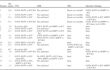
We identified 16 patients who met our diagnostic criteria. They had a mean age of 7.7 years, ranging from 2.7 to 15 years, and 10 of them were males (Table 1). In 13 patients, the defect was related to the superior caval vein and in two to the inferior caval vein. In the remaining patient, there was isolated connection of a right superior pulmonary vein to the superior caval vein in the absence of an interatrial communication. All the patients with the superior variant had anomalous connection of a right superior pulmonary vein, or veins, to the superior caval vein or to its junction with the morphologically right atrium. The connections of the right pulmonary veins were also anomalous in both the patients with inferior defects. The arrangement, however, was subtle in one of the patients. In this instance, the right lower pulmonary vein opened in very close proximity to the mouth of the inferior caval vein. The defect itself was proximal to the inferior caval vein, with the caval venous orifice overriding the intact posterior rim of the oval fossa. The pulmonary venous orifice opened predominantly to the left atrium, but its rightward margin was in continuity with the walls of the right atrium, thus providing the extraseptal veno-venous conduit that permitted interatrial shunting.
Patient characteristics.

Anomalous pulmonary vein(s) inferred to drain below the level of RPA unless otherwise specified. “Same” denotes that the study or surgical findings demonstrated the same findings as the previous study, with studies listed in temporal order from left to right
ASD=atrial septal defect; CMR=cardiac magnetic resonance; ICV-RA=inferior caval vein–right atrial junction; I-SVD=inferior-type sinus venosus defect; RLPV=right lower pulmonary vein; RMPV=right middle pulmonary vein; RPA=right pulmonary artery; RUPV=right upper pulmonary vein; SCV=superior caval vein; SCV-RA=superior caval vein–right atrial junction; S-SVD=superior-type sinus venosus defect; TEE=transoesophageal echocardiogram; TTE=transthoracic echocardiogram
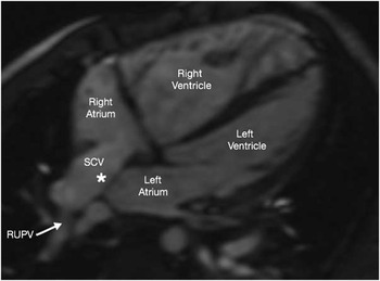
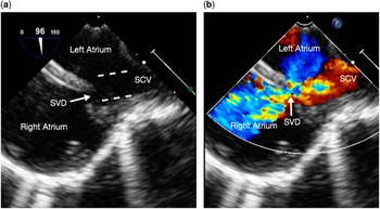
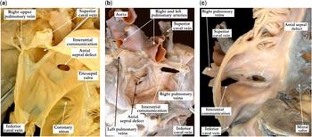
Transoesophageal echocardiographic reports were available for 12 patients. The transoesophageal echocardiographic findings were in keeping with the additional findings obtained in seven patients who also underwent cardiac magnetic resonance. In those who did not undergo cardiac magnetic resonance, transoesophageal echocardiography identified an additional anomalous pulmonary vein not appreciated on the transthoracic echocardiogram in one patient, with only two patients having pertinent findings at the time of surgery not previously appreciated. In six of the seven who underwent cardiac magnetic resonance, additional findings were discovered that had not been appreciated on transthoracic echocardiography (Table 1). In one patient, isolated connection of a solitary right pulmonary vein to the superior caval vein was revealed in the absence of an interatrial communication; this patient was erroneously diagnosed on the basis of transthoracic echocardiography as having a superior sinus venosus defect (Fig 1). In two of the patients, one from each subtype, the initial preoperative diagnosis, based on the transthoracic echocardiographic interrogation, had been that of an “ostium secundum” defect, in other words a defect within the oval fossa. There was suspicion, nonetheless, that the defect may have been of the sinus venosus variety. Both these patients were also investigated using cardiac magnetic resonance, revealing the presence of anomalously connected pulmonary veins in both. In one patient, the vein was connected to the superior caval vein and in the other to the inferior caval vein. In both instances, the anomalously connecting pulmonary veins had retained their left atrial connections. The findings were then confirmed in both patients, first using transoesophageal echocardiography and subsequently during surgical repair. Of the remaining four patients with superior defects, for whom preoperative transoesophageal echocardiographic reports were unavailable, the surgeon had noted the presence of additional anomalously connecting right pulmonary veins in two during the operative repair. At the time of surgical repair, the surgeon confirmed that all defects were outside the confines of the oval fossa, which exhibited intact rims. In all patients, there was also overriding of the mouth of the corresponding caval vein (Fig 2). These anatomical concepts, which serve to define the sinus venosus defect, are demonstrated in the autopsy specimens displayed in Figure 3.
Cardiac magnetic resonance axial imaging obtained in patient 14 demonstrating no superior sinus venosus defect (asterisk) as previously believed by transthoracic echocardiogram imaging, with the right upper pulmonary vein (RUPV) draining into the posterior aspect of the superior caval vein (SCV).

( a ) Transoesophageal echocardiogram (TEE) bicaval two-dimensional view demonstrating the superior caval vein (SCV) overriding the intact primary atrial septum (white dashed lines demonstrating the plane of anterior and posterior SCV walls), with a superior sinus venosus defect (SVD) located at the SCV–right atrial junction. ( b ) TEE bicaval color Doppler view demonstrating left-to-right flow across the SVD.

( a ) The right atrium is opened to show the superior caval vein–right atrial junction, the atrial septum, and the tricuspid valve guarding the inlet to the right ventricle. The coronary sinus is in its usual position, and there is a small atrial septal defect within the floor of the oval fossa. A probe is passed through the fibrofatty tissue of the superior interatrial fold, illustrating that the sinus venosus defect or interatrial communication is outside the confines of the true atrial septum. The superior caval vein overrides the defect, and the right upper pulmonary vein joins the superior caval vein at the roof of the defect. ( b ) The images shown in ( b ) and ( c ) are from the same heart. In ( b ) the left atrial aspect of the sinus venosus is demonstrated. The right and left pulmonary veins drain to the left atrium, with the right upper and middle pulmonary veins overriding the sinus venosus defect or interatrial communication. At the floor of the oval fossa, there is an atrial septal defect, which is well away from the sinus venosus defect. ( c ) The right atrioventricular junction of this heart with discordant atrioventricular connections is shown with the mitral valve guarding the inlet. There is a true atrial septal defect within the floor of the oval fossa, and the sinus venosus defect or interatrial communication is well away from the confines of the true atrial septum. This interatrial communication is also farther away from the mouth of the superior caval vein than usual. Although not clearly demonstrated in this image, the right upper and middle pulmonary veins override the mouth of the sinus venosus defect connecting in part to the right atrium but still maintaining their connection to the left atrium.

Discussion
Even with advances in modern-day imaging, it remains difficult preoperatively, when the oval fossa has deficient posterior–superior or inferior borders, to distinguish sinus venosus interatrial communications from so-called “ostium secundum” defects. In one recent retrospective review of transoesophageal echocardiographic and surgical operative reports of 24 patients initially diagnosed with some form of sinus venosus defect, for example, one-quarter were instead found to have an “ostium secundum” defect.Reference Oliver, Gallego, Gonzalez, Dominguez, Aroca and Mesa 1 Similarly, in another retrospective review, both patients initially thought to have the inferior-type of defect, and one-quarter of the patients initially thought to have the superior-type defects, were re-classified as having “ostium secundum” defects at the time of surgical repair.Reference Al Zaghal, Li, Anderson, Lincoln, Shore and Rigby 2 These difficulties, particularly those encountered in distinguishing inferior defects from the holes within the oval fossa that extend inferiorly towards the mouth of the inferior caval vein, highlight the need to employ strict anatomical criteria when defining the sinus venosus defects. Thus, the defects must be located at the mouth of either the superior or the inferior caval vein and extend into the cavity of the left atrium. They must be outside the confines of the intact rims of the oval fossa. Previous use of this strict definition has already been shown to markedly improve the accuracy of distinction between these two defects when using either transthoracic or transoesophageal echocardiography.Reference Plymale, Kolonski, Frommelt, Bartz, Tweddell and Earling 10 Our current findings lend support to all these previous investigations. Of our patients, two were initially diagnosed as having defects within the oval fossa, but one each was subsequently shown to have a superior and an inferior sinus venosus defect, respectively. An additional patient, furthermore, initially thought to have a superior sinus venosus defect, was shown only to have partially anomalous pulmonary venous connection in absence of an interatrial communication. In each instance, because of suspicions raised on the basis of poor anatomical understanding, the patients underwent cardiac magnetic resonance, which revealed the correct diagnosis. Our experience, therefore, suggests that, despite the known limitations of both transthoracic and transoesophageal echocardiographic techniques, a combination of these modalities is fully capable of providing images that fully delimit both variants of the sinus venosus defect, providing it is appreciated that the essential feature is anomalous connection of a pulmonary vein, or veins, which retains its left atrial connection. Cardiac magnetic resonance need be employed only in those rare instances in which the anatomical detail remains poorly understood.
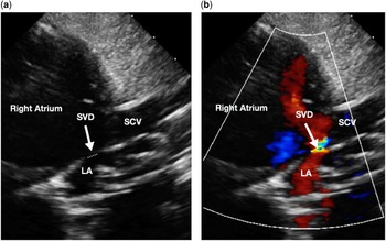
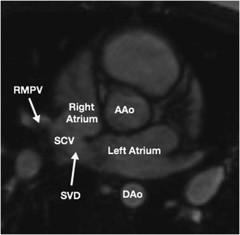
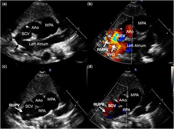
Our report does emphasise the ongoing difficulty in fully defining the extent of the anomalous connection of the right pulmonary veins. In a significant number of our patients, the transthoracic echocardiographic examination identified only the anomalous connection of the right upper pulmonary vein, failing to reveal the anomalously connected right middle pulmonary veins. Transthoracic interrogation has classically depended on the use of imaging in the sagittal plane to define superior sinus venosus defects, usually obtained through subcostal short-axis and high right parasternal views (Fig 4).Reference Plymale, Kolonski, Frommelt, Bartz, Tweddell and Earling 10 These views are adequate to assess the interatrial communication, but they place the defect in a more perpendicular plane relative to the ultrasound beam; because of this, they are limited in their evaluation of the associated anomalous pulmonary venous connections. These veins typically enter along the rightward aspect of the superior caval vein or at its rightward junction with the right atrium. When using standard views, these anomalous right pulmonary veins are insonated in an orthogonal plane. Visualisation depends on the sonographer sweeping rightward, which is limited by lung artefacts and by the pulmonary venous inflow entering perpendicular to the color Doppler signal. Our own institutional review revealed little focus on the use of axial transthoracic echocardiographic imaging when assessing sinus venosus defects, despite its recognised importance when using cardiac magnetic resonance (Fig 5). Use of the orthogonal plane places the superior sinus venosus defect in a right–left axis, with the additional benefit of sweeping in the superior direction to reveal any pulmonary vein connecting anomalously to the superior caval vein at any point from its junction with the right atrium (Fig 6). We propose, therefore, that those using transthoracic echocardiography to provide a precise diagnosis of sinus venosus defects, and in particular when seeking to define the essential associated anomalous pulmonary venous connections, should focus on using short-axis imaging from the parasternal window in addition to the standard sagittal views.
( a ) Transthoracic echocardiogram (TTE) high right parasternal two-dimensional (2D) view demonstrating a superior sinus venosus defect (SVD) (actual diameter marked by the white dotted line, more evident by color Doppler) located at the superior caval vein (SCV)–right atrial junction. Override of the SCV with the intact primary atrial septum is not apparent, however, was reported at the subsequent surgical repair. ( b ) TTE high right parasternal color Doppler view demonstrating left-to-right flow across the superior SVD, suggesting a smaller defect than that seen on 2D imaging. LA=left atrium.

Cardiac magnetic resonance axial imaging demonstrating a large superior sinus venosus defect (SVD) with anomalous drainage of the right middle pulmonary vein (RMPV) into the lateral aspect of the superior caval vein (SCV) at its junction with the right atrium. AAo=ascending aorta; DAo=descending aorta.

( a ) Transthoracic echocardiogram (TTE) parasternal short-axis (PSAx) two-dimensional (2D) view demonstrating a superior sinus venosus defect (SVD), with right middle pulmonary vein (RMPV) anomalously draining into the posterolateral aspect of the superior caval vein (SCV). ( b ) TTE PSAx color Doppler demonstrating left-to-right flow across the superior SVD, with flow from the RMPV into the SCV as described. ( c ) TTE PSAx 2D view, sweeping superiorly from the previous images, demonstrating an additional anomalous vein, the right upper pulmonary vein (RUPV) draining into the posterolateral aspect of the SCV. ( d ) TTE PSAx color Doppler confirming anomalous drainage of the RUPV into the SCV. AAo=ascending aorta; MPA=main pulmonary artery; RPA=right pulmonary artery.

Developmental concepts
An understanding of the mechanisms of normal atrial septation and establishment of the venoatrial connections is paramount to the understanding of the morphology of the sinus venosus defects. This is more important in light of the recognised need to have anomalous pulmonary venous connections so as to produce these particular interatrial communications. It has been known since the turn of the 19th century that the so-called “septum secundum” is not really a septum at all. Instead, it is an infolding of the superior and posterior atrial walls between the attachments of the pulmonary veins to the left atrium and the caval veins to the right atrium.Reference Rose 4 Understanding the true nature of this superior interatrial fold, which makes up the cranial margin of the oval fossa, is key to appreciating the nature of the sinus venosus defects. If guided by the illustrations in many current textbooks, the surgeon or interventionalist would find himself or herself outside of the heart if dissecting or puncturing through the alleged “septum secundum”. The sequence of events occurring during normal development as revealed by episcopic microscopyReference Anderson, Mohun and Brown 7 has now served to demonstrate the formation of the superior interatrial fold. These findings also serve to reveal the pitfalls in previous hypotheses used to explain the morphogenesis of the sinus venosus defects.
The evidence derived from the episcopic data sets shows in the first place that it is not possible to recognise the boundaries of the systemic venous sinus until after the formation of the venous valves. The primary pulmonary vein, furthermore, does not canalise within the mid-pharyngeal strand until after the appearance of the venous valves.Reference Anderson, Brown and Moorman 11 This progression has been well demonstrated in the avian heart.Reference Webb, Brown, Anderson and Richardson 12 It is the comparisons between mouse and man, however, that are more relevant. This is because in man, as in the mouse, the pulmonary vein is first formed adjacent to the left atrioventricular junction.Reference Webb, Brown, Wessels and Anderson 13 In the human heart, the folding of the superior interatrial walls to produce the alleged “septum secundum” cannot take place until after the right pulmonary veins have migrated to reach the atrial roof, a process that only occurs subsequent to 8 weeks of development.Reference Webb, Kanani, Anderson, Richardson and Brown 14 When using valid markers to distinguish between the pulmonary venous and the systemic venous myocardium in the developing human heart, furthermore, it has now been shown that the developing pulmonary vein in man is always discrete from the developing tributaries of the systemic venous sinus.Reference Sizarov, Anderson, Christoffels and Moorman 15 This finding revealed the deficiencies of previous studies that used markers such as HNK-1, CCS-LacZ, and podoplanin. Investigators using these agents argued that they were “transient” markers of the sinus venosus myocardium. They interpreted the findings to suggest that a common wall was shared between the caval and pulmonary veins.Reference Van Praagh, Carrera, Sanders, Mayer and Van Praagh 3 , Reference Blom, Gittenberger-de Groot, Jongeneel, DeRuiter, Poelmann and Ottenkamp 16 – Reference Mahtab, Vicente-Steijn and Hahurij 18 These immunochemical markers, however, demonstrate features such as migrations of cells in general, rather than demonstrating cellular lineages. They do not distinguish between the systemic and the pulmonary myocardium, as was achieved in the investigation of the developing human heart.Reference Sizarov, Anderson, Christoffels and Moorman 15
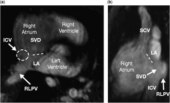
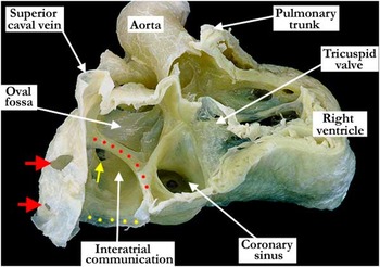
By combining the information gained from the study of autopsied specimens with sinus venosus defectsReference Butts, Crean and Hlavacek 5 with the knowledge of the changes occurring during normal development,Reference Anderson, Mohun and Brown 7 we can now show that the sinus venosus defects are formed when there is anomalous connection of one or more pulmonary veins to a systemic venous channel, but with the anomalously connected pulmonary vein or veins retaining its or their left atrial connection. This process produces a veno-venous connection, which then provides the conduit for interatrial shunting. This abnormal process can involve either the superior or the inferior cavoatrial junctions.Reference Anderson, Brown and Mohun 19 In some instances, however, as in our one patient in whom there seemingly was no associated anomalous pulmonary venous connection, the pulmonary vein involved can retain its connection almost exclusively with the cavity of the left atrium, so that its anomalous connection may not immediately be appreciated. The posterior wall of such a defect, nonetheless, remains formed by the union of the inferior caval vein with the pulmonary vein, thus producing the veno-venous conduit, which permits the interatrial shunting (Fig 7). The autopsy specimen displayed in Figure 8 also demonstrates this concept. A comparable but opposite arrangement is seen in the setting of the superior sinus venosus defect, which usually involves overriding of the rim of the oval fossa by the mouth of the superior caval vein. If the anomalous pulmonary vein is connected at a distance relative to the cavoatrial junction, the superior caval vein itself can drain exclusively to the cavity of the right atrium.
( a ) Cardiac magnetic resonance (CMR) axial imaging obtained in patient 12 demonstrating where the inferior caval vein (ICV) (dashed circle) enters just inferior to this imaging plane into the right atrium, overriding the interatrial septum (dashed line). The right lower pulmonary vein (RLPV) appears to enter the left atrium (LA); however, the posteroinferior wall of the inferior sinus venosus defect (SVD) is formed by the union of the ICV and RLPV. ( b ) CMR sagittal image of the same patient demonstrating the ICV entering the right atrium, overriding the interatrial septum (dashed line) and inferior SVD, with the RLPV entering just superior to the ICV into the LA, with the continuity of the ICV and RLPV forming the posteroinferior border of the SVD. SCV=superior caval vein.

The free walls of the right atrium and ventricle in this heart with double-outlet right ventricle have been removed. Within the right atrium, the flap valve at the floor of the oval fossa is well formed, and the coronary sinus is large secondary to a persistent left superior caval vein draining to it. The superior caval vein enters the roof of the right atrium in the usual manner and the inferior caval vein (yellow dots) is dilated. There is a sinus venosus defect or interatrial communication associated with the inferior caval vein. Its superior-most rim marked with red dots is well circumscribed and is clearly separated from the true atrial septal structures. The right middle and lower pulmonary veins are marked with the red arrows and connect with the walls of the inferior caval vein but also maintain a connection with the left atrium. Within the left atrium, the yellow arrow marks the left lower pulmonary vein.

We recognise that our clinical study is limited by its small sample size and by its retrospective nature, depending much on the interpretation of written reports and surgical operative notes. It was also the case that preoperative reports of transoesophageal echocardiographic interrogations were not available for a small portion of the study cohort, and that interpretations of some of the available preoperative transoesophageal interrogations may have been influenced by features evident from previous cardiac magnetic resonance.
Conclusions
Our study has shown that a proper understanding of cardiac development is crucial to the appreciation of the anatomical variability to be found in the spectrum of sinus venosus defects, which represent retained veno-venous connections. We believe that knowledge of such information will improve the interpretation of the images obtained subsequent to clinical investigation, particularly the most appropriate planes to use for imaging. In this way, it will be possible to provide the surgeon with an appropriate description of the problem at hand, guiding the correct surgical approach.
Acknowledgements
None.
Financial Support
This research received no specific grant from any funding agency, commercial or not-for-profit sectors.
Conflicts of Interest
None.