Case description
A 26-year-old woman was referred for a foetal echocardiography due to an abnormal three-vessel-trachea view on a mid-trimester ultrasound. In the 22nd week of pregnancy, the diagnosis of transposition of the great arteries with small ventricular septal defect was made. At that time, as well as in the 30th week, the flow through the foramen ovale was bidirectional with freely moving thin septum primum, and the ductus arteriosus shunt was unrestrictive right to left.
In the 32nd week of pregnancy, the patient was admitted to the regional hospital with threatened pre-term labour. She received tocolysis with atosiban (oxytocin and vasopressin antagonist) and steroids for foetal lung maturation (2 × 12 mg of betamethasone). She was discharged home after 4 days. No foetal echocardiogram was performed at admission or before discharge.
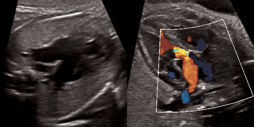
The next foetal echocardiographic examination in 34th week showed markedly dilated pulmonary arteries, pulmonary valve regurgitation, impaired function of the left ventricle (visually decreased contractility, Tei index > 0.5), and the complete closure of the ductus arteriosus (Fig 1 and Supplementary Fig S1). The foramen ovale flap was thick and hypermobile, enabling limited, bidirectional flow. The reason for ductal closure was unclear, with the prophylactic steroid course as the most probable factor and undetermined effect of atosiban.
Foetal echocardiography. Left panel – dilated pulmonary arteries. Right panel – pulmonary regurgitation, no flow at the expected site of ductus arteriosus (*).

Because the echocardiographic findings were consistent with long-standing, irreversible ductal closure, indicating a high risk of severe pulmonary hypertension in the newborn, the decision to perform an urgent caesarean section was made. The girl was born, weighing 2200 g, and received an 8–9 Apgar score. However, her condition rapidly deteriorated, and she became severely cyanotic and required intubation in the fifth minute of life. The blood oxygen saturation, initially around 45% on ventilation with oxygen, increased to 60% after initiation of inhaled NO (20 ppm). The bedside echocardiography showed redundant interatrial septum with limited left-to-right flow and a narrow vessel – ductus arteriosus or collateral – therefore, the prostaglandin E1 infusion at high dose (0.05 µg/kg per minute) was also started.
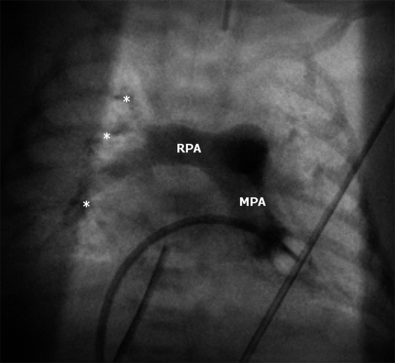
The newborn was immediately transported to the catheterisation lab and had an atrioseptostomy performed, with no further increase in saturation (at that time of about 50%). Pulmonary angiography showed typical findings for pulmonary vascular disease – dilation of the central pulmonary arteries and attenuation of the peripheral vessels (Fig 2). No patent ductus arteriosus was seen despite prostaglandin infusion.
Angiogram (0º) showing dilation of the central pulmonary arteries (RPA and MPA) and attenuation of the peripheral vessels (*). RPA = right pulmonary artery; MPA = main pulmonary artery.

Additionally to inhaled NO, epoprostenol infusion (20–40 ng/kg per minute) was started. During the following days, the condition of the child remained very poor and unstable, with blood saturation of 50–75% and systemic hypotension requiring catecholamines. Attempts to decrease the doses of pulmonary vasodilating agents led to a desaturation below 50%, with no improvement after restoring the initial doses – likely due to the opening of the arteriovenous shunts in the lungs. Extracorporeal membrane oxygenation was considered, but eventually not used due to the small size of the baby and pre-maturity, sonographic signs of hypoxic-ischaemic encephalopathy (high risk of bleeding), and, most likely, the irreversible character of pulmonary hypertension. The child developed multi-organ failure, with anuria, cholestasis, and generalised oedema. On the 16th day of life, the multi-specialist team deemed further extension of the treatment as futile therapy. The child received comfort care and died 2 days later. At the request of the family, an autopsy was not performed.
Discussion
Transposition of the great arteries is one of the most common cyanotic heart malformations, accounting for 5–7% of CHDs. It is well tolerated haemodynamically in the prenatal period, because of foetal shunts: the foramen ovale and the ductus arteriosus. After birth, the situation changes dramatically, as two separate parallel blood circuits are created, which is incompatible with life.Reference Wernovsky, Allen, Driscoll, Shaddy and Feltes1
The current management of newborns with transposition of the great arteries includes the prostaglandin E1 infusion for ductal patency, and, if needed, restoring adequate flow at the level of interatrial septum with balloon atrioseptostomy procedure. These interventions usually allow the stabilisation of the newborn’s condition until the corrective operation.Reference Wernovsky, Allen, Driscoll, Shaddy and Feltes1
There is, however, a small subgroup of patients with transposition who, despite prostaglandin infusion and adequate-sized interatrial communication, develop intractable hypoxemia, acidosis, and often die pre-operatively.Reference Karimi, Kirshbom, Kopf, Steele and Sullivan2 This phenomenon is attributed to persistent pulmonary hypertension of the newborn, defined as failure of normal pulmonary vascular resistance to fall after birth and usually presenting with varying degrees of respiratory failure.Reference Mathew and Lakshminrusimha3 In newborns with transposition of the great arteries, persistent pulmonary hypertension, especially with restrictive interatrial communication, is deemed a critical combination.Reference Karimi, Kirshbom, Kopf, Steele and Sullivan2,Reference Roofthooft, Bergman, Waterbolk, Ebels, Bartelds and Berger4 Management strategies of these patients include balloon atrioseptostomy, administration of pulmonary vasodilators, and extracorporeal membrane oxygenation in refractory cases, in addition to the appropriate ventilation, sedation, and inotropic support.Reference Roofthooft, Bergman, Waterbolk, Ebels, Bartelds and Berger4 The data on extracorporeal membrane oxygenation use in either pre- or post-operative period are not extensive; however, they show a possibility to stabilise the patient until the corrective surgery and reverse the pulmonary hypertension in some cases. Still, the mortality among patients requiring extracorporeal membrane oxygenation remains the highest. Timing of arterial switch operation is controversial, and some authors suggest early surgery with post-operative extracorporeal membrane oxygenation support as a preferred option.Reference Sallaam, Natarajan and Aggarwal5
The pathophysiology of persistent pulmonary hypertension in newborns with transposition is complex and not yet fully understood. It is suggested that the prenatal restriction of the ductus arteriosus increasing the pulmonary blood flow may lead to anatomical changes in pulmonary arterioles, which were demonstrated in histological studies.Reference Maeno, Kamenir, Sinclair, van der Velde, Smallhorn and Hornberger6,Reference Kumar, Taylor, Sandor and Patterson7 Prenatal ductal constriction has long been related to maternal intake of non-steroidal anti-inflammatory drugs in the third trimester of pregnancy, which inhibit the prostaglandin synthesis pathway. However, there is a growing area of evidence that many other substances, especially polyphenol-rich foods and potentially corticosteroids, may have a similar effect. The pathophysiologic mechanisms are yet to be elucidated; however, disruption of arachidonic acid signalling and modulating effect on ductal wall reactivity to different stimuli are implied.Reference Zielinsky and Busato8 This fact is of great clinical importance, since corticosteroids are often used in threatened pre-term labour, and many foods generally considered as healthy (e.g., green tea, oranges, grapes, berries) are rich in polyphenols.
In our patient, the reason for ductal closure was unclear; however, since there was no history of exposure to non-steroidal anti-inflammatory drugs or polyphenol-rich food, prophylactic steroid course seemed the most probable factor. There is no existing data on the influence of atosiban on the ductus arteriosus,9 and therefore, we cannot exclude its co-effect – it should be subject to further research. In the literature, we found just one description of complete closure of the ductus arteriosus in a foetus with transposition, also with no apparent cause.Reference Donofrio10
Conclusions
Restriction of the ductus arteriosus in a foetus with transposition of the great arteries can lead to severe pulmonary hypertension in the newborn, which in some cases can be irreversible despite aggressive postnatal management.
It is crucial to inform pregnant patients with transposition of the great arteries in their foetuses to avoid substances with possible ductal-constricting properties, and, if they are used, to serially assess the blood flow in the ductus arteriosus.
In foetuses with prenatally diagnosed ductal-dependent lesions, corticosteroids should be administered with caution, preferably under echocardiographic monitoring.
Supplementary Material
To view supplementary material for this article, please visit https://doi.org/10.1017/S1047951120000529
Acknowledgements
The authors thank Grażyna Brzezińska-Rajszys, Paweł Własienko, Agata Serwatowska-Bargieł, Eliza Michalska, and Anna Migdał for their contribution.
Financial Support
This research received no specific grant from any funding agency, commercial or not-for-profit sectors.
Conflicts of Interest
None.
Ethical Standards
The authors assert that all procedures contributing to this work comply with the ethical standards of the national guidelines on human experimentation and with the Helsinki Declaration of 1975, as revised in 2008.