Introduction
A vascular ring refers to encirclement of the trachea and oesophagus by an abnormal combination of derivatives of the aortic arch system. Vascular rings represent approximately 1 per cent of congenital cardiovascular anomalies. They can be wholly vascular, or a combination of vascular and ligamentous elements. These malformations can cause variable compression of the oesophagus, trachea or both. Thus, symptoms can range from no effect to severe stridor, dyspnoea, dysphagia and/or cyanotic episodes.
In the past, the main radiological investigations used to identify and define the anatomy of a suspected vascular ring included plain chest radiography and barium swallow studies. These were often supplemented with angiography.
However, with the advent of magnetic resonance imaging (MRI) and multidetector computed tomography (CT), it is now easy to obtain a comprehensive image of the anatomical components of a vascular ring, and to assess its effect on the trachea and/or oesophagus.
Methods
We present a case series of six paediatric patients managed over a six-year period (2003–2009), who exhibited the features of four different types of vascular ring.
These patients were investigated using microlaryngoscopic bronchoscopy. A vascular ring was suspected on discovery of a pulsatile indentation or constriction of the tracheobronchial wall, with reduction of cross-sectional area. Microlaryngoscopic bronchoscopy was performed under spontaneous ventilation, using either a Parsons or Benjamin laryngoscope, through which a 0° rigid Hopkins endoscope was introduced.
Following microlaryngoscopic bronchoscopy, those children with a suspected vascular malformation (e.g. those in whom the investigation had identified pulsation associated with tracheal narrowing) underwent either multidetector CT or MRI to confirm the diagnosis.
Results
Our patient series comprised six children (four girls and two boys) aged from 37 weeks to four years. Using these cases, we herein present the typical clinical scenario, microlaryngoscopic bronchoscopy findings and radiological features of each of the four different types of vascular ring, i.e. pulmonary sling (patient A), double aortic arch (patient B), right aortic arch with anomalous left subclavian artery (patient C), and aberrant right subclavian artery (patients D, E and F).
Pulmonary sling
The clinical scenario of a child with a pulmonary sling is illustrated by the case of patient A.
This boy was delivered at 39 weeks' gestation by emergency lower segment caesarean section due to fetal distress, at a peripheral maternal unit. Apgar scores at one and five minutes were normal.
Twenty-four hours post-delivery, the infant developed cyanotic episodes, stridor and tachypnoea which were worse during feeding. A lower respiratory infection was initially suspected, and the infant was commenced on intravenous antibiotics. However, he failed to improve, and was transferred to a tertiary paediatric centre for further investigation.
A vascular ring was suspected, and microlaryngoscopic bronchoscopy was thus performed. The endoscopist noted external compression with pulsation of the lower trachea, and a segment of complete tracheal rings.
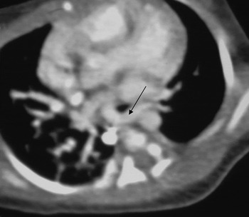
Computed tomography angiography confirmed the presence of a pulmonary sling (Figure 1).
Axial computed tomography pulmonary angiogram demonstrating a pulmonary sling (arrow).

A cardiothoracic opinion was requested.
This infant underwent surgical division of his vascular ring and repair of his tracheal abnormality via slide tracheoplasty.
On review at the out-patient clinic one month post-operatively, the child's symptoms had resolved. At the time of writing, he was doing well.
Double aortic arch
Patient B illustrates the clinical scenario of a child with a double aortic arch.
This patient, a six-month-old girl with Down's syndrome, was referred to our paediatric otorhinolaryngology service with stridor.
Microlaryngoscopic bronchoscopy demonstrated a moderate degree of laryngomalacia, and an aryepiglottoplasty was performed. In addition, there was significant external compression of the lower trachea, with pulsation.
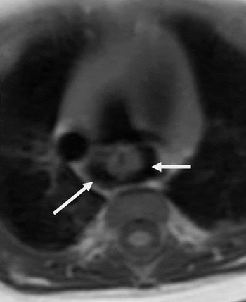
Magnetic resonance angiography demonstrated a double aortic arch, with a right descending aorta compressing the trachea (Figure 2).
Axial magnetic resonance angiogram demonstrating a double aortic arch (arrows).

The infant was referred to our cardiothoracic colleagues, who performed a division and repair of her vascular ring.
Repeated microlaryngoscopic bronchoscopy three months later showed significant improvement in the child's airway, with no compression or collapse of the lower trachea.
At the time of writing, this child was well with no symptoms.
Right aortic arch with anomalous left subclavian artery
The clinical scenario of a child with a right aortic arch with anomalous left subclavian artery is illustrated by patient C.
This female infant was delivered at 37 weeks' gestation by lower segment caesarean section. She was diagnosed with DiGeorge syndrome (22q deletion).
On day 13, the infant developed stridor and cyanotic episodes which were exacerbated during feeding.
Microlaryngoscopic bronchoscopy demonstrated a type 1 laryngeal cleft with a normal trachea.
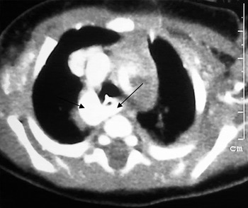
Computed tomography angiography was performed, and showed a right aortic arch with anomalous left subclavian artery (Figure 3).
Axial computed tomography angiogram demonstrating a right aortic arch with aberrant left subclavian artery (arrows).

A cardiothoracic opinion was sought. However, no surgery was performed, due to this infant's underlying medical condition.
Aberrant right subclavian artery
The clinical scenario of a child with an aberrant right subclavian artery is illustrated by our last three patients.
Patient D was a four-year-old girl with Down's syndrome, who was referred to the paediatric otorhinolaryngology service with stridor. The child had suffered stridor since birth, with associated feeding difficulties. Microlaryngoscopic bronchoscopy demonstrated a 1.5 cm segment of complete tracheal rings at the mid-trachea. Magnetic resonance angiography identified the presence of an aberrant right subclavian artery. In addition, the MRI also confirmed the presence of a segment of complete tracheal rings. A cardiothoracic opinion was sought, and this child underwent a tracheoplasty using the slide tracheoplasty technique. A recent follow up out-patient appointment indicated that this child's symptoms had improved significantly, with only occasional stridor persisting.
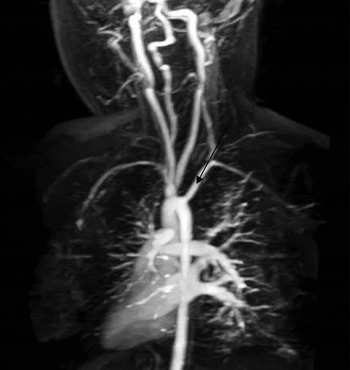
Patient E was a female infant delivered at full term by normal vaginal delivery. At three months of age, the child was referred to the paediatric otolaryngology service with a history of stridor, exacerbated by feeding, since birth. Flexible nasal endoscopy showed mild laryngomalacia. However, on follow up in the out-patient clinic this child's symptoms had failed to improve. Microlaryngoscopic bronchoscopy demonstrated mild laryngomalacia and tracheobronchomalacia, with pulsation of the lower end of the trachea, and an aryepiglottoplasty was performed. Magnetic resonance angiography showed the presence of an aberrant right subclavian artery (Figure 4). The child's symptoms improved following microlaryngoscopic bronchoscopy and aryepiglottoplasty, and she required no further surgical intervention.
Three dimensional reconstructed magnetic resonance angiogram demonstrating an aberrant right subclavian artery (arrow).

Finally, patient F was a male infant delivered at 38 weeks' gestation by lower segment caesarean section. Shortly after delivery, he was diagnosed with Peter's anomaly. (This is a rare form of anterior segment dysgenesis, in which abnormal cleavage of the anterior chamber of the eye results in abnormalities of the cornea; the condition occurs in utero during the first trimester, and is associated with other ocular and systemic abnormalities.)Reference Harissi-Dagher and Colby1 Soon after delivery, the infant developed cyanotic episodes exacerbated by feeding, which did not improve. A paediatric otorhinolaryngology opinion was sought, and microlaryngoscopic bronchoscopy was performed. This demonstrated pulsation of the lower third of the trachea and mild bronchomalacia. Magnetic resonance angiography demonstrated an aberrant right subclavian artery. The child was managed conservatively.
Discussion
A vascular ring is a congenital disorder in which anomalous configurations of the aortic arch and/or associated vessels surround the trachea and oesophagus. Malformations of the aortic arch and its branches are caused by errors in the development of the primitive aortic arches: for example, the persistence of parts which normally regress, or the regression of parts that normally develop.Reference Oddone, Granata, Vercellino, Bava and Toma2 The result is the formation of a complete or incomplete ring around these structures.
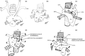
Historically, the first vascular ring to be described was a double aortic arch reported by Hommel in 1737. In 1794, Bayford described a retro-oesophageal right subclavian artery, following the autopsy of a woman who had experienced dysphagia for many years.Reference Turner3, Reference Gross4 Dysphagia due to compression from an aberrant right subclavian artery is also known as ‘dysphagia lusoria’. Although usually asymptomatic, patients may develop symptoms later in life due to the development of atherosclerotic rigidity, tortuosity or aneurysmal dilatation of the aberrant vessel.Reference Gomes, Bernatz and Forth5 Maude Abbott described five cases of double aortic arch in 1932, and suggested that surgical intervention should be undertaken in such patients. Finally, in 1945 Gross was the first to use the term ‘vascular ring’, in the New England Journal of Medicine, after performing the first successful division of a double aortic arch (Figure 5).Reference Backer and Mavroudis6
Diagrams of vascular ring anomalies (a) normal anatomy: (b) double aortic arch; (c) pulmonary sling; (d) right aortic arch with aberrant left subclavian artery; and (e) aberrant right subclavian artery. RCC = right common carotid artery; LCC = left common carotid artery; O = oesophagus; RSA = right subclavian artery; LSA = left subclavian artery; T = trachea; AA = aortic arch; PA = pulmonary artery; LPA = left pulmonary artery; RPA = right pulmonary artery; DA = descending aorta; RCA = right coronary artery; LCA = left coronary artery

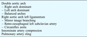
Vascular rings are uncommon and probably make up less than 1 per cent of all congenital cardiac defects. They occur equally in both sexes, with no geographical or racial predominance. Some vascular rings are associated with other cardiac abnormalities, while others occur in isolation. The International Congenital Heart Surgery Nomenclature and Database Committee has recommended the vascular ring classification shown in Table I.Reference Backer, Mavroudis, Rigsby and Hollinger7
A Vascular ring classification*

* As defined by the International Congenital Heart Surgery Nomenclature and Database Committee.
The two most common types of anatomically complete vascular rings are double aortic arch and right aortic arch with left ligamentum arteriosum. Other anomalies that produce symptoms but do not form complete rings (i.e. they constitute anatomically incomplete rings) include the anomalous innominate artery and the right subclavian artery with left-sided aorta and left ligamentum arteriosum. A double aortic arch is formed when the embryonic fourth aortic arches and dorsal aortic roots persist and remain patent on both sides. The resultant ring is tight, and patients generally present early. The development of a right aortic arch is similar to that of a double aortic arch, but the left arch regresses. Several variations exist, including a right aortic arch with aberrant left subclavian artery and mirror image branching.Reference Backer, Mavroudis, Rigsby and Hollinger7, Reference Ruzmetov, Vijay, Rodefield, Turrentine and Brown8
The anomalous left pulmonary artery, also known as the pulmonary sling, accounts for approximately 10 per cent of vascular rings. It is not associated with the aortic arch or its branches, but rather arises from an abnormality of the sixth branchial arch, and produces a complete ring. This abnormality occurs when the left pulmonary artery arises as a branch of the right pulmonary artery, instead of originating from the main pulmonary artery. Most patients are symptomatic shortly after birth and present with respiratory symptoms, as this vascular ring causes significant compression of the trachea. These infants frequently have other tracheobronchial anomalies, such as tracheomalacia, stenosis, webs or complete tracheal rings. The combination of an anomalous left pulmonary artery plus one of these latter lesions is also referred to as a ‘ring-sling complex’.
The most common clinical symptoms of patients with anatomically complete vascular rings are stridor, cough, recurrent upper respiratory tract infections and cyanotic episodes. Oesophageal compression is usually posterior, so slow feeding or fatigue during feeding are among the symptoms that may occur in infancy. The introduction of solid food often causes more pronounced dysphagia. However, some patients with a complete vascular ring have minimal symptoms or are asymptomatic. In such patients, the abnormal arch anatomy is usually discovered incidentally.Reference Bakker, Berger, Witsenburg and Bogers9, Reference Malik, Bruce, Kaushik, Willatt, Wright and Rothera10
There has been a major shift in the last 20 years with regards to investigating children with a suspected vascular ring. Multidetector CT or MRI is the first-line imaging modality for diagnostic investigation of paediatric airway obstruction suspected to be due to a vascular ring. The multiplanar and three-dimensional imaging capabilities of CT and MRI angiography allow excellent delineation of the aortic arch and its branches. If assessment of the airway is important, CT is currently more reliable than MRI for definition of the airways. Advocates of MRI maintain that it is noninvasive and avoids the use of ionising radiation.Reference Schulman-Hernanz11–Reference Saleh, Patel, Lee, Boechat, Ratib and Saraiva13
The preference of multidetector CT over MRI is based on the speed with which the scan can be obtained. Due to the development of multidetector technology, the speed of CT scans has now increased such that images can be obtained in minutes, and usually require only mild sedation. Neonates may be imaged with a ‘feed and wrap’ approach, without sedation. State-of-the-art multidetector scanners now allow imaging during quiet breathing, without appreciable respiratory artefact. In comparison, an MRI can take up to 30 minutes and thus will usually require general anaesthetic in young children, as controlled breath-holding is essential for achieving high quality contrast-enhanced images.Reference Backer, Mavroudis, Rigsby and Hollinger7, Reference Schulman-Hernanz11–Reference Saleh, Patel, Lee, Boechat, Ratib and Saraiva13 In children with an associated cardiac defect, MRI has the advantage of being able to provide additional information on ventricular function and intracardiac blood flow. In some institutions, MRI is the preferred modality, and CT is performed to evaluate aortic arch anomalies only if MRI is contraindicated, for example in unstable patients or cases of suspected thoracic or lung abnormality.Reference Lee, Siegel, Hildebolt, Gutierrez, Bhalla and Fallah14
Direct microlaryngoscopic bronchoscopy is an ideal first-line investigation as it allows the surgeon to view the entire airway. Microlaryngoscopic bronchoscopy can diagnose a variety of airway problems which may be associated with a vascular ring, and allows functional assessment of a child's overall airway. Typical findings in patients with vascular rings include extrinsic tracheal compression, abnormal pulsation and complete tracheal rings.
Our institution uses a combination of microlaryngoscopic bronchoscopy and either MRI or multidetector CT to investigate and diagnose vascular rings, and this approach is supported by other centres.Reference Backer, Mavroudis, Rigsby and Hollinger7, Reference Schulman-Hernanz11
• A vascular ring refers to encirclement of the trachea and oesophagus by an abnormal combination of derivatives of the aortic arch system
• Multidetector computed tomography or magnetic resonance imaging is the first-line imaging modality for investigation of paediatric airway obstruction suspected to be due to a vascular ring
• Direct microlaryngoscopic bronchoscopy is an ideal first-line investigation, as it allows the surgeon to view the entire airway
• The paediatric otolaryngologist plays a key role in identifying cases in children who present with airway symptoms
However, despite all the available methods of investigation, a high degree of clinical suspicion remains the most important factor in the diagnosis of paediatric airway obstruction due to a vascular ring.
Surgical division of symptomatic vascular rings is the only appropriate form of therapy.
Any child who presents with a symptomatic vascular ring, especially with airway symptoms, should be referred to cardiothoracic surgical colleagues for further management.
Conclusion
Vascular rings are relatively rare, and very interesting, vascular anomalies. The majority will present early on in life, due to symptoms caused by compression of the trachea and/or oesophagus, particularly in patients with complete vascular rings. Incomplete rings may produce mild symptoms or none at all, and thus are sometimes incidental findings.
In general, the ‘gold standard’ for investigating children with suspected vascular rings includes microlaryngoscopic bronchoscopy used in combination with either multidetector CT or MRI. Patients with vascular rings require a high index of clinical suspicion in order to establish the diagnosis. The paediatric otolaryngologist plays a key role in identifying cases in children who present with airway symptoms.