Introduction
The first synthetic uranium oxalates were reported in 1842 by Eugène-Melchior Péligot (e.g. Péligot, Reference Péligot1842). From these, he was able to show that the material that Martin Heinrich Klaproth discovered and named ‘urane’ in 1789 was actually an oxide, rather than a pure element. Péligot obtained green and yellow uranium oxalates by adding oxalic acid to uranium chloride in aqueous solution. He proposed the name ‘uranium’ for the metallic element and ‘uranyl’ for the yellow salts of uranium. As early as 1930, synthetic uranyl oxalate was being used in chemical actinometers. Today, actinide oxalates have important applications (Abraham et al., Reference Abraham, Arab-Chapelet, Rivenet, Tamain and Grandjean2014), particularly in the separation reprocessing of spent nuclear fuel, because of the chelating ability of the oxalate group with actinides. In the last several decades, there have been numerous synthetic and structural studies of uranyl oxalates because of the unusual array of linking geometries that they demonstrate (Abraham et al., Reference Abraham, Arab-Chapelet, Rivenet, Tamain and Grandjean2014; Giesting et al., Reference Giesting, Porter and Burns2006; Loiseau et al., Reference Loiseau, Mihalcea, Henry and Volkringer2014); however, until now, uranyl oxalates have not been found in Nature.
Herein, we describe the first two uranyl oxalate minerals, uroxite and metauroxite. The name ‘uroxite’ is based on the mineral being a uranyl (UR) oxalate (OX). The name ‘metauroxite’ is based on the compositional similarity of the mineral to uroxite, with one less H2O group per formula unit (pfu). These minerals correspond to known synthetic phases and both were reported with their structures by Duvieubourg et al. (Reference Duvieubourg, Nowogrocki, Abraham and Grandjean2005). The new minerals and their names were approved by the Commission on New Minerals, Nomenclature and Classification of the International Mineralogical Association based upon proposals IMA2018-100 (Kampf et al., Reference Kampf, Plášil, Nash, Němec and Marty2018) for uroxite and 2019-030 (Kampf et al., Reference Kampf, Plášil, Nash and Marty2019) for metauroxite. The type specimens, all micromounts, have been deposited in the collections of the Natural History Museum of Los Angeles County, 900 Exposition Boulevard, Los Angeles, CA 90007, USA. Four uroxite cotypes are assigned catalogue numbers 73514 (Burro), 73515 (Burro), 73516 (Markey) and 73517 (Markey). Two metauroxite cotypes are assigned catalogue numbers 67289 and 67290.
Occurrence
Uroxite and metauroxite were first collected on September 22, 2016 underground in the Burro mine, Slick Rock district, San Miguel County, Colorado, USA (38°2′42″N, 108°53′23″W). Uroxite was collected on the following day, underground in the Markey mine, Red Canyon, White Canyon mining district, San Juan County, Utah (37°32′57″N, 110°18′08″W).
The Burro mine is near the southern end of the Uravan Mineral Belt, in which uranium and vanadium minerals occur together in bedded or roll-front deposits in the sandstone of the Salt Wash member of the Jurassic Morrison Formation (Carter and Gualtieri, Reference Carter and Gualtieri1965; Shawe, Reference Shawe2011). The uranium and vanadium ore mineralisation was deposited where solutions rich in U and V encountered pockets of strongly reducing solutions that had developed around accumulations of carbonaceous plant material. Mining operations have exposed both unoxidised and oxidised U and V phases. Under ambient temperatures and generally oxidising near-surface conditions, water reacts with pyrite to form aqueous solutions with relatively low pH, which then react with the earlier-formed phases, resulting in diverse suites of secondary minerals.
The Markey mine is located ~1 km southwest of the Blue Lizard mine, on the east-facing side of Red Canyon, ~72 km west of the town of Blanding, Utah, and ~22 km southeast of Good Hope Bay on Lake Powell. The geology of the Markey mine is quite similar to that of the Blue Lizard mine (Chenoweth, Reference Chenoweth1993; Kampf, et al., Reference Kampf, Plášil, Kasatkin, Marty and Čejka2017), although the secondary mineralogy of the Markey mine is notably richer in carbonate phases. The information following is taken largely from Chenoweth (Reference Chenoweth1993). Mineralised channels are in the Shinarump member of the Chinle Formation. The Shinarump member consists of medium- to coarse-grained sandstone, conglomeratic sandstone beds and thick siltstone lenses. Ore minerals were deposited as replacements of wood and other organic material and as disseminations in the enclosing sandstone (Chenoweth, Reference Chenoweth1993). Since the mine closed in 1978, oxidation of primary ores in the humid underground environment has produced a variety of secondary minerals, mainly sulfates, as efflorescent crusts on the surfaces of mine walls (Kampf, et al., Reference Kampf, Plášil, Kasatkin, Marty and Čejka2017).
Uroxite and metauroxite are both rare and occur on asphaltum–quartz matrix. At the Markey mine, uroxite is associated with feynmanite and gypsum. At the Burro mine, both minerals are associated with abernathyite, gypsum, tyuyamunite, uranopilite and one other unidentified uranyl oxalate. At both the Burro and Markey mines, the oxalate presumably derives from the decomposed/fossilised organic matter noted above, most likely directly from the asphaltum on which the new minerals have formed.
Physical and optical properties
Uroxite
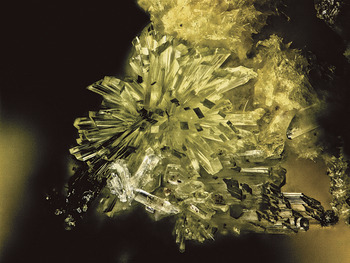
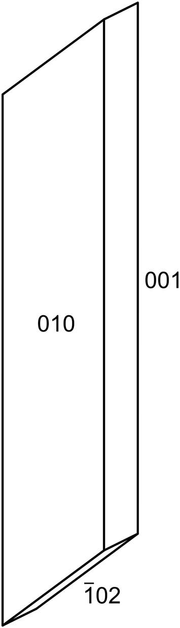
Uroxite occurs in radiating sprays of striated prisms or blades, up to ~1 mm long, elongate and striated on [100], more or less flattened on {010} and with steeply sloping terminations (Figs 1 and 2). The prism forms are {010} and {001} and the termination is { ${\bar 1}$02}. The colour is light yellow, the lustre is vitreous and the streak is very pale yellow. Crystals are brittle, with Mohs hardness of ca 2, uneven fracture, one perfect cleavage on {
 ${\bar 1}$02} and one fair cleavage on {001}. Moderate neon-green fluorescence is observed under 405 nm laser illumination. The density could not be measured with available density liquids; uroxite appears to gradually dissolve in Clerici solution. The calculated density is 4.187 g cm−3 for both the empirical and ideal formulas. Uroxite is optically biaxial (–) with α = 1.602(2), β = 1.660(2) and γ = 1.680(2), determined in white light. The 2V measured directly on a spindle stage is 59(1)°; the calculated 2V is 59.1°. There is moderate r > v dispersion. The optical orientation is Y = b and Z ∧ a = 35° in obtuse β. No pleochroism was observed. The Gladstone–Dale compatibility, 1 – (K p/K c), is 0.005, in the superior range (Mandarino, Reference Mandarino1981) for both the empirical and ideal formulas. At room temperature, uroxite is insoluble in H2O and slowly soluble in dilute HCl.
Uroxite crystals from the Burro mine (cotype #73514). The field of view is 0.68 mm across.

Crystal drawing of uroxite crystal; clinographic projection.

Metauroxite
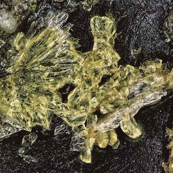
Metauroxite occurs as crude blades and lozenge-shaped tablets, up to ~100 μm in maximum dimension; commonly in irregular and bowtie-like intergrowths to ~200 μm across (Fig. 3). No crystal forms could be measured because faces are generally poorly formed; the tablets are apparently flattened on {011}. Twinning is ubiquitous, by 180° rotation around the perpendicular to {011} (indicated by single-crystal diffraction data); observation of crystals under crossed polars suggests that the twinning is commonly polysynthetic. The colour is light yellow, the lustre is vitreous and the streak is very pale yellow. Crystals are brittle, with Mohs hardness of ca 2, uneven fracture and two good cleavages probably on {101} and {010} on the basis of the structure. Weak green–grey fluorescence is observed under 405 nm laser illumination. The density could not be measured with available density liquids. The calculated density is 4.403 g cm−3 for both the empirical and ideal formulas. Optical determinations were problematic because of poor crystal quality and polysynthetic twinning. It was only possible to obtain approximate minimum (α′) and maximum (γ′) indices of refraction: α′ = 1.615(5) and γ′ = 1.685(5) measured in white light. The average of the estimated minimum and maximum indices of refraction is 1.65, which gives a Gladstone-Dale compatibility, 1 – (K p/K c), of 0.0195, in the superior range (Mandarino, Reference Mandarino1981) for the empirical formula. At room temperature, metauroxite is insoluble in H2O and slowly soluble in dilute HCl.
Yellow, bowtie-like intergrowths of metauroxite crystals (right-centre), yellow radiating uroxite prisms (left) and colourless gypsum blades on asphaltum matrix (cotype #67289). The field of view is 0.50 mm across.

Raman and infrared spectroscopy
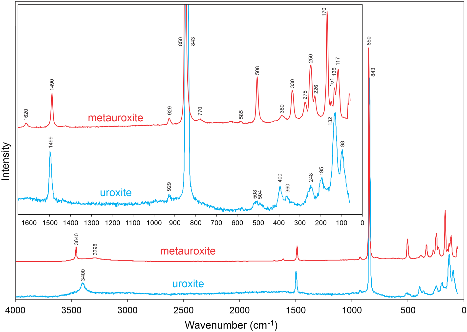
Raman spectra for both minerals were recorded from 4000 to 60 cm−1 using a 532 nm diode laser on a Horiba XploRA PLUS. Uroxite suffered quick decomposition at laser power >1%; however, metauroxite was less susceptible to decomposition, perhaps because its structure does not include an isolated H2O group (see below). The uroxite spectrum was recorded at 1% power and that of metauroxite was recorded at 10% power. The intensity of the uroxite spectrum shown in Fig. 4 was factored by ×6 relative to that of metauroxite.
Raman spectra of uroxite and metauroxite recorded using a 532 nm diode laser. The inset shows the expanded 1650–0 cm−1 region. Note that the uroxite spectrum had to be recorded at much lower laser intensity and has been scaled by ×6 relative to that of metauroxite, which accounts for the lower signal-to-noise ratio for the uroxite spectrum.

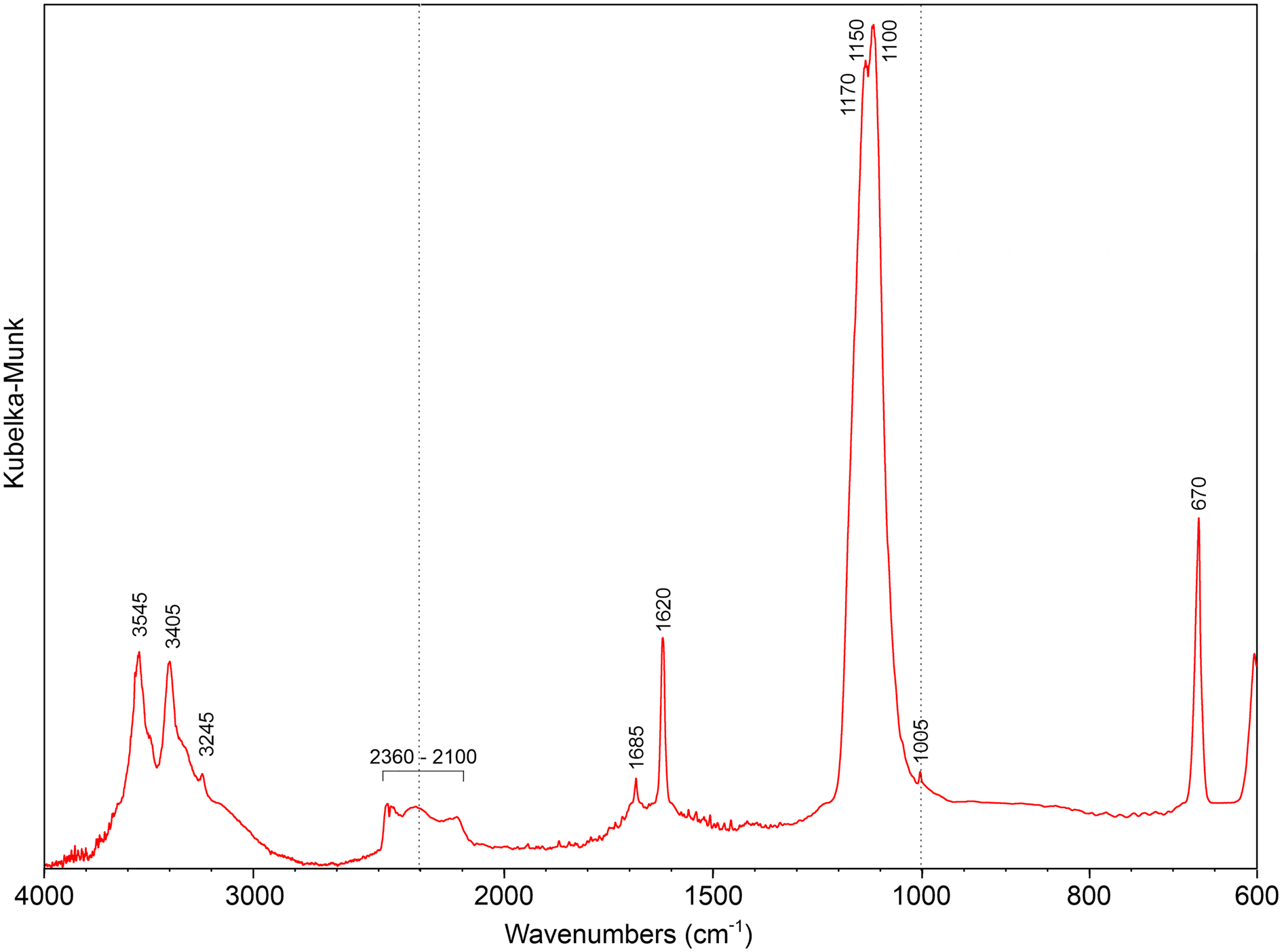
Relatively large crystals of uroxite enabled us to perform micro-diffuse reflectance infrared Fourier transform spectroscopy (micro-DRIFTS) using a Thermo Nicolet 6700 FTIR spectrometer (range 4000–600 cm−1, resolution 4 cm−1, 128 scans, Happ–Genzel apodisation) equipped with a Spectra Tech InspectIR Plus micro-spectroscopic accessory (liquid N2 cooled Hg–Cd–Te detector). Uroxite crystals were pulverised, mixed with KBr and analysed immediately without preparation of the pellet. Pure KBr was taken as the blank reference. The infrared spectrum for uroxite is shown in Fig. 5.
Infrared spectrum (micro-DRIFTS) of uroxite. Note changes in horizontal scale at dotted lines.

The band assignments for the infrared and Raman spectra shown in Table 1 are based on papers by Bartlett and Cooney (Reference Bartlett and Cooney1989), Čejka (Reference Čejka, Burns and Finch1999), Colmenero (Reference Colmenero2019), Mancilla et al. (Reference Mancilla, D'Antonio, González-Baróc and Baranc2009), Peterson and Pullman (Reference Peterson and Pullman2016) and Shippey (Reference Shippey1980). According to the correlation given by Libowitzky (Reference Libowitzky1999), the bands in the spectra attributed to ν (O–H) correspond to hydrogen bonds ranging from ~3.2 to 2.8 Å. Bartlett and Cooney (Reference Bartlett and Cooney1989) provided an empirical relationship to derive the approximate U–OUr bond lengths from the band position assigned to the ν1 (UO2)2+ stretching vibration, which gives 1.77 Å for uroxite and 1.76 Å for metauroxite, in excellent agreement with U–OUr bond lengths from the X-ray data: 1.763 and 1.767 Å for uroxite and 1.775 Å for metauroxite.
Band assignments for the Raman and infrared spectra of uroxite and metauroxite.

Chemical composition
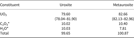
Chemical analyses were performed at the University of Utah on a Cameca SX-50 electron microprobe with four wavelength dispersive spectrometers and using Probe for EPMA software. Analytical conditions were 15 kV accelerating voltage, 10 nA beam current and beam diameters of 15 μm (for uroxite from the Burro mine) and 5 μm (for metauroxite). Counting times were 30 s on peak and 15 s on background. Raw X-ray intensities were corrected for matrix effects with a φρ(z) algorithm (Pouchou and Pichoir, Reference Pouchou and Pichoir1991); ideal formula concentrations of O and C were used for the matrix calculations. Synthetic UO2 was used as the probe standard for UO3. A time-dependent intensity correction was applied for ingrowth of U. Because insufficient material is available for a direct determination of C2O3 or H2O, they are calculated based upon the structure determination. For uroxite from Burro, eight points on four crystals were analysed and, for metauroxite, eight points on five crystals were analysed. The results are given in Table 2.
Chemical compositions (in wt.%) for uroxite and metauroxite.

* Based on the structure.
For uroxite, the empirical formula (calculated on the basis of 13 O atoms pfu) is [(U1.00O2)2(C2O4)(OH)2(H2O)2]⋅H2O and the ideal formula is [(UO2)2(C2O4)(OH)2(H2O)2]⋅H2O, which requires UO3 79.88, C2O3 10.06, H2O 10.06, total 100.00 wt.%.
For metauroxite, the empirical formula (calculated on the basis of 12 O atoms pfu) is (U1.00O2)2(C2O4)(OH)2(H2O)2. The ideal formula is (UO2)2(C2O4)(OH)2(H2O)2, which requires UO3 81.94, C2O3 10.32, H2O 7.74, total 100.00 wt.%.
X-ray crystallography and structure refinement
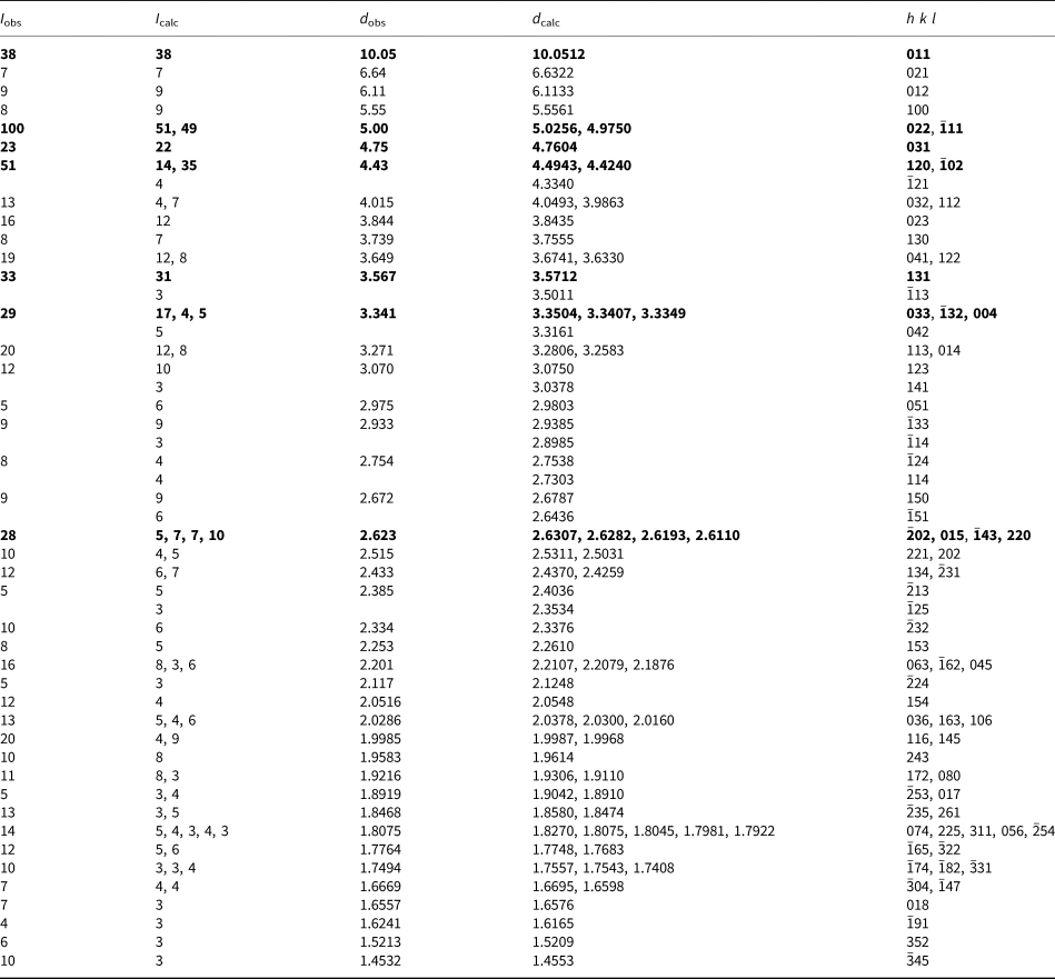
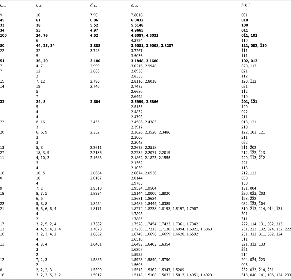
Powder X-ray studies were done using a Rigaku R-Axis Rapid II curved imaging plate microdiffractometer, with monochromatised MoKα radiation (λ = 0.71075 Å). A Gandolfi-like motion on the φ and ω axes was used to randomise the samples and observed d values and intensities were derived by profile fitting using JADE 2010 software (Materials Data, Inc.). The powder data for uroxite (Table 3) and those for metauroxite (Table 4) show good agreement with the patterns calculated from the structure determinations.
Powder X-ray diffraction data (d in Å) for uroxite*.
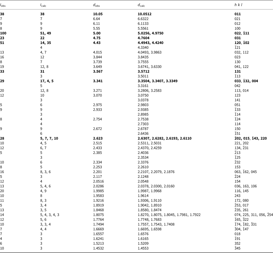
* The calculated intensities have been scaled such that the intensities of the adjacent 022 and  ${\bar 1}$11 lines total 100. After scaling, only calculated lines with I ≥ 3 are included. Further data are in the Supplementary material. The strongest lines are given in bold.
Powder X-ray diffraction data (d in Å) for metauroxite.

* The calculated intensities have been scaled such that the intensities of the adjacent 0 ${\bar 1}$1 and 101 lines total 100. After scaling, only calculated lines with I ≥ 2 are included. The strongest lines are given in bold.
For both minerals, the single-crystal structure data were collected at room temperature using the same diffractometer and radiation noted above. The Rigaku CrystalClear software package was used for processing the structure data, including the application of an empirical absorption correction using the multi-scan method with ABSCOR (Higashi, Reference Higashi2001). For uroxite, a single crystal from the Burro mine was used for the data collection. For metauroxite, the crystal used was twinned by 180° rotation around the perpendicular to {011}, as indicated by the TwinSolve program within the Rigaku CrystalClear software package. Initial structure models were obtained using SIR2011 (Burla et al., Reference Burla, Caliandro, Camalli, Carrozzini, Cascarano, Giacovazzo, Mallamo, Mazzone, Polidori and Spagna2012). SHELXL-2016 (Sheldrick, Reference Sheldrick2015) was used for the refinement of the structures.
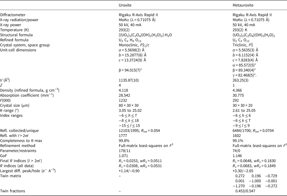
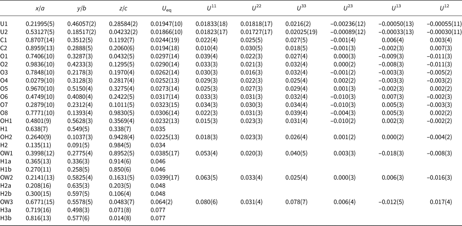
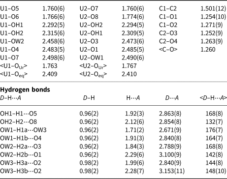
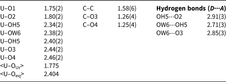
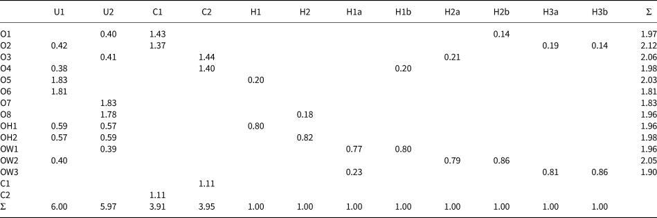
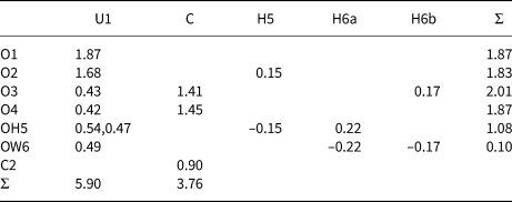
For uroxite, difference-Fourier syntheses located all non-hydrogen atoms not located in the original structure solution, and subsequent cycles located all H sites. The structure was found to be identical to that of synthetic compound (#3 of Duvieubourg et al., Reference Duvieubourg, Nowogrocki, Abraham and Grandjean2005), which lacks the positions of H atoms. The H atoms were refined with soft constraints of 0.97(2) Å on the O–H distances and with the U eq of each H set to 1.5 times that of the donor OH and 1.2 times that of the donor OW atom. For metauroxite, difference-Fourier syntheses failed to locate H atoms. The high residuals are attributed to the relatively poor crystal quality, to the difficulty in integrating the twinned reflections and to the inability of the ABSCOR program to completely correct for the absorption by the twinned crystal. The metauroxite structure was found to be identical to that of synthetic compound #1 of Duvieubourg et al. (Reference Duvieubourg, Nowogrocki, Abraham and Grandjean2005). For both structures, data collection and refinement details are given in Table 5. Atom coordinates and displacement parameters for uroxite are in Table 6 and those for metauroxite in Table 7. Selected bond distances for uroxite are in Table 8 and those for metauroxite are in Table 9. A bond-valence analysis for uroxite is in Table 10 and that for metauroxite is in Table 11. The crystallographic information files have been deposited with the Principal Editor of Mineralogical Magazine and are available as Supplementary material (see below).
Data collection and structure refinement details for uroxite and metauroxite.
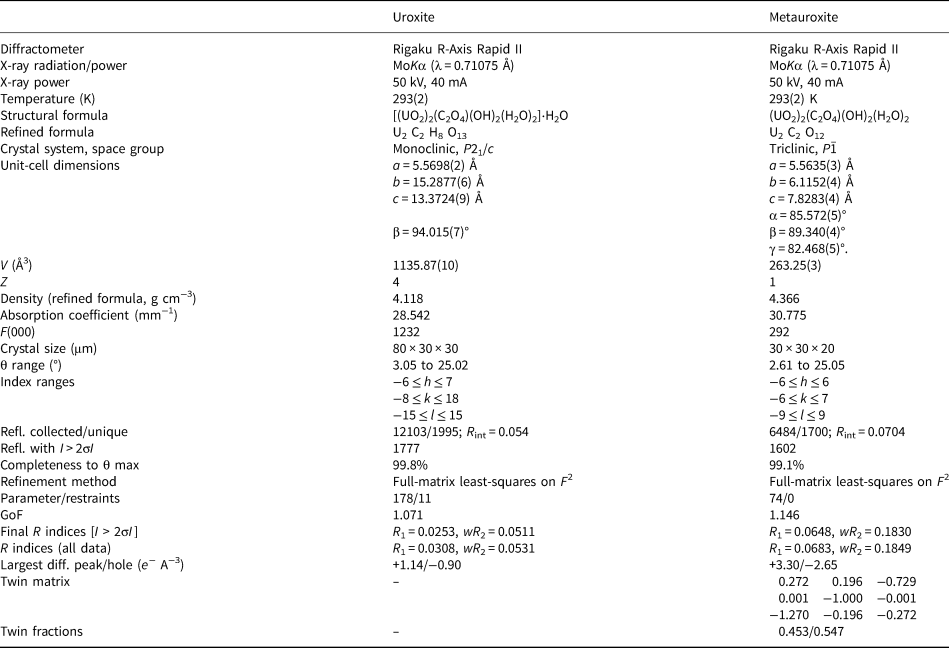
R int = Σ|F o2–F o2(mean)|/Σ[F o2]. GoF = S = {Σ[w(F o2–F c2)2]/(n–p)}1/2. R 1 = Σ||F o|–|F c||/Σ|F o|. wR 2 = {Σ[w(F o2–F c2)2]/Σ[w(F o2)2]}1/2; w = 1/[σ2(F o2)+(aP)2+bP] and P is [2F c2+Max(F o2,0)]/3; for uroxite a is 0.0169 and b is 6.577; for metauroxite a is 0.0746 and b is 28.4245.
Atom coordinates and displacement parameters (Å2) for uroxite.

Atom coordinates and displacement parameters (Å2) for metauroxite.

Selected bond distances (Å) for uroxite.

Selected bond distances (Å) for metauroxite.

Bond-valence analysis for uroxite. Values are expressed in valence units.*

* Bond-valence parameters are from Gagné and Hawthorne (Reference Gagné and Hawthorne2015). H+–O bond valence is based on the graphical relation from Brown (Reference Brown2002).
Bond-valence analysis for metauroxite. Values are expressed in valence units.*

* Bond-valence parameters are from Gagné and Hawthorne (Reference Gagné and Hawthorne2015). Hydrogen-bond strengths are based on O–O bond lengths from Ferraris and Ivaldi (Reference Ferraris and Ivaldi1988).
Descriptions of the structures
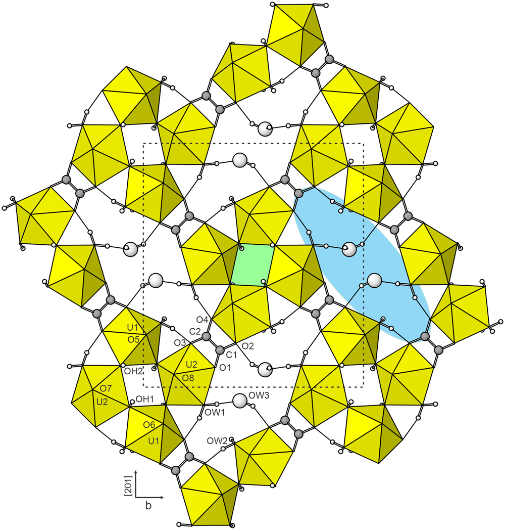
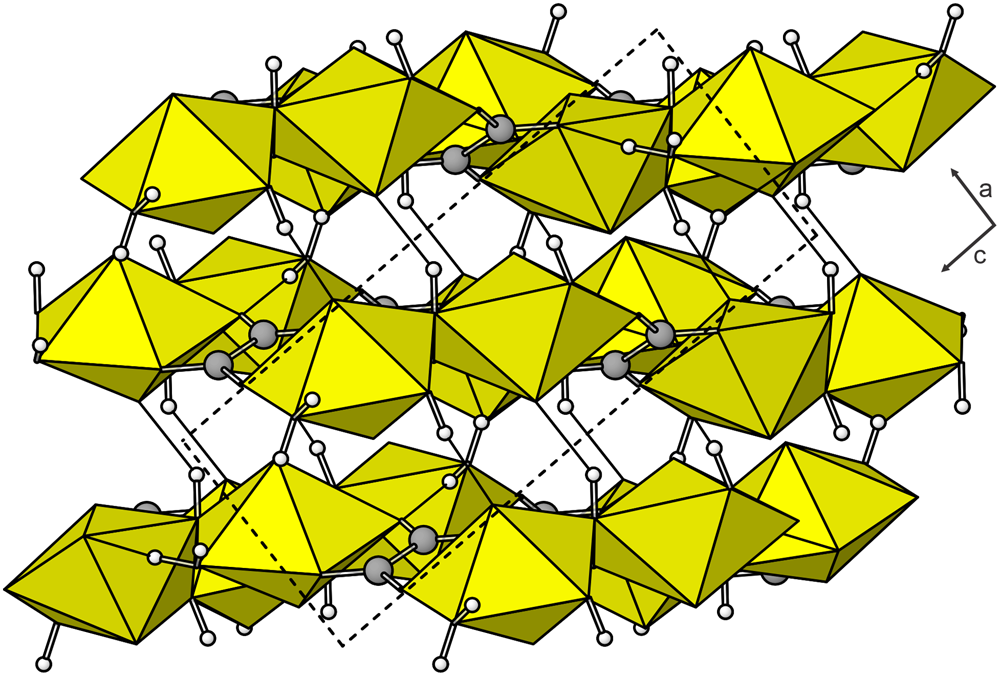
In the uroxite structure, there are two U, two C, 13 O sites (including two OH and three H2O sites) and eight H sites. Both U atoms in the structure of uroxite are surrounded by seven O atoms forming a UO7 pentagonal bipyramid. This is the most typical coordination for U6+ in the solid state, where the two short apical bonds of the bipyramid constitute the UO2 uranyl group (Burns et al., Reference Burns, Ewing and Hawthorne1997; Burns, Reference Burns2005). Two carbon atoms and four equatorial O atoms (Oeq: O1, O2, O3 and O4) of the UO7 bipyramids form oxalate groups. The basic connectivity of cation polyhedra is via sharing of two OH vertices (OH1 and OH2) of each bipyramid with other two UO7 bipyramids; this results in a tetrameric fundamental building block (FBB), [U4O24]. Oxalate groups connect one tetrameric FBB to four others to form corrugated layers parallel to { ${\bar 1}$02} (Fig. 6). There are two distinct types of ring linkages in the structure of uroxite, four-membered rings (U–OH)4 creating an unoccupied diamond-shaped void and a much larger eight-membered ring (U–OH–U–Ox)4, in which uranium atoms are alternately connected by OH and oxalate groups (Fig. 7). These large voids are occupied by the OW3 H2O groups that are linked strongly by hydrogen bonds to the uranyl oxalate network (Table 6). Adjacent sheets of U polyhedra are linked only via hydrogen bonds (Table 6; Fig. 7) and there are no interlayer atoms, resulting in a rather short spacing of adjacent sheets, as seen by the (
 ${\bar 1}$02) powder X-ray diffraction line of 4.424 Å (Table 3).
The layer in the structure of uroxite viewed perpendicular to the { ${\bar 1}$02} sheets. Hydrogen bonds are shown as slender black lines. The unit cell outline is shown by dashed lines. Two coloured areas highlight voids at the centres of two different types of ring linkages: the small vacant area (lime green) built by tetramers of UO7 polyhedra and the large area (sky blue) formed by four pairs of UO7 polyhedra alternating with four oxalate groups. Each large area accommodates two OW3 groups.

Stacking of [(UO2)2(C2O4)(OH)2(H2O)2] sheets (parallel to { ${\bar 1}$02}) in the structure of uroxite. The OW3 groups are omitted. The sheets are seen to be linked to one another by hydrogen bonds (single black lines). The unit cell outline is shown by dashed lines.

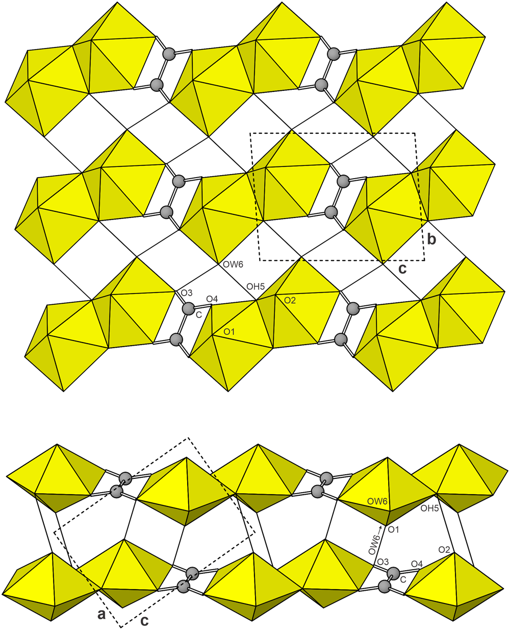
The metauroxite structure has one U, one C and six O sites (including one OH and one H2O site). As in the structure of uroxite, the U atom is surrounded by seven O atoms forming a squat UO7 pentagonal bipyramid, with the two short apical bonds of the bipyramid (Oap O1 and O2) constituting the UO2 uranyl group. The carbon atom and two equatorial O atoms (Oeq: O3 and O4) of the UO7 bipyramids form C2O4 oxalate groups. Two equivalent UO7 bipyramids share an equatorial OH–OH edge to form a dimer. Oxalate groups connect adjacent uranyl dimers to form chains along [10 ${\bar 1}$]. Uranyl oxalate chains are linked to one another only via hydrogen bonds (Fig. 8).
The structure of metauroxite viewed along [100] (top) and [010] (bottom). Hydrogen bonds are shown with single solid black lines. Note that the hydrogen bond that appears to be between O1 and O3 actually is from an OW6 in a chain behind the one shown. The unit-cell outline is shown by dashed lines.

The C2O4 groups link the UO7 bipyramids in the same way in the structures of both uroxite and metauroxite; however, the UO7 bipyramids link to one another in quite different ways in the two structures. In the structure of uroxite, each UO7 bipyramid shares two equatorial OH corners with two other UO7 bipyramids, resulting in a corner-linked uranyl tetramer, instead of the edge-linked dimer in metauroxite. The quite different structural topologies suggest that it is unlikely that the structures convert from one to the other. In fact, their close association on some specimens indicates that they form under similar conditions, more or less contemporaneously.
Discussion
The extensive body of research on the synthesis, structural characterisation and properties of actinide oxalates has been driven by the selective affinity of the oxalate group for the actinides in their various oxidation states and, as a consequence, by the wide-ranging assortment of important applications including (but not limited to) radioactive waste management, selective actinide separation and recovery, nuclear fuel recycling and actinide isotope production (cf. Abraham et al., Reference Abraham, Arab-Chapelet, Rivenet, Tamain and Grandjean2014). Given the pronounced affinity of oxalate for uranyl cations and the intimate association of uranium mineralisation and carbonaceous plant material in the uranium deposits of the Colorado Plateau, it seems surprising that naturally occurring uranyl oxalates have not been discovered until now. There are two likely reasons: (1) the oxalate group is quite ephemeral in natural systems; its presence in the fossilised carbonaceous plant material would require it to have been quite recently generated and probably only locally in very small amounts; (2) most syntheses of actinide oxalates reported in the literature have utilised conditions quite different from those currently prevailing in the Colorado–Plateau uranium deposits.
As an example of the latter, Duvieubourg et al. (Reference Duvieubourg, Nowogrocki, Abraham and Grandjean2005) synthesised the equivalent of uroxite using (UO2)(NO3)2⋅6H2O, KNO3, oxalic acid and water, maintained at 120°C for four days. They synthesised the equivalent of metauroxite using U3O8, oxalic acid and water, maintained at 190°C for two weeks under autogeneous pressure.
At both the Burro and Markey mines, it is likely that the oxalate, C2O42–, was derived from the asphaltum matrix (probably fossilised wood), while the uranyl, UO22+, was derived from earlier-formed primary (esp. uraninite) and/or secondary uranium-bearing minerals. Based upon secondary minerals associated intimately with uroxite and metauroxite and presumably contemporaneously formed, additional cations and anions were also present in the solutions: Na+, Ca2+ and SO42– at the Markey mine and K+, Ca2+, SO42–, AsO43– and VO43– at the Burro mine. It is also clear from the modes of occurrence in post-mining secondary assemblages that these minerals formed under ambient conditions: atmospheric pressure, moderate relative humidity and temperatures probably not exceeding 30°C.
Acknowledgements
Structures Editor Peter Leverett and an anonymous reviewer are thanked for their constructive comments on the manuscript. A portion of this study was funded by the John Jago Trelawney Endowment to the Mineral Sciences Department of the Natural History Museum of Los Angeles County. JP acknowledges support through project GACR 17–09161S of the Czech Science Foundation.
Supplementary material
To view supplementary material for this article, please visit https://doi.org/10.1180/mgm.2019.57.