Klippel–Trenaunay syndrome (KTS) is a rare congenital vascular disorder characterized by capillary malformations, venous malformations and soft tissue or osseous hypertrophy, typically affecting a lower extremity. The condition belongs to the PIK3CA-related overgrowth spectrum and results from somatic activating mutations affecting vascular and tissue development. Reference Wang, Drucker, Gupta, Marshalleck and Shortell1
A 39-year-old woman with a known diagnosis of KTS underwent longitudinal imaging follow-up for cervicofacial soft tissue lesions. Her medical history was notable for congenital venous and lymphatic malformations, left lower extremity hypertrophy and previous sclerotherapy treatment (Figure 1).
Photographs showing erythematous port-wine stains over the left cervicothoracic transition (red arrows), as well as lower limb hypertrophy – slightly more pronounced on the left – accompanied by multiple varicosities bilaterally (A, B, C and D). Note the soft tissue overgrowth of the great toes (black arrows – C). Clinical examination of the posterior scalp. Despite active inspection for vascular abnormalities, hair coverage limited visualization and the ectoscopic evaluation was unremarkable (E, F, G and H).

Figure 1. Long description
Panel A: A close-up photo of the left cervicothoracic transition area with a red arrow pointing to an erythematous port-wine stain. Panel B: Another close-up photo of the same area from a slightly different angle, also marked with a red arrow. Panel C: A full view of the person’s lower limbs standing, showing hypertrophy more pronounced on the left leg with multiple varicosities bilaterally and black arrows pointing to the overgrowth of the great toes. Panel D: A rear view of the person’s lower limbs, showing the same hypertrophy and varicosities. Panel E: A photo of the back of the person’s head, showing hair with a visible patch of lighter hair. Panel F: A closer view of the same area, focusing on the lighter hair patch. Panel G: Another close-up view of the lighter hair patch from a different angle. Panel H: A final close-up view of the lighter hair patch.
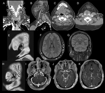
Serial ultrasound examinations demonstrated stable minimally complex cystic lesions in the right periauricular region and a midline suprahyoid lesion with a spongiform appearance, consistent with veno-lymphatic malformations. Contrast-enhanced CT of the neck revealed extensive trans-spatial low-flow vascular malformations involving the cervical and suprahyoid regions without enhancing soft tissue components, aggressive imaging features or pathologic lymphadenopathy (Figure 2).
Contrast-enhanced CT of the neck. Coronal (A), sagittal (B) and axial (C, D) images demonstrate multiple low-flow vascular malformations, predominantly venous, with trans-spatial distribution involving the neck and thoracic inlet, more extensive on the left side. Brain MRI. Sagittal T1-weighted images without contrast (E and H), post-contrast FLAIR sequences (F and G) and volumetric gradient-echo images (I, J and K) demonstrate prominent, tortuous vessels along the posterolateral aspect of the scalp, more evident on the left, consistent with scalp varices. No intracranial vascular abnormalities were identified.
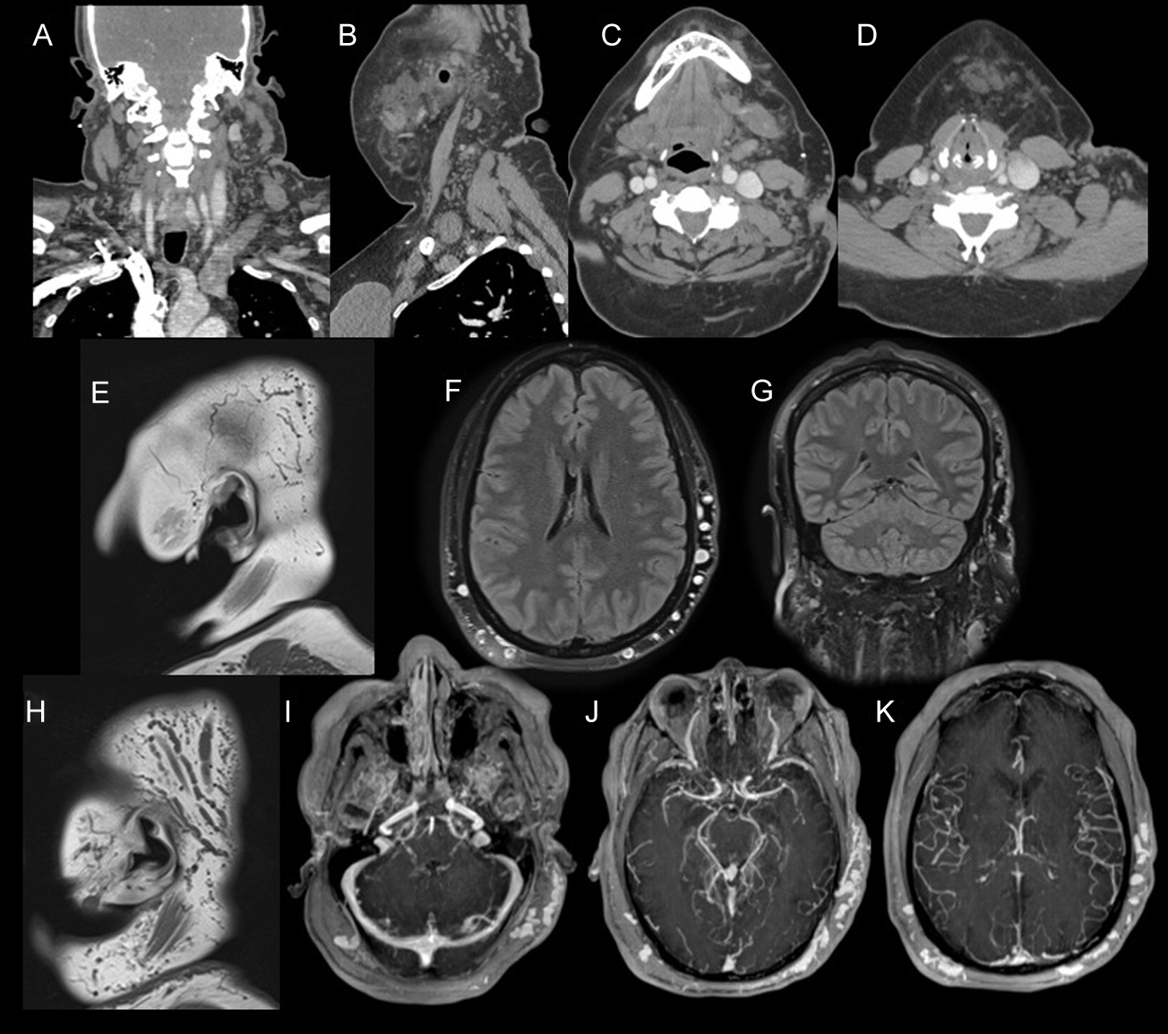
Brain MRI demonstrated multiple enlarged and tortuous vessels along the posterolateral scalp, more prominent on the left side, consistent with scalp varices (Figure 2). No intracranial vascular abnormalities were identified. Clinically, the patient denied pain, dysphagia, voice changes, neurologic symptoms or compressive complaints. Physical examination was largely unremarkable because hair coverage limited visualization of the scalp lesions.
Given the benign imaging appearance, long-term stability and known diagnosis of KTS, conservative management with imaging surveillance was recommended.
Although extremity involvement is the hallmark of KTS, head and neck manifestations are increasingly recognized with the widespread use of cross-sectional imaging. Cervicofacial lesions most commonly represent venous, lymphatic or combined veno-lymphatic malformations and may mimic neoplastic or inflammatory masses when the syndromic diagnosis is not known. Reference Wang, Drucker, Gupta, Marshalleck and Shortell1,Reference Covington, Krishnamurthy and Wendel2
Neurovascular abnormalities have also been described in KTS, including developmental venous anomalies, cavernous malformations, venous sinus abnormalities, intracranial arteriovenous malformations and aneurysms. Reference Covington, Krishnamurthy and Wendel2–Reference Kim, Kim, Hwang, Choung and Khwarg4 In contrast, isolated scalp vascular malformations may remain clinically occult, particularly in women with dense hair coverage. Consequently, careful review of extracranial soft tissues during neuroimaging is important to avoid overlooking these lesions.
Radiologists should be aware that scalp vascular malformations may represent an underrecognized manifestation of KTS. Recognition of their characteristic imaging appearance and benign behavior may prevent unnecessary biopsy or intervention while supporting appropriate long-term surveillance.
Author contributions
Laura Ziemba Araujo: (1) conception and design, or acquisition of data, or analysis and interpretation of data, (2) drafting the article or revising it critically for important intellectual content, (3) final approval of the version to be published and (4) agree to be accountable for all aspects of the work if questions arise related to its accuracy or integrity. Liaydis D. Martin Valdes: (1) conception and design, or acquisition of data, or analysis and interpretation of data, (2) drafting the article or revising it critically for important intellectual content, (3) final approval of the version to be published and (4) agree to be accountable for all aspects of the work if questions arise related to its accuracy or integrity. Kevin J Abrams: (1) conception and design, or acquisition of data, or analysis and interpretation of data, (2) drafting the article or revising it critically for important intellectual content, (3) final approval of the version to be published and (4) agree to be accountable for all aspects of the work if questions arise related to its accuracy or integrity. Márcio Luís Duarte: (1) conception and design, or acquisition of data, or analysis and interpretation of data, (2) drafting the article or revising it critically for important intellectual content, (3) final approval of the version to be published and (4) agree to be accountable for all aspects of the work if questions arise related to its accuracy or integrity. Leonardo Furtado Freitas: (1) conception and design, or acquisition of data, or analysis and interpretation of data, (2) drafting the article or revising it critically for important intellectual content, (3) final approval of the version to be published and (4) agree to be accountable for all aspects of the work if questions arise related to its accuracy or integrity.
Funding statement
No specific funding was received for this work.
Competing interests
The authors declare that there are no conflicts of interest relevant to this work.
Ethical standards
-
– Name of the institutional review board or ethics committee that approved the study: No institutional review board (IRB) was requested for this case report.
-
– Declaration of patient consent: Informed patient consent was not necessary for this work.
-
– We confirm that we have read the Journal’s position on issues involved in ethical publication and affirm that this work is consistent with those guidelines.
Disclosure
The authors declare that there are no additional disclosures to report (financial disclosures for the previous 12 months).