The introduction of Fontan-type procedures into clinical practice resulted in a great progress in treatment of patients with different variants of the so-called single-ventricle physiology.Reference Gewillig 1
The idea of this operation is to restore almost proper physiology without restoration of correct anatomy of the heart. This may cause many adverse consequences. Changed anatomical relations are often responsible for increase of venous pressure and, as a consequence, lead to creation of additional pathways for blood flow, which can result in the intensification of desaturation and worsening of exercise tolerance.Reference Ghanayem, Berger and Tweddell 2 , Reference McCrindle, Williams and Mitchell 3 The quite frequent complication is formation of systemic to pulmonary venous collaterals. In most cases, these vessels are small and can be closed by routine coil technique.Reference Sugiyama, Yoo, Williams and Benson 4 Much less, as a result of the recanalisation of left superior caval vein, is developing much more pronounced cyanosis. After analysing our clinical material, it turned out that the complication is not so rare.
Materials and methods
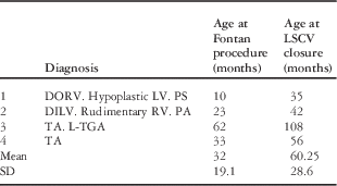
We analysed 19 patients catheterised in the last 27 months because of the growth of the desaturation after a previously performed total cavopulmonary connection. The reasons of this state were: fenestration, systemic to pulmonary venous fistula, intrapulmonary arterio-venous fistula. In four patients (21%), the only or main reason of increasing cyanosis was recanalisation of left superior caval vein draining into the coronary sinus. The average age of the children was 60.25±28.6 months. The mean value of haemoglobin was 18.4±1.4 mg/dl. The diagnosis or confirmation of diagnosis – only two patients had previous echocardiographic suspicion – of recanalisation of the left superior caval vein was made during catheterisation, which took place 28.25 months after the last stage of the Fontan procedure – intracardiac tunnel (Table 1). During this period of time, the oxygen saturation decreased from 95.25±1.1 to 82.25±4.7%. In all cases, the patients were earlier catheterised but none of them had persistent left superior caval vein.
Demographic date

DILV=double-inlet left ventricle; DORV=double-outlet right ventricle; LSCV=left superior caval vein; LV=left ventricle; PA=pulmonary atresia; PS=pulmonary stenosis; RV=right ventricle; TA=tricuspid atresia; l-TGA=l-transposition of the great arteries.
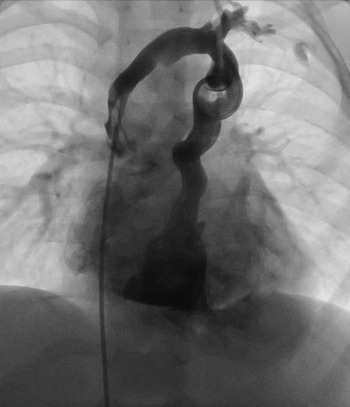
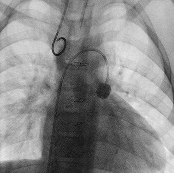
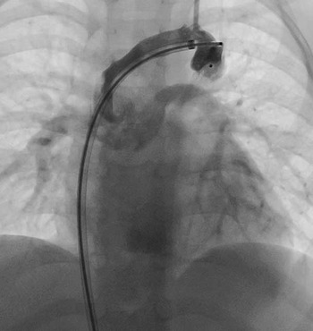
Cardiac catheterisations were performed under general anaesthesia, after antibiotic and heparin injection. Vascular access was achieved via right femoral vessels (artery and vein), and with a second venous line from the right jugular vein – for the pressure measurement during interventional procedures. In all four patients, the venography revealed patent both superior caval veins connected with the innominate veins (Fig 1). It was decided to attempt percutaneous closure of the additional left superior caval vein draining into coronary sinus in all cases. In order to evaluate the safety of this procedure, before the transcatheter treatment, a balloon occlusion test was done. For this purpose, wedge-type catheter or angioplasty balloon was introduced through the inferior caval vein, superior caval vein, innominate vein to the left superior vena cava, where the balloon was filled, closing the lumen of the vessel completely for 15 minutes (Fig 2).
Venography revealed superior caval veins connected by innominate vein.

Temporary closure of the left superior caval vein.

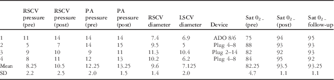
During this time, the vital parameters of children were carefully monitored, as well as the pressure that was measured invasively in the superior caval vein, the pulmonary artery, and in the aorta (Table 2). During this observation, there was no deterioration in the measured parameters, and arterial oxygen saturation increased from 82±4.0 to 93.5±1.1%.
Haemodynamic parameters of the patients

ADO=Amplatzer Duct Occluder; LSCV=left superior caval vein; PA=pulmonary artery; Plug=Amplatzer Vascular Plug; post=after closure; pre=before closure; RSCV=right superior caval vein.
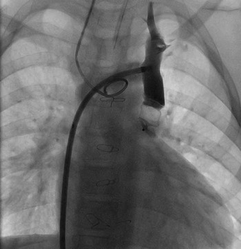
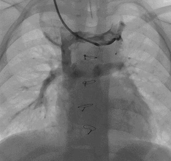
As a result of obtaining good outcomes in the occlusive test, the interventional procedures were started. A multipurpose catheter was placed in the left superior caval vein through the right femoral vein, inferior and superior caval vein, and later after the introduction of a super-stiff 0.035×260 cm wire – Johnson & Johnson, New Jersey, United States of America – into the coronary sinus the catheter and the sheath were withdrawn. Through the same wire, the sheaths were replaced by the delivery systems, through which the right Amplatzer devices – St. Jude Medical, Plymouth, Minnesota, United States of America – (Table 2) were introduced into the target vessels (Fig 3), and after that were released in the left superior caval veins (Fig 4). In patients number 2 and 4, Amplatzer Vascular Plugs – St. Jude Medical – were implanted directly through the diagnostic catheter.
The Amplatzer Duct Occluder in the left superior caval vein just before the releasing.

The Amplatzer Vascular Plug 2 in the left superior caval vein after the releasing.

Results
Implants were positioned in such a way that did not cause narrowing or closure of the hemiazygos accessory vein to left superior caval vein ostium, and still allowed the flow of venous blood from lower to upper part of the body. Control venographies revealed complete closure of all veins without disturbances of the flow. Three different types of devices were used: one designed for ductus arteriosus closure and two Amplatzer Vascular Plugs – St. Jude Medical – (Fig 5). The type and size of devices were selected according to the shape and size of the vessel (landing zone). Vascular plugs 4 – St. Jude Medical – were applied in the vessels of smaller calibre.
The Amplatzer Vascular Plug 4 just after releasing in the left superior caval vein.

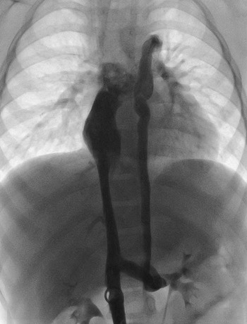
In patient number 1, venography in inferior caval vein – performed at the beginning of the procedure – revealed the presence of inferior caval vein stenosis (diameter of the narrowest part – 4 mm, diameter of remaining part of this vessel – 8 mm). Angiography showed venous collaterals (venous to venous connections) draining into hemiazygos vein, hemiazygos accessory vein, and next into an additional left superior caval vein (Fig 6). A balloon angioplasty with a dilation balloon catheter (12 mm in diameter and 4 cm in length) was performed; the position of the balloon catheter was changed twice. Treatment resulted in an effective dilation of the size of the vessel diameter up to 8.2 mm – the same as the non-stenotic part of the inferior caval vein with normalisation of venous flow. During the procedure, a short-time bradycardia occurred, but disappeared quickly after resuscitation. In all cases, a significant increase in oxygen saturation was noted (Table 2). At 18 months’ follow-up, the effectiveness of treatment has been confirmed, and oxygen saturation remains at the post-operative level.
Venous flow from the lower part of the body.

Discussion
The principal purpose of Fontan operation is to reduce the overload of the ventricle and complete separation of the pulmonary and systemic circulations, by direct connection of the systemic venous return with the pulmonary artery. The palliative nature of this treatment is responsible for the appearance of several adverse effects and complications, such as: thromboembolism, protein-losing enteropathy, plastic bronchitis, liver failure, immune disorders, dysrhythmias, heart failure, and the most often progressive increasing cyanosis.Reference Ghanayem, Berger and Tweddell 2 , Reference Motoki, Ohuchi, Miyazaki and Yamada 5 , Reference Menon, Dearani and Cetta 6 Moderate cyanosis is often seen, even in patients with normal Fontan circulation. This is due to venous return from the coronary sinus to the pulmonary venous atrium and is a natural consequence of the surgically performed fenestration between the tunnel and atrium. In other cases, it results from abnormal formation of patency of vascular connections, as, for example, pulmonary arterio-venous fistulas or less frequently – collaterals between systemic veins and pulmonary veins or pulmonary venous atrium.Reference McCrindle, Williams and Mitchell 3 , Reference Sugiyama, Yoo, Williams and Benson 4 , Reference Karur, Mahima and Nanjappa 7 , Reference Madan, Ellozy and Love 8
During the embryological development of the venous system, the involution of left cardinal vein occurs, resulting in an exclusively right-sided superior caval vein. However, in some subjects, the persistence of left superior caval vein occurs: in 0.3% of the population with normal cardiovascular system and in about 5% of the population with congenital cardiac anomalies.Reference Hass, Shreef, Bumgarner, Gillespie and Stouffer 9
None of our analysed patients had a patent left superior caval vein in the previous catheterisation. Some months after surgery, owing to changes in the physiology of venous flow, there has been an increase in the desaturation. During repeated catheterisation – after 28.25 months – recanalisation of these veins was confirmed. In the literature, we did not find an important percentage of such recanalisations. In our series, desaturation happened as much as in 21% of catheterised patients. It is likely that in some patients this anomaly is not recognised, as happened in two of our patients who had catheterisation in another hospital because of a sudden increase in cyanosis, but an adequate venography had not been performed. It is clear that before the Glenn or hemi-Fontan operation is necessary to exclude or determine the presence of additional left superior caval vein, because it implies enhanced cardiosurgical procedure. However, exclusion at this stage does not release us from the search for recanalisation in the future. Therefore, the search for left superior caval vein as a source of cyanosis should be part of every echocardiogram in these patients. However, angiography remains the best diagnostic tool. For this purpose, it is necessary to introduce the diagnostic catheter through the innominate vein into the left jugular vein and perform the venography. Only this method clearly allows us to diagnose this cause of desaturation.
There are 54 patients after Fontan procedure who are observed in our department and outpatient clinic. The vast majority of them are doing well and the level of the oxygen saturation is stable, higher than 91%. An increase in cyanosis was observed in 19 of them during follow-up. In the majority of these patients, the decrease in oxygen saturation was slow by 2–3% in a period of 3–6 months – according to the rules of our institution, the stable patients after Fontan operation are examinated every 3–6 months – and did not reach a level lower than 85%. However, in three patients a decrease of at least 5% was noted at 3-month intervals and, consequently, after several months they reached an oxygen saturation lower than 85% – in one patient with additional left superior caval vein the decrease was less pronounced. In these patients, the recanalisation of the left superior caval vein was diagnosed. For this reason, we put forward a hypothesis that such fast development of cyanosis – “sudden desaturation” – in patients after Fontan operation is likely to indicate a recanalisation of the left superior caval vein. The desaturation had a progressive course but the rate of rise of cyanosis was significantly faster than in the other patients. The oxygen saturation decreased by about 14% (from 95.25 to 82.75%) during post-surgical follow-up (28.25 months). The data may suggest that recanalisation of the left superior caval vein after Fontan procedure is not such a rare complication because in our material it is 7.4% of all patients after Fontan operation and as many as 21% of these patients with increasing cyanosis.
Implementation of the palliative Fontan operation in clinical practice resulted in a progress in the treatment of patients in whom biventricular correction was not possible. Unfortunately, even this method is not available for all the patients with different variants of single-ventricle heart. In order to perform it, a precise evaluation of the entire circulatory system must have been done, before the particular stages of this palliative treatment.
Nevertheless, most of the information on this topic is provided by a diagnostic cardiac catheterisation with haemodynamic and morphological evaluation. However, even the fulfilment of the classic Choussat’s criteria does not guarantee a good long-term result. At different times, in the post-operative period, various complications can occur. In addition to the widely described ones, the above-mentioned one can also occur, either sporadically or even incidentally.
Therefore, patients who have experienced disturbing symptoms, of unclear cause, require precise diagnostic evaluation. Despite the development of new excellent diagnostic methods, the traditional cardiac catheterisation provides the highest value in examination of patients after completion of Fontan surgery. Therefore, when cyanosis appears or is getting more intensive, these patients should undergo precise angiography, in the search for causes of desaturation. However, it is also very important to emphasise the role of echocardiography in the diagnosis of dilated additional left superior caval vein. The availability of new-generation, high-resolution echo-machines allows non-invasive diagnosis of the left superior caval vein, especially in patients with enlargement of the coronary sinus. The final echocardiographic confirmation of the diagnosis is the injection of contrast medium in a left antecubital vein in order to confirm the passage of the medium first into the coronary sinus and then in the atrium.Reference Recupero, Pugliatti and Rizzo 12 Currently, this procedure was introduced for all patients with increasing cyanosis in order to exclude the possibility of misdiagnosis.
The development of new methods of percutaneous treatment allows to perform procedures that, not far until recently, were only possible by surgical means.Reference Girisch, Sieverding and Rauch 10 It is worth mentioning that in some centres these vessels are still occluded surgically.Reference Baslaim and Hussain 11
The progress appears to be particularly important in patients with single-ventricle physiology. Owing to the necessity of multi-staged treatment, they require many thoracotomies, which are responsible for the development of scarring and adhesions. This means that each subsequent treatment becomes more difficult and burdened with a great probability of complications. In some situations, surgical treatment of complications of Fontan circulation becomes an extremely large procedure. Therefore, interventional options of treatment are extremely attractive for these patients. The variety of available devices allows almost all anomalous vascular connections to be closed percutaneously. Regardless of the morphology, diameter, length, or vessel tortuosity, it is necessary to select the appropriate implant, which optimally adapts to the shape of the vessel. For this reason, we used three different types of devices. However, not every abnormal vascular connection closure is safe in these patients. Therefore, in these particular circumstances, before making a decision of performing such a procedure, a balloon occlusion test should be carried out, to simulate the state after closing the vascular connection. In our cases, balloon catheters were introduced temporarily into the left superior vena cava, placed in such a way so as not to close the connections of hemiazygos accessory veins with the caval vein.
Conclusion
Meticulous investigation of unclear causes of desaturation in cyanotic patients after Fontan completion is necessary. Almost all causes of desaturation, including recanalised additional left superior caval vein, can be effectively treated percutaneously.
Acknowledgements
None.
Financial Support
None.
Conflicts of Interest
None.