INTRODUCTION
Trypanosoma cruzi is the only generalist species of the subgenus Schizotrypanum; all other species of this subgenus are restricted to Chiroptera. T. cruzi is transmitted by triatomine insects and is the agent of human Chagas disease, a major public health problem of the American continent. Schizotrypanum trypanosomes restricted to bats may occur exclusively in the Americas (T. c. marinkellei), or they may be widespread in the New and Old Worlds. Brazilian bats are commonly infected by Schizotrypanum spp., and the bat-restricted species are more prevalent than T. cruzi (Marinkelle, Reference Marinkelle, Lumsden and Evans1976; Pinto and da Costa Bento, Reference Pinto and da Costa Bento1986; Molyneux, Reference Molyneux, Kreier and Baker1991; Fabián, Reference Fabián1991; Cavazzana et al. Reference Cavazzana, Marcili, Campaner, Veludo, Takata, Paiva, Takeda and Teixeira2003; Maia da Silva et al. Reference Maia da Silva, Marcili, Lima, Cavazzana, Ortiz, Campaner, Takeda, Paiva, Nunes, Camargo and Teixeira2009).
Different populations of T. cruzi circulate in enzootic cycles from the southern half of North America to southern South America, infecting species of virtually all mammalian orders (Gaunt and Miles, Reference Gaunt and Miles2000). T. cruzi comprises highly phenotypic and genotypic heterogeneous populations classified as T. cruzi I (TCI) and T. cruzi II (TCIIa–e) lineages, which have been defined based on zymodemes, RAPD, ribosomal, mini-exon, and cytochrome b gene markers (Miles et al. Reference Miles, Povoa, De Souza, Lainson, Shaw and Ketteridge1981; Souto et al. Reference Souto, Fernandes, Macedo, Campbell and Zingales1996; Brisse et al. Reference Brisse, Verhoef and Tibayrenc2001; Marcili et al. Reference Marcili, Valente, Valente, Junqueira, Maia da Silva, Naiff, Campaner, Coura, Camargo, Miles and Teixeira2008). In the southern cone of South America, isolates from humans and vectors of domestic and peridomestic transmission cycles are predominantly of lineages TCIIb and TCIId/e. TCI has been reported in the sylvatic cycle throughout Latin America, and predominantly infects humans in endemic areas northwest of the Amazon basin. TCIIa has been sporadically isolated from humans and occurs mainly in the Brazilian Amazonia. TCIIc is widespread in South America and also is mostly sylvatic and sporadically found in humans (Miles et al. Reference Miles, Povoa, De Souza, Lainson, Shaw and Ketteridge1981; Brisse et al. Reference Brisse, Henriksson, Barnabé, Douzery, Berkvens, Serrano, De Carvalho, Buck, Dujardin and Tibayrenc2003; Yeo et al. Reference Yeo, Acosta, Llewellyn, Sánchez, Adamson, Miles, López, González, Patterson, Gaunt, de Arias and Miles2005; Martins et al. Reference Martins, Marcili, Castanho, Therezo, Oliveira, Suzuki, Teixeira, Rosa and Sperança2008; Maia da Silva et al. Reference Maia da Silva, Naiff, Marcili, Gordo, D'Affonseca Neto, Naiff, Franco, Campaner, Valente, Valente, Camargo, Teixeira and Miles2008; Marcili et al. Reference Marcili, Valente, Valente, Junqueira, Maia da Silva, Naiff, Campaner, Coura, Camargo, Miles and Teixeira2009).
Ecobiological and phylogenetic analyses have suggested that ecotopes and preferential mammalian hosts and vectors may be determining factors of T. cruzi lineages in sylvatic cycles. Although lineage association with both mammals and vectors is far from absolute some relevant correlations have been observed. TCI is strongly associated with opossums of Didelphis and with vectors of the genus Rhodnius. Nevertheless, isolates of this lineage also infect other didelphids, wild primates, rodents and carnivores, and can be found in other genera of triatomines. TCIIc is associated mainly with armadillos and few other terrestrial mammals, and is transmitted by triatomines of terrestrial ecotopes. Natural hosts and ecotopes of TCIIa are not clearly resolved, with recent reports of this lineage in wild primates and in Rhodnius spp. from the Brazilian Amazonia. TCIIa has been also reported in racoons and dogs in North America (Miles et al Reference Miles, Povoa, De Souza, Lainson, Shaw and Ketteridge1981; Gaunt and Miles, Reference Gaunt and Miles2000; Yeo et al. Reference Yeo, Acosta, Llewellyn, Sánchez, Adamson, Miles, López, González, Patterson, Gaunt, de Arias and Miles2005; Roellig et al. Reference Roellig, Brown, Barnabé, Tibayrenc, Steurer and Yabsley2008; Marcili et al. Reference Marcili, Valente, Valente, Junqueira, Maia da Silva, Naiff, Campaner, Coura, Camargo, Miles and Teixeira2009).
Several studies have reported Brazilian bats infected with T. cruzi, from the Amazonia rainforest to urban areas and including roofs of human dwellings in Central, Northeast and Southeast Brazil (Funayama and Barretto, Reference Funayama and Barretto1970a, Reference Funayama and Barrettob, Reference Funayama and Barreto1973; Barretto et al. Reference Barreto, Ribeiro and Filho1974; Fabián, Reference Fabián1991; Cavazzana et al. Reference Cavazzana, Marcili, Campaner, Veludo, Takata, Paiva, Takeda and Teixeira2003; Lisboa et al. Reference Lisboa, Pinho, Herrera, Gerhard, Cupolillo and Jansen2008; Maia da Silva et al. Reference Maia da Silva, Marcili, Lima, Cavazzana, Ortiz, Campaner, Takeda, Paiva, Nunes, Camargo and Teixeira2009). Although the genetic diversity of T. cruzi isolates from bats is virtually unknown, recent studies showed that they can be infected by TCI, TCII and Z3 isolates (Lisboa et al. Reference Lisboa, Pinho, Herrera, Gerhard, Cupolillo and Jansen2008; Maia da Silva et al. Reference Maia da Silva, Marcili, Lima, Cavazzana, Ortiz, Campaner, Takeda, Paiva, Nunes, Camargo and Teixeira2009; Añez et al. Reference Añez, Crisante and Soriano2009). Furthermore, the phylogenetic relationships between T. cruzi isolates from bats and those from other mammals have not yet been addressed.
Several mammalian and triatomine species sustain domestic and sylvatic transmission cycles of T. cruzi, while domestic (dogs and cats) and peridomestic (opossums and rodents) animals are responsible for the interaction between these two cycles (Yeo et al. Reference Yeo, Acosta, Llewellyn, Sánchez, Adamson, Miles, López, González, Patterson, Gaunt, de Arias and Miles2005; Gürtler et al. Reference Gürtler, Cecere, Lauricella, Cardinal, Kitron and Cohen2007; Marcili et al. Reference Marcili, Valente, Valente, Junqueira, Maia da Silva, Naiff, Campaner, Coura, Camargo, Miles and Teixeira2009). Although poorly investigated, bats may play an important role as a risk factor for human Chagas disease. Bats harbouring T. cruzi have been observed in various sylvatic niches, as well as roosting in buildings, human dwelling lofts, and peridomiciliary environments, where they can attract triatomines from nearby ecotopes and serve as a source of infected blood for these vectors. Precipitin tests and experimental studies confirmed that triatomines feed on bats, which can be infected through contamination with feces and by ingestion of T. cruzi-infected bugs (Barreto et al. Reference Barreto, Ribeiro and Filho1974; Thomas et al. Reference Thomas, Rasweiler and D'Alessandro2007). It was recently reported that bats can be infected with T. cruzi by congenital transmission (Añez et al. Reference Añez, Crisante and Soriano2009).
Identification of T. cruzi from bats requires careful analysis because Schizotrypanum species are morphologically indistinguishable and generically named as T. cruzi-like. In addition, more than one species of trypanosome can infect a given bat species, and mixed infections are common. Methods employed for distinguishing species of Schizotrypanum such as zymodemes (Baker et al. Reference Baker, Miles, Godfrey and Barrett1978; Barnabé et al. Reference Barnabé, Brisse and Tibayrenc2003) and restriction analysis of kDNA (Teixeira et al. Reference Teixeira, Gonçalves, Romanha, Steindel and Pinto1993; Steindel et al. Reference Steindel, Grisard, de Carvalho Pinto, Cordeiro, Ribeiro-Rodrigues and Romanha1998) are time-consuming, require a large number of parasites, and cannot detect mixed infections. Moreover, these methods have not been evaluated for T. cruzi isolates from bats. Due to difficulties in the identification of Schizotrypanum species using methods employed for the diagnosis of T. cruzi, there are few unquestionable reports of T. cruzi in bats. T. cruzi can be convincingly confirmed by the ability to infect mice, a feature shared by all T. cruzi lineages but lacked by all T. cruzi-like trypanosomes (Funayama and Barretto, Reference Funayama and Barretto1970a, Reference Funayama and Barrettob, Reference Funayama and Barreto1973; Fabian, Reference Fabián1991; Maia da Silva et al. Reference Maia da Silva, Marcili, Lima, Cavazzana, Ortiz, Campaner, Takeda, Paiva, Nunes, Camargo and Teixeira2009).
A survey of trypanosomes infecting Brazilian bats disclosed several isolates of the subgenus Schizotrypanum. Analysis of SSU rDNA sequences from these isolates allowed separation of T. cruzi from other trypanosomes found infecting Brazilian bats: T. c. marinkellei, T. dionisii-like and T. rangeli (Cavazzana et al. Reference Cavazzana, Marcili, Campaner, Veludo, Takata, Paiva, Takeda and Teixeira2003; Maia da Silva et al. Reference Maia da Silva, Marcili, Lima, Cavazzana, Ortiz, Campaner, Takeda, Paiva, Nunes, Camargo and Teixeira2009). In this study, we gathered a multigene data set from T. cruzi isolates from Brazilian bats to infer phylogenetic relationships among isolates from bats and other hosts representative of established lineages of T. cruzi. In addition, behavioural features of bat isolates were evaluated in culture and in experimentally infected mice and triatomines.
MATERIALS AND METHODS
Study areas and selection of T. cruzi isolates
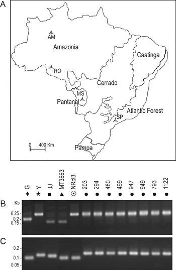
T. cruzi isolates characterized in this study are from bats captured in the Brazilian States of Amazonia (North region, Amazonia), Mato Grosso do Sul (Center region, Cerrado/Pantanal) and São Paulo (Southeast region, the Atlantic Forest) (Fig. 1). Bats were captured using appropriated nets, anaesthetized and manipulated for blood-sample collection according to permits of the IBAMA (Instituto Brasileiro do Meio Ambiente). Trypanosomes were isolated by haemoculture (HE), and purified cultures of T. cruzi from HE mixed with other Schizotrypanum spp. were obtained by HE of experimentally infected mice, approximately 30 days after inoculation of mixed cultures (Maia da Silva et al. Reference Maia da Silva, Rodrigues, Campaner, Takata, Brigido, Junqueira, Coura, Takeda, Shaw and Teixeira2004a). Isolates were cryopreserved in the Trypanosomatid Culture Collection of the Department of Parasitology, University of São Paulo. Brazilian T. cruzi isolates included in this study were from the following Brazilian States: PA, Pará; AC, Acre; AM, Amazonas; SP, São Paulo; BA, Bahia; RO, Rondônia; MG, Minas Gerais; RN, Rio Grande do Norte; MS, Mato Grosso do Sul; GO, Goiás.
(A) Geographical origin ( ) of isolates of Trypanosoma cruzi from bats captured in the following States of different Brazilian biomes: Amazonas (AM) and Rondonia (RO) in Amazonia biome; Mato Grosso do Sul (MS) in the Pantanal/Cerrado; and São Paulo (SP) in the Atlantic Forest. DNA profiles generated by genotyping of isolates of T. cruzi using PCR assays based on mini-exon (B) and ribosomal (C) markers. Controls were performed using DNA from reference strains/isolates of T. cruzi lineages: TCI, G (◆); TCIIb, Y (
 ); TCIIa, JJ (▪); TCIIc, MT3663 (▴); TCIId, NRcl3 (
 ) and TCbat (•). Isolates of TCbat: TryCC 203-1112.

Molecular diagnosis and genotyping of T. cruzi isolates
DNA from cultured trypanosomes was extracted using the traditional phenol/chloroform method. Diagnosis of T. cruzi isolates was done by PCR based on kDNA sequences and SSU rDNA sequence analysis as before (Maia da Silva et al. Reference Maia da Silva, Naiff, Marcili, Gordo, D'Affonseca Neto, Naiff, Franco, Campaner, Valente, Valente, Camargo, Teixeira and Miles2008, Reference Maia da Silva, Marcili, Lima, Cavazzana, Ortiz, Campaner, Takeda, Paiva, Nunes, Camargo and Teixeira2009). Genotyping of T. cruzi was done using PCRs based on mini-exon (Fernandes et al. Reference Fernandes, Santos, Cupolillo, Mendonça, Derre, Junqueira, Santos, Sturm, Naiff, Barrett, Campbell and Coura2001) and LSU 24Sα-rRNA (Souto et al. Reference Souto, Fernandes, Macedo, Campbell and Zingales1996) gene sequences. Reference strains of major T. cruzi lineages were used as controls: TCI (G), TCIIa (JJ), TCIIb (Y), TCIIc (MT3663) and TCIId (NRcl3).
PCR amplification, sequencing and data analysis of SSU rDNA, cytochrome b and Histone H2B genes
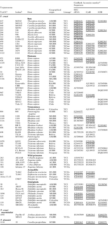
PCR amplification of a 900 bp DNA fragment corresponding to partial sequence of SSU rDNA (V7–V8 region) was performed using primers and PCR reactions previously described (Maia da Silva et al. Reference Maia da Silva, Noyes, Campaner, Junqueira, Coura, Añez, Shaw, Stevens and Teixeira2004b). Amplification of 450 bp sequences of Histone H2B (H2B) gene was performed as described by Sturm et al. (Reference Sturm, Vargas, Westenberger, Zingales and Campbell2003). Sequences of mitochondrial cytochrome b (Cyb) were amplified (500 bp) using primers described by Brisse et al. (Reference Brisse, Henriksson, Barnabé, Douzery, Berkvens, Serrano, De Carvalho, Buck, Dujardin and Tibayrenc2003). New sequences from nuclear SSU rDNA (38 sequences), mitochondrial (Cyb) (33 sequences), and H2B (13 sequences) genes determined in this study were aligned with corresponding sequences from reference strains of T. cruzi and other bat trypanosomes from GenBank (Table 1). Alignments were made using ClustalW and manually refined. Phylogenetic inferences were done by parsimony (P) (PAUP*4.0b10, Swofford, Reference Swofford2002) and maximum likelihood (ML) (RAxML, Stamatakis, Reference Stamatakis2006), with bootstrap analyses performed with 100 replicates, as previously described (Rodrigues et al. Reference Rodrigues, Paiva, Campaner, Stevens, Noyes and Teixeira2006; Ferreira et al. Reference Ferreira, De Souza, Freitas, Campaner, Takata, Barrett, Shaw and Teixeira2008).
Trypanosoma cruzi isolates, host and geographical origin, lineages and sequences of SSU rDNA, cytochrome b and Histone H2B genes employed in the phylogenetic analyses performed in this study

a TryCC, Code number of the isolates/strains cryopreserved in the Trypanosomatid Culture Collection (TCC), Department of Parasitology, University of São Paulo, São Paulo, SP, Brazil.
b Original codes of isolates.
c Lineages determined based on mini-exon markers (Fernandes et al. Reference Fernandes, Santos, Cupolillo, Mendonça, Derre, Junqueira, Santos, Sturm, Naiff, Barrett, Campbell and Coura2001) and phylogenetic analyses inferred in this study.
d Sequences determined in this study and deposited in the GenBank are underlined.
Brazilian States: PA, Pará; AC, Acre; AM, Amazonas; SP, São Paulo; BA, Bahia; RO, Rondônia; MG, Minas Gerais; RN, Rio Grande do Norte; MS, Mato Grosso do Sul; GO, Goiás; BR, Brazil.
PCR-RFLP analysis of ITS1 rDNA from T. cruzi and other Schizotrypanum species
The primers and PCR conditions employed for amplification of ITS1 rDNA have been described previously (Maia da Silva et al. Reference Maia da Silva, Noyes, Campaner, Junqueira, Coura, Añez, Shaw, Stevens and Teixeira2004b; Rodrigues et al. Reference Rodrigues, Paiva, Campaner, Stevens, Noyes and Teixeira2006). Amplified ITS1 rDNA was digested with several restriction enzymes. The enzyme Bsh 1236 was selected to standardize a PCR-RFLP assay able to separate lineages of T. cruzi and to distinguish T. cruzi from T. c. marinkellei and T. dionisii. Length and restriction profiles of amplified ITS1 rDNA were analysed by electrophoresis in 2·5% agarose gels stained with ethidium bromide.
RAPD fingerprinting and karyotyping
RAPD profiles from T. cruzi isolates were assessed using 5 decameric oligonucleotide primers to amplify DNA from all T. cruzi isolates from bats and reference strains/isolates of all lineages (Maia da Silva et al. Reference Maia da Silva, Rodrigues, Campaner, Takata, Brigido, Junqueira, Coura, Takeda, Shaw and Teixeira2004a). The amplified DNA fragments were separated on 2·0% agarose gels and stained with ethidium bromide.
For comparison of karyotyping patterns, chromosome blocks prepared by embedding 107 epimastigotes of isolates belonging to several lineages in 1·2% low-melting agarose were submitted to Pulsed Field Gel Electrophoresis (PFGE) in a CHEF Mapper apparatus (Bio-Rad) as described previously (Cano et al. Reference Cano, Gruber, Vazquez, Cortés, Levin, González, Degrave, Rondinelli, Zingales and Ramirez1995).
Growth and morphology of T. cruzi isolates from bats
Isolates of T. cruzi from bats were cultivated in LIT medium supplemented with 10% or 3–5% fetal bovine serum (FBS), at 28°C over a period of 15 days. Smears of logarithmic- (5 days) and stationary-phase (after 12 days) cultures were fixed in methanol and Giemsa stained. Cultures containing metacyclic trypomastigotes were transferred to 24-well plates containing glass cover-slips with monolayers of HeLa cells cultivated in RPMI medium with 5% FBS at 37°C in a 5% CO2 humid atmosphere (1×10⁵ cells/well and 1×10⁶ parasite/well). After 1 h, 24 h, 4 and 7 days, the cover-slips were washed 3 times in phosphate-buffered saline, fixed in methanol and Giemsa-stained for light microscopy.
Infectivity analysis of T. cruzi isolates from bats to mice and triatomine bugs
For evaluation of infectivity and virulence, 2 selected bat isolates of each lineage found in bats, TCI and TCII, were employed to infect Balb/c mice (6 for each isolate) by intra-peritoneal inoculation of cultures containing metacyclic forms (~106/animal). Mice blood samples were examined weekly from 7 to 30 days post-inoculation (p.i.) by the microhaematocrit method and chronic infection was confirmed by haemoculture after the 30th day p.i. Smears from the blood of experimentally infected mice were Giemsa-stained for light microscopy. Purified T. cruzi cultures were recovered from mice infected with trypanosomes from mixed cultures containing T. c. marinkellei or T. dionisii-like besides T. cruzi as described previously (Maia da Silva et al. Reference Maia da Silva, Rodrigues, Campaner, Takata, Brigido, Junqueira, Coura, Takeda, Shaw and Teixeira2004a, Reference Maia da Silva, Marcili, Lima, Cavazzana, Ortiz, Campaner, Takeda, Paiva, Nunes, Camargo and Teixeira2008b).
Eight species of triatomines were used for behavioural analysis of selected bat isolates: Rhodnius prolixus, R. robustus (genetic population II), R. pictipes, R. domesticus, R. neglectus, Triatoma infestans, T. vitticeps and Panstrongylus megistus. Mice infected with bat isolates of T. cruzi were used for xenodiagnosis with 20–30 fifth instar nymphs of each species. The infected triatomines were fed on normal mice every 15 days, dissected 15, 30, and 60 days p.i., and their guts examined for the presence of trypanosomes.
RESULTS
Genotypes of T. cruzi isolates from bats
The 4 bat isolates of T. cruzi from Amazonia were assigned to lineage TCI, and all isolates from Central and Southeast Brazil were ascribed to TCIIb using the traditional genotyping method based on mini-exon gene (Fernandes et al. Reference Fernandes, Santos, Cupolillo, Mendonça, Derre, Junqueira, Santos, Sturm, Naiff, Barrett, Campbell and Coura2001). Genotyping based on ribosomal (LSU 24Sα rRNA) markers (Souto et al. Reference Souto, Fernandes, Macedo, Campbell and Zingales1996) confirmed all the TCI isolates. However, using this method all the 11 bat isolates from Pantanal/Cerrado and the Atlantic Forest genotyped as TCIIb by mini-exon markers yielded DNA fragments slightly larger (~140 bp) than that generated for TCIIb isolates (125 bp), and different from those of other lineages (Fig. 1C, Table 1).
T. cruzi isolates from bats characterized in this study were recovered mainly from insectivorous bats of Myotis spp. (Vespertilionidae) (9 isolates), Noctilio albiventris (Noctylionidae) (2 isolates), and 1 was from Thyroptera tricolor (Tryropteridae). Three isolates from Amazonian bats were from frugivorous/insectivorous Carollia perspicillata (Phyllostomidae) (Table 1).
Phylogenetic relationships among T. cruzi isolates from bats and other hosts based on SSU rDNA, cytochrome b and Histone H2B gene sequences
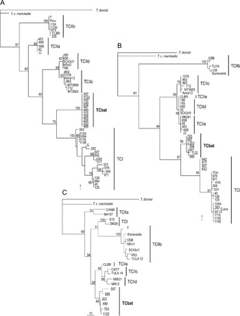
Phylogenetic relationships and degrees of genetic relatedness were inferred by comparing sequences from genes V7–V8 SSU rDNA, Cyb and H2B of T. cruzi from bats aligned with corresponding sequences of isolates from humans, other wild mammals and triatomine bugs belonging to previously described lineages. Positioning of bat isolates in phylogenetic analysis of SSU rDNA and Cyb sequences corroborated 2 groups as previously evidenced by genotyping based on ribosomal and mini-exon markers. One clade comprised only TCI isolates from bats that cluster with TCI from other hosts forming a monophyletic assemblage showing a complex branching pattern. However, analysis based on SSU rDNA demonstrated that TCI from bats form a homogeneous subclade within clade TCI, differing from all other TCI isolates, which also formed subclades according to vertebrate host and/or geographical origin (Fig. 2).
Phylogenetic trees inferred by parsimony analyses based on (A) V7–V8 SSU rDNA sequences (825 characters, 108 parsimony informative) of 54 Trypanosoma cruzi isolates, (B) cytochrome b sequences (490 characters, 84 parsimony informative) of 52 isolates, and (C) Histone H2B partial sequence (457 characters, 104 parsimony informative) of 24 isolates. T. cruzi isolates from bats are underlined. The numbers at the nodes correspond to parsimony percentage bootstrap values derived from 100 replicates.

The 11 bat isolates genotyped as TCII all nested into a well-supported assemblage composed exclusively of bat isolates from SP and MS, clearly separated from all previously established T. cruzi lineages in all analyses. Therefore, data provide evidence that these bat isolates belong to a new genotype that we designated TCbat. The clade TCbat was separated from TCIIb by the largest sequence divergences of all genes examined: ~5·7%, 11% and 12·3% for SSU rDNA, Cyb and H2B gene, respectively. Similar sequence divergences of these three genes separated TCIIb from TCI (~5·5%, 11·2% and 11%). Divergences between TCbat and TCI were also large for these genes (~6·2%, 4·2% and 8·4%), and similar to those separating this new genotype from TCIIa (~5·0%, 5·5% and 9·5%), TCIIc (~5·5%, 5·5%, 6·0%), and TCIId (~4·7%, 5·1% and 10·7%). TCIIe was represented in the phylogenetic analyses by the hybrid CL Brener strain, which showed sequences positioned within TCIIb (SSU rDNA) or closest to TCIIc/d (Cyb and H2B) (Fig. 2).
Therefore, the bat isolates of the new genotype clustered tightly together forming a clade well supported in all analyses and clearly different from all known lineages (Fig. 2). Despite being closest to TCI in analyses using SSU rDNA and Cyb sequences, positioning of TCbat was only well supported using Cyb sequences (bootstrap of 97%). In analysis using H2B sequences, placement of the new genotype was weakly supported close to clade comprising TCIIc/d/e. Therefore, analyses based on separated nuclear (SSU rDNA and H2B) and mitochondrial (Cyb) genes were unable to clearly resolve the relationships between TCbat and conventional T. cruzi lineages (Fig. 2). We found no evidence of hybrid characteristics in the new lineage by sequencing 5–8 clones of each SSU rDNA, Cyb and H2B genes from 2 bat isolates (TryCC 793 and 1122).
All phylogenetic analyses included T. c. marinkelei and T. dionisii-like, which enabled confirmation that isolates of TCbat genotype indeed belong to T. cruzi and not to other very closely related Schizotrypanum species from bats (T. cruzi-like) (Fig. 2).
Phylogenetic relationships among T. cruzi and T. cruzi-like from bats (Schizotrypanum) inferred using a combined data set of SSU rDNA, cytochrome b and Histone H2B sequences
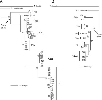
Phylogenetic analyses were performed using 2 data sets of concatenated aligned sequences aiming to better resolve the phylogenetic relationships of the new T. cruzi genotype from bats in relation to previously established lineages of T. cruzi as well as of T. cruzi-like trypanosomes. Alignment 1 consists of SSU rDNA plus Cyb sequences from 40 T. cruzi isolates plus T. c. marinkelei and T. dionisii-like. Alignment 2 comprises sequences from SSU rDNA, Cyb and H2B from 11 T. cruzi isolates plus T. c. marinkelei and T. dionisii-like. Phylogenetic trees based on both alignments were inferred by P, Bayesian (data not shown) and ML methods (Fig. 3). In trees generated by the two alignments, the clade TCbat was always well supported and positioned closest to TCI. TCIIb was positioned as the basal lineage of the clade T. cruzi, while the other lineages were distributed in 5 major clades. Clade TCI and TCbat formed a monophyletic assemblage in all analyses using the two combined data sets (Fig. 3). TCbat was separated from the subclade formed by bat isolates assigned to TCI by large genetic distances. Independent of alignments and analytical methods, T. cruzi-like, T. dionisii and T. c. marinkellei, were always positioned as outgroups of the clade harbouring all T. cruzi isolates (Fig. 3).
Phylogenetic trees inferred by ML analyses using combined data sets: (A) SSU rDNA and cytochrome b sequences from 42 isolates (1315 characters, −Ln=3044·252544), and (B) SSU rDNA, cytochrome b and Histone H2B sequences from 11 isolates (1791 characters, −Ln=4761·077323) of Trypanosoma cruzi isolates. T. cruzi isolates from bats are underlined. Numbers at nodes are bootstrap values derived from 100 replicates.

Analyses using combined data sets positioned TCbat closer to TCI than to any other lineage, and unequivocally within T. cruzi (Fig. 3). The assemblage formed by TCI isolates corroborated the major subclades revealed by SSU rDNA analysis (Figs 2 and 3). The 4 TCI isolates from bats generated a subclade formed exclusively by isolates from Amazonia whereas isolates from opossum, monkeys and humans from this region clustered separately. However, these isolates were all genotyped as typical TCI by traditional PCR methods and tightly clustered together forming a clade separated from TCbat. Although TCbat isolates were all from bats captured in MS and SP states, isolates from didelphids and triatomines from these states never nested within clade TCbat while they were segregated in 2 clades according to their geographical origin (Figs 2 and 3).
Polymorphism analysis of bat isolates by RAPD and karyotype patterns
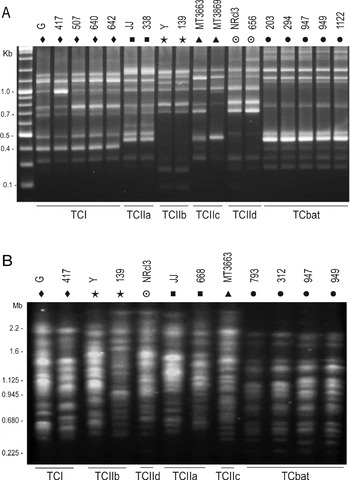
For additional detection of inter- and intralineage polymorphisms among bat trypanosomes, we compared RAPD patterns generated with 5 selected primers using DNA of all bat isolates assigned to both TCbat and TCI. RAPD profiles generated using all primers were shown to be almost identical for bat isolates within the same clade (data not shown). For illustrative purpose we selected 1 primer (672) that yielded a distinct pattern for TCbat genotype, allowing its separation from all T. cruzi lineages (Fig. 4A), and generating similar RAPD profiles shared by bat isolates assigned to the same clade (TCI and TCbat).
Genetic polymorphism of Trypanosoma cruzi isolates analysed through RAPD and karyotype patterns selected to illustrate inter-lineage genetic variability, and small polymorphism of TCbat isolates. Agarose gel stained with ethidium bromide showing (A) RAPD profiles generated using primer 672, and (B) PFGE chromosome band profiles. Isolates included in these analyses were from TCbat (•) and lineages TCI (◆), TCIIa (▪), TCIIb (), TCIIc (▴) and TCIId ().

Despite relevant heterogeneity of karyotyping patterns generated by PFGE among isolates belonging to different lineages, and even within the same lineage, chromosome band profiles separate TCbat isolates from T. cruzi of established lineages. Similar patterns of chromosome bands are shared by the 4 isolates of TCbat examined, with small differences between isolates TryCC 793/312 and 947/949. Interestingly, karyotyping profiles of TCbat isolates apparently lack the largest chromosome bands and are less complex than those from isolates of the other lineages (Fig. 4B).
Taken together, similar RAPD and karyotyping patterns, which are highly sensitive tools for polymorphism analyses, besides high conservation of SSU rDNA, Cyb and H2B sequences, indicated that TCbat isolates from SP and MS formed a highly homogeneous group different from other lineages of T. cruzi.
Development of a PCR-RFLP method targeting ITS1 rDNA sequences for T. cruzi genotyping and identification of Schizotrypanum species
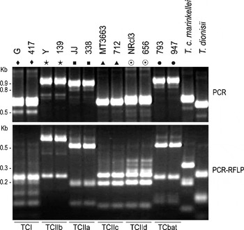
Analyses of PCR-amplified DNA fragments corresponding to ITS1 rDNA revealed length polymorphism among T. cruzi isolates. However, this polymorphism did not allow either a clear separation between TCIIb and TCbat or identification of T. c. marinkellei and T. dionisii-like. However, restriction patterns generated by PCR-RFLP analysis of representative isolates of each T. cruzi lineage distinguished TCbat. Moreover, this method also allowed identification of T. cruzi lineages and separation of T. cruzi from T. c. marinkellei and T. dionisii-like (Fig. 5). In addition, these methods confirmed the existence of mixed-infected bats harbouring different combinations of TCI or TCbat together with T. c. marinkellei and/or trypanosomes closely related to European isolates of T. dionisii (data not shown).
(A) PCR-amplified DNA fragments corresponding to ITS1 rDNA. (B) Genotyping of Trypanosoma cruzi isolates based on restriction fragment length polymorphism (RFLP) of PCR amplified ITS1 rDNA digested with the restriction enzyme Bsh 1236. Lineages TCI (◆), TCIIa (▪), TCIIb (), TCIIc (▴), TCIId () and TCbat (•). Brazilian isolates of T. dionisii-like and T. c. marinkeleii were used as controls of T. cruzi-like trypanosomes from bats. Agarose gels (2·5%) stained with ethidium bromide.

Behavioural analyses of T. cruzi isolates from bats in culture, in mice and in triatomines
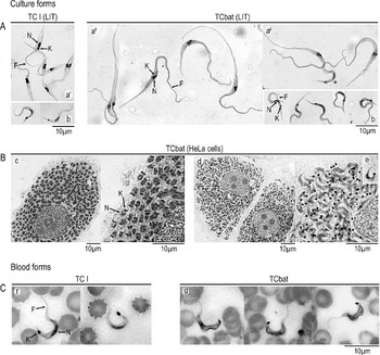
Analyses of culture behaviour of bat isolates in LIT medium supplemented with 10% FBS showed morphologically similar epimastigotes for isolates of TCI and TCbat (data not shown). However, in LIT containing 3–5% FBS, epimastigotes of TCbat isolates became very long, with several large dividing forms. Stationary cultures showed only forms with a long and pointed posterior extremity, in addition to metacyclic forms (Fig. 6). In contrast, in all culture media and growth conditions, TCI isolates showed epimastigotes that resembled isolates of other T. cruzi lineages. Metacyclic trypomastigotes from TCI and TCbat appeared similar (Fig. 6), despite lower numbers of these forms in TCbat compared to TCI. Bat isolates of TCI (TryCC 507 and 417) (data not shown) and TCbat (TryCC 204 and 793) developed inside HeLa cells (Fig. 6) and other mammalian cells similar to isolates of other T. cruzi lineages. Invasion by metacyclic trypomastigotes and multiplication inside mammalian cells as amastigotes, which differentiate to trypomastigotes that invade new cells, are features shared by all trypanosomes of Schizotrypanum (Molyneux, Reference Molyneux, Kreier and Baker1991).
Photomicrographs (Giemsa-staining) selected to illustrate morphological and growth features of bat trypanosomes. (A) Morphology of selected bat Trypanosoma cruzi isolates assigned to TCbat (TryCC 793) and TCI (TryCC 507) cultivated in LIT medium: epimastimastigotes of logarithmic (a1) and stationary phase (a2), and metacyclic trypomastigotes* (b). (B) Development within HeLa cells showing intracellular amastigotes (c) and trypomastigotes (d) 4 and 7 days post-infection, respectively, and trypomastigotes released from HeLa cells (e). (C) Trypomastigotes in blood smears from mice experimentally infected with bat T. cruzi isolates of TCI (f) and TCbat (g). N, nucleus; K, kinetoplast; F, flagellum.

Isolates assigned to TCI and TCbat were infective to mice. Despite the fact that bat isolates from these two lineages yielded low parasitaemias, blood trypomastigotes could be detected in blood smears (Fig. 6). The bat isolates lacked virulence to mice as assessed by mortality rates. Chronic infections in mice inoculated with these isolates were confirmed by haemocultures performed within approximately 120 days of infection.
Isolates of TCbat did not develop in 8 triatomine species investigated: R. prolixus, R. neglectus, R. robustus (genetic populations II); R. pictipes; R. domesticus; T. vitticeps; T. infestans and P. megistus. Results showed that TCbat isolates survived in the digestive tube of bugs from the triatomine for up to ~20 days, following complete elimination of the flagellates. In contrast, TCI isolates from bats developed in all these triatomines, which showed metacyclic trypomastigotes in their guts after 30 days of infection.
DISCUSSION
Data regarding prevalence rates, distribution, genetic diversity and phylogenetic relationships of T. cruzi isolates from bats are scarce. Comparative studies of T. cruzi with its very closely related bat-restricted trypanosomes may be helpful to the understanding of host-parasite relationships of the Schizotrypanum species. Moreover, a detailed understanding of the genetic diversity, ecobiology and phylogeography of Schizotrypanum trypanosomes is crucial for an understanding of the evolutionary history of T. cruzi. With these aims, we recently carried out a large survey of trypanosomes in Brazilian bats and obtained isolates of T. cruzi, T. c. marinkellei and T. dionisii-like (Cavazzana et al. Reference Cavazzana, Marcili, Campaner, Veludo, Takata, Paiva, Takeda and Teixeira2003; Maia da Silva et al. Reference Maia da Silva, Marcili, Lima, Cavazzana, Ortiz, Campaner, Takeda, Paiva, Nunes, Camargo and Teixeira2009). T. cruzi was isolated mainly from Vespertilionidae and Phyllostomidae bats from Central and Southeast Brazil, besides some isolates from Amazonia, mostly from Phyllostomidae. Bats of Vespertilionidae (Myotis) and Phyllostomidae (Phyllostomus, Artibeus, Desmodus and Anoura) were implicated as important reservoirs of T. cruzi. These bats exhibit varied alimentary habits, including insectivorous, frugivorous, haematophagous and omnivorous species, and can be infected by T. cruzi in sylvatic and anthropic environments (Barreto et al. Reference Barreto, Ribeiro and Filho1974; Funayama and Barretto, Reference Funayama and Barretto1970a, Reference Funayama and Barrettob, Reference Funayama and Barreto1973; Fabián Reference Fabián1991; Lisboa et al. Reference Lisboa, Pinho, Herrera, Gerhard, Cupolillo and Jansen2008; Maia da Silva et al. Reference Maia da Silva, Marcili, Lima, Cavazzana, Ortiz, Campaner, Takeda, Paiva, Nunes, Camargo and Teixeira2009; Añez et al. Reference Añez, Crisante and Soriano2009).
In this study, we characterized T. cruzi isolates from Brazilian bats genotyped as TCI and TCIIb using PCR based on mini-exon gene markers (Fernandes et al. Reference Fernandes, Santos, Cupolillo, Mendonça, Derre, Junqueira, Santos, Sturm, Naiff, Barrett, Campbell and Coura2001). However, genotyping of 15 bat isolates using ribosomal markers (Souto et al. Reference Souto, Fernandes, Macedo, Campbell and Zingales1996) confirmed 4 isolates as TCI, whilst amplified DNA fragments shown by isolates assigned to TCII were not compatible with any previously defined TCII sublineages (TCIIa–e), indicating that these isolates belong to a distinct genotype.
Analysis of phylogenetic relationships was carried out with the aim of understanding the phylogenetic relationships between the new isolates from bats and isolates from other hosts previously assigned to established T. cruzi lineages. For this purpose, we employed independent and concatenated SSU rDNA, Cyb and H2B sequences. Isolates from Amazonian bats always nested within TCI, as did the majority of isolates from this region (Miles et al. Reference Miles, Povoa, De Souza, Lainson, Shaw and Ketteridge1981; Fernandes et al. Reference Fernandes, Santos, Cupolillo, Mendonça, Derre, Junqueira, Santos, Sturm, Naiff, Barrett, Campbell and Coura2001; Maia da Silva et al. Reference Maia da Silva, Naiff, Marcili, Gordo, D'Affonseca Neto, Naiff, Franco, Campaner, Valente, Valente, Camargo, Teixeira and Miles2008; Marcili et al. Reference Marcili, Valente, Valente, Junqueira, Maia da Silva, Naiff, Campaner, Coura, Camargo, Miles and Teixeira2009). The 11 bat isolates previously genotyped as TCIIb clustered tightly together constituting a new clade separated by relevant genetic distances from all known lineages, thus confirming that they belong to a new genotype (TCbat). All analyses performed using separated or combined data sets separated isolates of genotype TCbat from isolates of lineage TCIIb. However, results did not indicate an unquestionable positioning of TCbat within any established lineages of T. cruzi. Phylogenetic analysis of Schizotrypanum trypanosomes using concatenated alignment of SSU rDNA, Cyb and H2B genes positioned all bat isolates characterized in this study within the clade T. cruzi, which is more closely related to T. c. marinkellei than to T. dionisii clades.
Most inferred phylogenetic analyses suggested that TCbat genotype is closer to TCI than to any other lineage, although separated by large genetic distances. In contrast to TCbat, isolates of TCI shared amplified DNA fragments of the same length when genotyped using traditional methods. In this study, we demonstrated that TCbat isolates differed from TCI isolates from Amazonian bats, and also from TCI from didelphids and triatomines from the same regions where bats infected with TCbat have been found. TCbat is clearly different from any isolate assigned to the TCI lineage, even from bat isolates assigned to TCI by all molecular markers investigated in this study.
The monophyletic assemblage formed by TCI from bats, humans, wild monkeys and didelphids provided evidence of geographical clustering within TCI. Recent studies have evidenced subclusters within TCI that could be associated with both mammalian hosts and geographical origin. Analysis of the highly polymorphic intergenic region of the mini-exon gene separated North American from South American isolates, and disclosed a subclade related to Didelphis sp. (O'Connor et al. Reference O'Connor, Bosseno, Barnabé, Douzery and Brenière2007). Mini-exon markers revealed 4 haplotypes (Ia–Id) of Colombian TCI isolates related to distinct transmission cycles (Herrera et al. Reference Herrera, Bargues, Fajardo, Montilla, Triana, Vallejo and Guhl2007). Analysis of Cyb sequences of TCI isolates from Chile revealed a new genotype (DTU1b) associated with caviomorph rodents (Spotorno et al. Reference Spotorno, Córdova and Solari2008).
Comparative analyses of TCbat and TCI isolates from a range of hosts and geographical origin may help to define recommendations for the description of new genotypes/lineages closely related to TCI. Recently, isolates of lineage TCI have been investigated using polymorphic molecular markers, and results showed high intralineage genetic diversity and a complex populational structure of TCI populations (O'Connor et al. Reference O'Connor, Bosseno, Barnabé, Douzery and Brenière2007; Herrera et al. Reference Herrera, Bargues, Fajardo, Montilla, Triana, Vallejo and Guhl2007; Spotorno et al. Reference Spotorno, Córdova and Solari2008). Our data do not support TCI and TCII as 2 major lineages within T. cruzi since TCIIa–e sublineages are not monophyletic and varied according to the gene analysed, as previously demonstrated (Machado and Ayala, Reference Machado and Ayala2001; Brisse et al. Reference Brisse, Henriksson, Barnabé, Douzery, Berkvens, Serrano, De Carvalho, Buck, Dujardin and Tibayrenc2003; Sturm et al. Reference Sturm, Vargas, Westenberger, Zingales and Campbell2003; Westenberger et al. Reference Westenberger, Barnabé, Campbell and Sturn2005, Reference Westenberger, Sturm and Campbell2006). Therefore, relationships among lineages of T. cruzi are far from understood. Further analysis from more isolates from several lineages may help to resolve the phylogeny of T. cruzi. Until more data can be gathered, we have designated the new genotype of T. cruzi from bats characterized in this study as TCbat.
Isolates of TCbat were shown to be distinguishable by morphological and biological features. These isolates showed very large and pointed epimastigotes, different from those of all other T. cruzi lineages. In addition, TCbat isolates were unable to yield established infection in triatomines of genera Rhodnius, Triatoma and Panstrongylus, a behaviour shared by T. c. marinkellei and T. dionisii. T. c. marinkellei seems to be transmitted only by triatomines of genus Cavernicola, which are usually associated with bat colonies, whereas T. dionisii is transmitted by cimicids (Marinkelle, Reference Marinkelle, Lumsden and Evans1976; Molyneux, Reference Molyneux, Kreier and Baker1991). Triatomines that live in tree holes and caves, palms and house roofs can transmit T. cruzi among bats. The majority of T. cruzi-infected bats are insectivorous and likely to be infected by ingestion of triatomines (Marinkelle, Reference Marinkelle, Lumsden and Evans1976; Thomas et al. Reference Thomas, Rasweiler and D'Alessandro2007). Vector permissiveness to T. cruzi and association between lineages/strains and triatomine species depends on both vectors and parasite features, apparently, with superior vector competence of sympatric sylvatic species, as clearly demonstrated for T. rangeli (Maia da Silva et al. Reference Maia da Silva, Junqueira, Campaner, Rodrigues, Crisante, Ramirez, Caballero, Monteiro, Coura, Añez and Teixeira2007). Unfortunately, triatomines of Cavernicola and other sylvatic species from regions where TCbat isolates originated were not available for this study. In Central Brazil, several triatomine species, mainly those inhabiting palms such as R. neglectus, R. robustus and R. stali could transmit T. cruzi among bats (Gurgel-Gonçalves et al. Reference Gurgel-Gonçalves, Abad-Franch, Ferreira, Santana and Cuba2008). However, the genotypes of T. cruzi circulating in sylvatic triatomines from MS and SP were completely unknown. Isolates from R. stali (MS) and P. megistus (SP) included in this study were genotyped as TCI.
Altogether, sequence divergences and phylogenetic analysis of SSU rDNA, Cyb and H2B genes, morphology and behaviour in triatomines indicate that isolates of TCbat indeed belong to a new genotype of T. cruzi represented, so far, exclusively by bat isolates from antrophic areas of Central and Southeast Brazil. Both regions are endemic for Chagas disease. Distances separating these areas are easily crossed by bats found to be infected with TCbat. The limited data regarding T. cruzi genotypes in wild mammals are insufficient to rule out other mammals as hosts of TCbat, and also humans living in houses inhabited by bats.
The hypotheses that T. cruzi evolved from a trypanosome restricted to bats or vice versa remain to be elucidated. The present-day distribution of Schizotrypanum species and the ability of bats to disperse over long distances, including crossing oceans, are consistent with both hypotheses (Stevens et al. Reference Stevens, Noyes, Dover and Gibson1999; Barnabé et al. Reference Barnabé, Brisse and Tibayrenc2003). The evolutionary histories of T. cruzi lineages have been correlated with a long-standing association with vertebrate hosts. TCI and TCII have been associated respectively with marsupials of Didelphimorpha (opossums) and with placentals of Xenarthra (armadillos), the early mammals in South America (~65 mya). Primates and rodents entered South America from Africa during the Oligocene (~35 mya), whereas chiropterans dispersed from Africa in the Eocene (~45 mya), and arrived in the Americas via Beringia or by a transatlantic route (Eick et al. Reference Eick, Jacobs and Matthee2005). Bats are ancient hosts of T. cruzi-like species or lineages transmitted by triatomines in the Americas as indicated by the description of Trypanosoma antiquus in triatomine feces fossilized in Dominican amber, together with bat hairs (Poinar, Reference Maia da Silva, Junqueira, Campaner, Rodrigues, Crisante, Ramirez, Caballero, Monteiro, Coura, Añez and Teixeira2005). The low genetic divergence showed in this and in previous studies is compatible with a recent split between T. cruzi and the bat-restricted Schizotrypanum, as well as with a recent diversification of all T. cruzi lineages (Machado and Ayala, Reference Machado and Ayala2001; Brisse et al. Reference Brisse, Henriksson, Barnabé, Douzery, Berkvens, Serrano, De Carvalho, Buck, Dujardin and Tibayrenc2003; Barnabé et al. Reference Barnabé, Brisse and Tibayrenc2003).
Besides T. cruzi, other Schizotrypanum species such as T. c. marinkellei and T. dionisii-like, in addition to T. rangeli have also been found infecting bats in the same locations where bats infected with TCbat were captured (Cavazzana et al. Reference Cavazzana, Marcili, Campaner, Veludo, Takata, Paiva, Takeda and Teixeira2003; Lisboa et al. Reference Lisboa, Pinho, Herrera, Gerhard, Cupolillo and Jansen2008; Maia da Silva et al. Reference Maia da Silva, Marcili, Lima, Cavazzana, Ortiz, Campaner, Takeda, Paiva, Nunes, Camargo and Teixeira2009). Taking into account the ITS1 rDNA polymorphisms among T. cruzi lineages, we standardized a PCR-RFLP, targeting this gene that allowed separation of T. cruzi from other species of Schizotrypanum and to distinguish TCbat from other genotypes. This method was shown to be a sensitive tool for easy detection of new genotypes of T. cruzi and T. cruzi-like.
Data from this study corroborated the high complexity of T. cruzi, pointing towards the existence of distinct T. cruzi genotypes waiting to be described, and the method described in this study can be very helpful for this purpose. Our results provide evidence that the understanding of enzootic transmission cycles of T. cruzi can be improved with phylogenetic analysis of more isolates, especially from poorly investigated sylvatic vertebrate and invertebrate hosts of unexplored geographical regions and ecotopes.
We would like to thank all students and colleagues for their inestimable help in the fieldwork, and for providing us with blood samples of bats and reference strains of T. cruzi. We are grateful to Erney P. Camargo, Michel A. Miles, Martin Llewellyn and Michael Lewis for valuable comments on the manuscript. We are thankful to Valdir Tadei (in memoriam) and Caroline C. Aires for identification of bats. Work in Rondonia (Amazonia) was done in ICB5-USP. This work was supported by grants from the Brazilian agencies CNPq (UNIVERSAL) and FAPESP (PRONEX). F. M. da S. was sponsored by CAPES (PRODOC-PROTAX), and A. M., L. L. and A. C. V. J. were recipients of scholarships from CNPq.