Cardiac myxoma is the most common primary tumour of the heart,Reference Gavrielatos, Letsas, Pappas, Dedeilias, Sioras and Kardaras1 accounting for about half of all cardiac tumours,Reference Meng, Lai, Lima, Tong, Quian and Lai2 and being more frequent in females. Most tumours originate from the region of the oval fossa,Reference Rogov, Sheremet’eva and Nechaenko3, Reference Alter, Grimm and Rominger4 with about four-fifths occupying the left atrium, and most of the remainder found in the right atrium. Ventricular myxomas are rare.Reference Alter, Grimm and Rominger4, Reference Grebenc, Rosado-de-Christenson, Green, Burke and Galvin5 The tumours are also known to exist in a familial variant, showing multiple and genetic heterogeneity.Reference Rogov, Sheremet’eva and Nechaenko3, Reference Casey, Mah and Merliss6, Reference Basson, MacRae, Korf and Merliss7 Echocardiography is now well established as the most reliable diagnostic technique, having sensitivity which can reach to 95%.Reference Percell, Henning and Siddique Patel8 We describe here our experience with echocardiographic diagnosis of 2 familial cases of cardiac myxoma.
Case reports
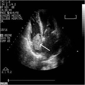
Our initial patient was a 10-year-old female, hospitalized for chronic coughing of nearly one year, which had become aggravated over the previous 3 days with chest pain and distress. On physical examination, she was found to have a regular cardiac rhythm, and a systolic murmur, graded at 2 from 6, was audible in the tricuspid area. The electrocardiogram revealed sinus tachycardia, while the chest radiographic showed an enlarged heart. Computed tomographic scanning of the lungs suggested pulmonary embolism. Echocardiographic examination revealed a smooth mass, initially thought to lack a pedicle, and measuring 7.2 centimetres by 5 centimetres. The mass filled the right atrium, obstructing the right atrioventricular orifice in diastole. Its upper pole was partially blocking the orifice of the superior caval vein, while inferiorly it encroached on the orifice of the inferior caval vein (Fig. 1). The maximal pulmonary arterial systolic pressure was calculated at 49 mmHg, so that the final echocardiographic diagnosis was a right atrial occupying lesion and moderate pulmonary hypertension. At surgery, the entirety of the right atrium was confirmed to be occupied by the tumour, which measured 8 by 5 centimetres. Surgical exploration revealed that the tumour did possess a pedicle, of 2 centimetres, and it was adherent not only to the base of the atrial septum, but extended into the right ventricle. Despite its soft and fragile texture, the tumour was successfully removed. The pathological findings confirmed the diagnosis of right atrial myxoma, and the patient recovered well.
The right atrial myxoma (arrow) as indetified in our first patient.

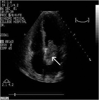
Our second patient was a 34-year-old female, the mother of our initial patient. She was hospitalized urgently for paroxysmal limitation over 1 day of movement of her right arm and leg. At physical examination, the cardiac rhythm was regular, and a diastolic murmur was heard at the cardiac apex, albeit without any palpable thrill. The muscle power and tone of the right limbs was degraded. The activity of the left limbs was normal, and the bilateral Babinski sign was positive. Inversion of the T waves was seen in leads II, III, and aVF. Injection of contrast showed embolization of the left middle cerebral artery. Echocardiography showed enlargement of the left atrium, which contained a pedicled mass of 2.7 by 5.2 centimetres, adherent to the base of the atrial septum (Fig. 2). With cardiac activity, part of the tumor intruded through the mitral orifice in diastole, returning to the left atrium in systole, and hence produced mechanical obstruction of the mitral valvar orifice. The ultrasonic diagnosis was a left atrial occupying lesion, and the patient was referred for surgical removal. At surgery, a tumour measuring 5 by 4 by 3 centimetres was found in the left atrium, attached by a pedicle of 1 centimetre to the base of the oval foramen. Pathological studies showed it to be a myxoma. The patient recovered well, and the power of the right limbs was restored at the time of discharge.
The left atrial myxoma (arrow) in our second patient, the mother of the index case.

Discussion
All familial cardiac myxomas have been shown to be associated with chromosomal abnormalities, the haploid cells having abnormal contents of deoxyribonucleic acid, with some showing heightened proliferative activity.Reference Kirklin9 Chromosomes 2 and 12 have been involved, and the tumour has been shown to be inherited in autosomal dominant fashion.Reference Stratakis10 Significantly, the familial form of cardiac myxoma is now considered potentially malignant.
Echocardiography is well recognized as being capable of showing the size of such tumours, their different positions during the cardiac cycle, and the degree of valvar stenosis and counter-flow that they can produce. Hence, echocardiography is the simplest, as well as the most reliable, means of early detection. Such early detection is important, since fragments of the tumours can embolise. Cerebral embolism is particularly dangerous. Once diagnosed, surgery is needed as soon as possible. Not only should the myxoma be thoroughly excised, but also the normal endocardium and cardiac muscle at the point of attachment of its pedicle. The familial myxoma is also known frequently to be multiple, and recurrences are well recognized. Thus, periodic follow-up is necessary. Should recurrences occur, the new tumours should again rapidly be removed.