




No CrossRef data available.
Article contents
Use of covered Cheatham-Platinum stents in aortic coarctation and recoarctation
Published online by Cambridge University Press: 20 January 2005
Abstract
We inserted covered Cheatham-Platinum stents in 4 patients, ranging in age from 12 to 19 years, who weighed between 45 and 94 kg. All the patients had aortic coarctation, with surgical repair having been attempted previously in one, and with balloon dilation having been performed as the primary treatment in two, resulting in formation of aneurysms. The fourth patient had not received any treatment. The gradients were reduced from 10 to 40 mmHg before insertion of the stent to 0 to 5 mmHg after stenting. No complications were encountered. All the patients are well at an interval of 3 to 14 months after stenting.
Information
- Type
- Original Article
- Information
- Copyright
- © 2004 Cambridge University Press
Covered stents have been used in the treatment of abdominal and thoracic atherosclerotic aneurysms in adults.1 In congenital heart disease, in contrast, they have hardly been used, and their role still remains to be defined.2–4 Theoretically at least, they could be used for the purposes of bailing out when patients develop aneurysms or dissections following an interventional procedure.
Normal uncovered stents, such as the Palmaz stents, have been used in all locations in children with congenital heart defects for the last few years.5 Usually these stents can be redilated at a later date to a size close to that needed in adulthood, allowing for growth of the children. The limitation of covered stents is that they can only be used in patients who are fully or nearly fully grown, as it may not be possible to redilate them without damaging the covering polytetrafluoroethylene material.
We report our early experience in four patients in whom custom-made covered Cheatham-Platinum stents were used in the treatment of either aortic coarctation or recoarctation.
Patients and methods
Since September 2001, we have implanted custom-made covered Cheatham-Platinum stents (NuMED Inc, Hopkinton, NY, USA) in four patients, 3 males and 1 female, for the treatment of aortic coarctation or recoarctation. Their ages ranged between 12 and 19 years, and their weights between 45 and 94 kg. The procedures were approved by the hospital ethics committee.
The clinical details are given in Table 1. The coarctation was isolated in 3 of the patients, while the fourth had additional patency of the arterial duct. The first two patients had previously undergone balloon dilation, and both had subsequently developed an aortic aneurysm. In the first of these, the aneurysm increased in size over 8 weeks after the dilation. The parents of the patient were Jehovah's Witnesses, and refused blood transfusions for the patient. The third patient had not undergone any previous procedures. This patient had a long hypoplastic transverse aortic arch in addition to localized coarctation. The fourth patient had previously undergone subclavian flap aortoplasty, combined with ligation of the arterial duct, but had developed aortic recoarctation and recanalization of the duct. Balloon dilation was unsuccessful in relieving the recoarctation.
Table 1.

All the patients were on antihypertensive treatment, consisting of 3 drugs in two patients, and 2 drugs in the other two. The combinations were acebutalol, amlodipine and enalapril in one, atenolol, nifedipine and enalapril in one, propranolol and amlodipine in one, and propranolol and metoprolol in one.
Technique of implantation
General anaesthesia was used in all the patients. Surgical femoral arteriotomy was performed for the insertion of sheaths and stents in 3 patients, while in the other, an introducer sheath was inserted percutaneously. In two of the patients, a brachial arterial sheath was also placed percutaneously, through which an angiographic pigtail catheter was positioned in the ascending aorta for aortography in order to define the anatomy, to perform measurements of the aorta in different positions, and in particular to aid in positioning of the stent. In two patients, a 12 french Mullins trans-septal sheath was placed in the femoral artery, and in two, a 14 french sheath was placed over a stiff exchange length 0.035″ guidewire positioned in the ascending aorta or the right subclavian artery. Heparin at a dose of 50 to 100 units/kg was given intravenously to maintain the activated clotting time above 200 s during the procedure. Flucloxacillin and gentamicin were given at the start of the procedure, and continued for 24 h afterwards.
After measurements of gradients and angiography, the diameters of the distal arch (if hypoplasia of the distal arch was present, then the diameter of the transverse arch was used) and the post-stenotic dilated segment were measured at maximum expansion (Table 2). The diameter of the stent was selected to be of a similar size to either the transverse arch or the distal arch, whichever was the greater. In the third patient, the diameter of the aorta at the level of the diaphragm was used for determining the diameter of the stent. The gradient across the site of the aortic coarctation prior to implantation ranged between 10 and 40 mmHg.
Table 2.

Covered Cheatham-Platinum stents were used in all four patients because of their specific problems (Fig. 1). The stents were custom made, and manufactured according to the diameters and lengths measured on the most recent angiogram. Usually the length of the stent that was required was determined by the distance from just beyond the left subclavian artery, or the left common carotid artery if the left subclavian artery had been used in previous surgery, to about 15 mm beyond the site of the coarctation. The stents are available in any length, and can be mounted on any balloon of diameter up to 25 mm. The whole length of the stent is covered with expanded polytetrafluoroethylene. The stent is premounted on a BIB balloon (NuMED Inc, Hopkinton, NY, USA), which consists of a balloon inside a balloon, in which the inner balloon is half the diameter and 1 cm shorter than the outer balloon. The advantage of using this balloon is that, during deployment of the stent, inflation of the inner balloon allows the position of the stent to be adjusted, before inflation of the outer balloon for the final deployment. The balloons were inflated to the pressure recommended by the manufacturer, usually up to 6 atmospheres, in order fully to expand the stent. At diameters of up to 22 mm, there can be shortening of the stent by up to one-eighth. Depending on the diameter of the outer balloon, the whole assembly can be passed through either a 12 or 14 french sheath and, depending on the size of the patients, the sheaths may be introduced either percutaneously or through a surgical arteriotomy.

Figure 1. A dilated covered Cheatham-Platinum stent. Note the round uncovered edges of the stent.
Angiography was performed through the pigtail catheter placed in the ascending aorta, or via the side-arm of the sheath during and after implantation. After the procedure, and subsequent to removal of the sheaths, haemostasis was achieved either by manual compression or surgical repair of the arteriotomy.
All the patients were maintained on aspirin at a dose of 3 to 5 mg/kg given once daily for 6 months after implantation.
Results
The stents ranged in length from 34 to 45 mm, and were deployed at diameters of 14 to 22 mm. After implantation, the gradient across the stent had been reduced to between 0 and 5 mmHg in all four patients. The stents were deployed in the correct position in all the patients. Angiography showed an excellent result, with no opacification of the arterial duct or the aneurysms (Figs 2 and 3).

Figure 2. An ascending aortogram (a) in the left anterior oblique projection demonstrates a small aneurysm (arrow) after balloon dilation of aortic coarctation in our first patient. An aortogram in the left anterior oblique projection after implantation of a covered stent (b) shows a good angiographic result. The aneurysm has been excluded. A spiral computerized tomographic scan 3 months after implantation (c) confirms the good result.

Figure 3. An ascending aortogram in the left anterior oblique projection (a) shows marked long segment distal arch hypoplasia with a localized severe coarctation of the aorta in our third patient. An aortogram (b) shows the covered stent mounted on the BIB balloon, with the inner balloon having been inflated allowing any repositioning of the stent if required. An aortogram (c) shows an excellent result after implantation.
The patients were discharged home 2 to 3 days after the procedure. Although it would have been possible to discharge the patients earlier than this, they were hospitalized for this period to assess the arteriotomy wound, any gastrointestinal symptoms, and any rebound hypertension, none of which occurred. There were no complications related to the procedure, nor were there any complications related to the sites of arteriotomy or puncture.
During the early follow up period of 3 to 14 months, all the patients have remained well. The antihypertensive medication was continued in all the patients, but the dosage was reduced with improved control of the blood pressure. In all the patients, the blood pressures were in the normal range, specifically below 140/90 mmHg.
Computerized tomographic scanning performed between 3 and 8 months after implantation revealed no aneurysmal formation, the stents being widely patent in all the patients.
Discussion
Stents have been used in many clinical situations as they reduce the possibility of disruption of a vessel by avoiding overdilation. Over the last few years, they have been increasingly used to treat aortic coarctation and recoarctation in an attempt to reduce the possibility of aneurysmal formation.5–13 Experience has shown that these stents, when used in smaller children, need to be, and can be, redilated without complications.12, 13 In fully grown adolescents and adults, they may not need redilation, and so may be used as an alternative to surgery for treating aortic coarctation. Although the main reason for using stents in aortic coarctation is to prevent aneurysms, aneurysms may occur, with a reported incidence of less than 5%.10, 13 Covered stents may avoid this complication, and so have a potential advantage over the uncovered stents, but they do have a limit to the extent to which they can be redilated. Consequently, their use is likely to be confined to patients who are fully or nearly fully grown.
Covered stents have been used in the treatment of aneurysms in the abdominal as well as thoracic aorta.1, 4 The main concern with covered stents is the possibility of occlusion of side branches and the complications arising from this. In the case of covered stents in the thoracic aorta, spinal arterial occlusion is the most catastrophic complication. The spinal artery usually originates below the level of the nineth thoracic vertebra. Whilst the incidence of paraplegia or paraparesis after surgery of the thoracic aorta is less than 1%, the incidence is greater than 10% when surgery is performed on the aorta just above the diaphragm.14, 15 In most patients with aortic coarctation or recoarctation in whom a covered stent may be contemplated, therefore, spinal arterial occlusion is unlikely to occur.
The Cheatham-Platinum stent is more radio-opaque than steel stents, and does not preclude future studies of the heart or other organs using resonance imaging. It has rounded edges, which are less traumatic to the native vessels. It can be implanted using any angioplasty balloon, or alternatively, the BIB balloon. This balloon allows the position of the stent to be adjusted after inflation of the inner balloon before the outer balloon is inflated. This combination of stent and balloon, therefore, has some important clinical advantages over other stents or techniques of implantation.
Our study reports our initial results using custom-made covered stents in aortic coarctation or recoarctation. The early results are encouraging. Two of the patients had had evidence of aneurysmal formation after balloon dilation. More importantly, one of them still had a residual coarctation, so further attempts at balloon dilation were contraindicated. One of these patients was also from a strict Jehovah's Witness family, and transfusion of blood products was to be avoided. In both of these patients, the stent covered the aneurysmal area completely, and produced a good haemodynamic and angiographic result. The third patient had recoarctation and residual patency of the arterial duct after previous surgical repair. By using a covered stent, it was possible to treat the combined lesions with one stent, rather than using separate devices to treat the two lesions by interventional techniques, as has been reported recently.16, 17 Furthermore, in patients with aneurysms and recoarctation, there are advantages of using covered stents over the use of coils to thrombose the aneurysm and an uncovered stent to treat the coarctation. There is a small possibility of this latter technique predisposing to rupture of the aneurysm. In our third patient, the decision was made to implant a covered stent electively, because of the presence of a severely hypoplastic aorta. There were concerns about development of an aneurysm with either balloon dilation or implantation of a conventional stent.
The limitations of or study include the small numbers stented, and the lack of availability of the data about the long-term fate of covered stents, especially with regard to neointimal formation. Thus, ours must be considered a preliminary study, and careful follow-up evaluation in a larger population of patients will be required to determine their role. Whilst in the majority of the patients, conventional stents will continue to be used to treat aortic coarctation, covered Cheatham-Platinum stents may have an important role in dealing with aneurysmal formation after previous surgery or balloon dilation.18 Indeed, they may also have a primary role in treating adults with combined lesions, or with associated cystic medial necrosis.
Acknowledgement
We thank Mr Allen Tower and Mr Doug Villnave, of NuMED Inc, Hopkinton, New York, United States of America, for the custom-made Cheatham-Platinum stents.
References
Taylor PR, Gaines PA, McGuinness CL, Beard JD, Cooper G, Reidy JF. Thoracic aortic stent grafts – early experience from two centres using commercially available devices. Eur J Vasc & Endovasc Surg 2001; 22: 70–76.Google Scholar
Gunn J, Cleveland T, Gaines P. Covered stent to treat coexistent coarctation and aneurysm of the aorta in a young man. Heart 1999; 82: 351.Google Scholar
de Giovanni JV. Covered stents in the treatment of aortic coarctation. J Intervent Cardiol 2001; 14: 187–190.Google Scholar
Khan MS, Moore JW. Treatment of abdominal aortic pseudoaneurysm with covered stents in a paediatric patient. Cathet & Cardiovasc Intervent 2000; 50: 445–448.Google Scholar
Cheatham JP. Stents and Amplatzers: what's an interventionalist to do? Catheter Cardiovasc Intervent 1999; 47: 39–40.Google Scholar
Magee AG, Brzezinska-Rajszys G, Qureshi SA, et al. Stent implantation for aortic coarctation and recoarctation. Heart 1999; 82: 600–606.Google Scholar
Brzezinska-Rajszys G, Qureshi SA, Ksiazyk J, et al. Middle aortic syndrome treated by stent implantation. Heart 1999; 81: 166–170.Google Scholar
Thanopoulous BD, Hadjinikolou L, Konstadopoulou GN, Tsaousis GS, Triposkiadis F, Spirou P. Stent treatment for coarctation of the aorta: intermediate term follow up and technical considerations. Heart 2000; 84: 65–70.Google Scholar
Hamdan MA, Maheshwari S, Fahey JT, Hellenbrand WE. Endovascular stents for coarctation of the aorta: initial results and intermediate term follow up. J Am Coll Cardiol 2001; 38: 1518–1523.Google Scholar
Ovaert C, Benson LN, Nykanen D, Freedom R. Transcatheter treatment of coarctation of the aorta: a review. Pediatr Cardiol 1998; 19: 27–44.Google Scholar
Ebeid MR, Prieto LR, Latson LA. Use of balloon-expandable stents for coarctation of the aorta: initial results and intermediate-term follow-up J Am Coll Cardiol 1997; 30: 1847–1852.Google Scholar
Suarez de Lezo J, Pan M, Romero M, et al. Balloon-expandable stent repair of severe coarctation of aorta. Am Heart J 1995; 129: 1002–1008.Google Scholar
Suarez de Lezo J, Pan M, Romero M, et al. Immediate and follow-up findings after stent treatment for severe coarctation of aorta. Am J Cardiol 1999; 83: 400–406.Google Scholar
Wada T, Yao H, Miyamoto T, Mukai S, Yamamura M. Prevention and detection of spinal cord injury during thoracic and thoraco-abdominal aortic repairs. Ann Thorac Surg 2001; 72: 80–84.Google Scholar
Connolly JE. Prevention of spinal cord complications in aortic surgery. Am J Surg 1998; 176: 92–101.Google Scholar
Hakim F, Hawelleh AA, Goussous Y, Hijazi ZM. Simultaneous stent implantation for coarctation of the aorta and closure of patent ductus arteriosus using the Amplatzer duct occluder. Cathet Cardiovasc Intervent 1999; 47: 36–38.Google Scholar
Galal O, Al-Fadley, Wilson N. Successful transcatheter closure of patent arterial duct six years after balloon dilation of coarctation of the aorta. Int J Cardiol 1992; 35: 123–125.Google Scholar
Cheatham JP. Stenting of coarctation of the aorta. Cathet Cardiovasc Intervent 2001; 54: 112–125.Google Scholar
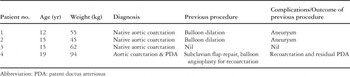
Table 1.

Table 2.

A dilated covered Cheatham-Platinum stent. Note the round uncovered edges of the stent.

An ascending aortogram (a) in the left anterior oblique projection demonstrates a small aneurysm (arrow) after balloon dilation of aortic coarctation in our first patient. An aortogram in the left anterior oblique projection after implantation of a covered stent (b) shows a good angiographic result. The aneurysm has been excluded. A spiral computerized tomographic scan 3 months after implantation (c) confirms the good result.
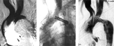
An ascending aortogram in the left anterior oblique projection (a) shows marked long segment distal arch hypoplasia with a localized severe coarctation of the aorta in our third patient. An aortogram (b) shows the covered stent mounted on the BIB balloon, with the inner balloon having been inflated allowing any repositioning of the stent if required. An aortogram (c) shows an excellent result after implantation.
 You have
Access
- 44
- Cited by