INTRODUCTION
Diatoms form an important but understudied component of coral reef biodiversity, where they are abundant as epiphytes on seaweeds (Lobban & Jordan, Reference Lobban, Jordan, Smol and Stoermer2010). A commonly seen genus is Licmophora C. Agardh, with L. flabellata (Carmichael ex Greville) C. Agardh emend Sar & Ferrario and L. remulus Grunow among the most common and easily recognized on Guam reefs (Jordan et al., Reference Jordan, Lobban and Theriot2009–2013; Lobban et al., Reference Lobban, Schefter and Ruck2011). The genus is characterized by heteropolar and heterovalvar frustules that are cuneate in both girdle and valve view, attached to substrata by mucilage pads or stalks (Hustedt, Reference Hustedt1931–1959; Round et al., Reference Round, Crawford and Mann1990). It is typified by L. flabellata [if, as pointed out by Sar & Ferrario (Reference Sar and Ferrario1990), Hustedt was right in placing L. argentescens C. Agardh into synonymy with L. flabellata]. However, this species is not typical of the genus in two significant features: it has large platelike plastids (the placochrome condition, sensu Williams & Kociolek, Reference Williams, Kociolek, Seckbach and Kociolek2011, whereas the usual condition is coccochrome), and it has many extra rimoportulae along the sternum (Sar & Ferrario, Reference Sar and Ferrario1990; Honeywill, Reference Honeywill1998; Lobban et al., Reference Lobban, Schefter and Ruck2011). However, until Sar & Ferrario pointed them out, diatomists did not mention or draw the extra rimoportulae, and Giffen (Reference Giffen1970) described L. pfannkucheae as distinguished from L. flabellata by a series of rimoportulae along the sternum (plastids unknown). Witkowski et al. (Reference Witkowski, Lange-Bertalot and Metzeltin2000) describe and illustrate both these species but mention the extra rimoportulae only for L. pfannkucheae. Most species of Licmophora have one rimoportula at the foot pole of one valve and one or two at the head pole; if there is only one at the head pole it is on the opposite valve from the foot-pole rimoportula. Most Licmophora species have many small discoid or irregular plastids without visible pyrenoids. Sar & Ferrario (Reference Sar and Ferrario1990) and Round et al. (Reference Round, Crawford and Mann1990) raised the question of whether the distinctive pattern of rimoportulae and plastids in L. flabellata was sufficient to warrant splitting the genus, but Honeywill (Reference Honeywill1998) considered all the British Licmophora species in her study, including L. flabellata, to be a single genus and the question remains open.
We have observed Licmophora in numerous collections over the last several years, including a new, unusually large fan-forming species, L. flucticulata Lobban, Schefter & Ruck, compared to L. flabellata and L. remulus (Lobban et al., Reference Lobban, Schefter and Ruck2011); Licmophora aff. ehrenbergii and L. abbreviata Agardh (Lobban, Reference Lobban2013). When a population of Licmophora was collected recently on a reef flat, the novelty of the species was quickly apparent in living material, because although colonies were similar to L. flabellata, the plastids were much smaller and more numerous. Whole mounts in a scanning electron microscope (SEM) quickly showed a row of rimoportulae and very fine striation, excluding it from all other Licmophora species, so acid-cleaned preparations were made. This paper describes the new species as L. comnavmaria.
MATERIALS AND METHODS
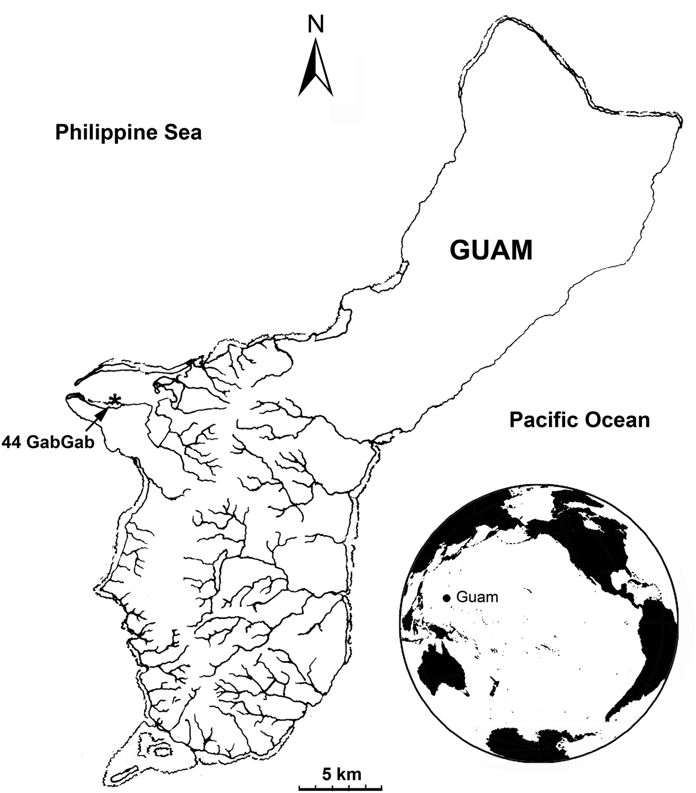
The diatom population was seen and collected in situ by snorkelling at GabGab, Apra Harbor, Guam, 13°26′33.63′′ N 144° 38′34.25′′ E (site GU44, Figure 1), 25 July 2011, on the shallow reef flat adjacent to a dredged swimming area on the US Naval Base, Guam. Guam is in the western Pacific Ocean, at the southern end of the Mariana Islands archipelago. The diatom sample was hand-collected in a quart Ziploc® bag and transported to the laboratory at ambient temperature. Living cells were observed and documented in a light microscope (LM) using a Nikon 80i microscope with DS-Fi1 colour camera head and L2 Controller. Live material was observed with differential interference contrast (DIC) but phase contrast was better for acid-cleaned material. The live sample observed in LM was transferred to a filter, dried and prepared for SEM as a whole mount. Standard acid cleaning with nitric acid was carried out on a further aliquot. For LM permanent mounts were made in Naphrax®; material for SEM was dried onto Millipore® cellulose nitrate filters (EMD Millipore Corporation, Billerica, MA) and fixed to stubs with carbon conductive tabs (Ted Pella Inc., Redding, CA). The samples were coated with Au/Pd in an SC7620 Mini Sputter Coater (Quorum Technologies, East Grinstead, West Sussex, UK) and observed with a PhenomWorld G2 Pro tabletop SEM (PhenomWorld US, Hillsboro, OR).
Map of Guam and its location in the western Pacific, showing the location of the collection site, GU44 GabGab.

SYSTEMATICS
Order licmophorales Round, 1990
Family licmophoraceae Kützing, 1844
Genus Licmophora C.A. Agardh, 1827
Licmophora comnavmaria Lobban et Schefter, sp. nov.
(Figures 2–6)
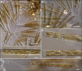
Licmophora comnavmaria, living material: (A, B) portions of L. comnavmaria colonies showing multistranded stalk with lateral individuals and terminal cluster; (C, D) cells in girdle view showing plastids, arrows indicate pyrenoids; (E) cell in valve view. Scale bars: A,B, 50 µm; C–E, 10 µm.

Licmophora comnavmaria, acid cleaned material in light microscope: (A–C) holotype valve, phase contrast, (A) whole valve imaged with 40× objective, (B, C) apical and basal portions imaged with 100× objective; on (B) white arrow marks the head pole rimoportula, arrowheads point to the first two intercalary rimoportula, black arrow indicates apical spine; (D) valve with basal rimoportula, phase contrast, 40× objective; (E) basal fragment with 100× objective, showing basal rimoportula (arrow). Scale bars, 10 µm.

Licmophora comnavmaria, in scanning electron microscope: (A, B) head pole and foot pole, respectively of frustules in a whole mount (live material dried directly onto filter), showing apical spines and basal multiscissura; (C) apical portion of acid-cleaned valve lying over a second valve and several girdle bands. Black arrow marks the apical rimoportula opening; white arrows indicate four intercalary rimoportulae along the sternum; double arrowheads point to area with apparent areolae (see text); (D, E) central region of valve in whole mount (external) and acid-cleaned (internal) respectively. Scale bars: A, B, D, 2 µm; C, D, 5 µm.

Licmophora comnavmaria, details of head and foot poles, SEM: (A) head pole exterior; (B) head pole interior, arrow indicates the most-apical intercalary rimoportula; (C) foot pole without rimoportula external; (D) foot pole without rimoportula internal, also showing the most-basal intercalary rimoportula (arrow); (E) foot pole with rimoportula external; (F) foot pole with rimoportula internal; (G) close-up of foot pole rimoportula; (H) close-up of foot pole showing multiscissura (external) and open ends of valvocopula. Scale bars: A–F, 5 µm; G, H, 2 µm.

Licmophora comnavmaria, details of copulae, SEM: (A) copulae, including valvocopula (V), two copulae (closed end of one, open end of other); (B) apex of valve with valvocopula in place showing absence of septum. Scale bars, 5 µm.

TYPE MATERIAL
Holotype: marked specimen on slide 861, deposited in the California Academy of Sciences, San Francisco, CA, USA, Accession no. 627416, CAS slide no. 223028 from GU44AJ, GabGab reef flat, 13°26′33.63′′N 144°38′34.25′′E, Apra Harbor, Guam, a population on Caulerpa racemosa (Forsskål) J. Agardh rhizomes collected 25 July 2011 by C.S. Lobban and M. Schefter. Figure 3A–C shows the holotype, a valve without a basal rimoportula.
Isotype: slides 860, 862, 863 and stubs 192, 213, 214 in the University of Guam Diatom Herbarium.
DIAGNOSIS
Colonies on extensive, branched, multistranded stalks, plastids numerous small plates with a pyrenoid. Frustules uniformly attenuate in both valve and girdle views. Valves 215–285 µm long, tapering from 13–14 µm to 8 µm wide, head pole broadly rounded with two small spines, foot pole barely inflated 8–9 µm wide. Striae 36–40 in 10 µm near foot pole, elsewhere 53–55 in 10 µm. Wide rimoportula and one isolated pore on foot pole of one valve, ~10–15 rimoportulae along sternum plus one apical rimoportula on both valves. Multiscissura with ~50 slits. Girdle bands open, with two rows of transapical slits; septa absent.
ETYMOLOGY
From COMNAVMAR, the military acronym for the department (Commander Naval Forces Mariana Islands) that runs the US Naval Base Guam, in appreciation for their long-standing support of our studies at GabGab and other reefs on the base.
DESCRIPTION
Colonies were large and bushy, with cells borne separately on branched, multistranded stalks (Figure 2A, B). Cells slender, clavate, weakly inflated at the foot pole (Figure 2E); narrowly clavate in girdle view (Figure 2C, D). Plastids numerous, irregularly oval, 13–19 µm long × 4–7 µm wide and with a faint pyrenoid (Figure 2C, D). Valves are naturally straight but acid-cleaned valves often appear curved or sinuous because they are very weakly silicified (Figures 3A, D & 4C). Striae are barely resolved in LM but a row of 10–15 (most commonly 12) rimoportulae along the entire sternum is clearly visible (Figure 3A–D). These intercalary rimoportulae (i.e. intercalated between the usual apical and basal rimoportulae) occur on both sides of the sternum with no consistent pattern or spacing. In SEM the valves appear shredded except at the base and around the head pole (Figures 4C–E & 5A–H). The basal 40–50 µm of the valve appears more strongly silicified; it remains straight and does not shred, and here the striae are divided into areolae by vimines (Figure 5C–H). For the rest of the valve length, except around the head pole itself, there are no vimines (Figures 4A, C–E, 5A, B & 6B), a situation that also occurs in distal striae of L. hyalina (Kützing) Grunow and in L. flucticulata. [There appears to be a patch of areolae in Figure 4C (double arrowhead) but this is an artefact caused by the underlying valve.] Near the foot pole, stria density is 36–40 in 10 µm and areolae are ~44 in 10 µm. For the majority of the valve length stria density is 53–55 in 10 µm (Figures 4E & 5A, B). There are two small spines on the head pole (Figures 3B, 4A, C & 5A). There is an apical rimoportula on both valves. The apical rimoportulae and the intercalary rimoportulae along the sternum are oval, sessile, and appear to consist of a simple short tube internally (Figures 4E & 5D). The rimoportula at the foot pole, present on only one valve (Figure 3A, C vs 3D, 4E), is larger than the others but otherwise similar, sessile and at 90° to the valve plane (Figure 5E–G). The basal sternum is deflected to the side away from the basal rimoportula and similarly deflected on the valve without a basal rimoportula (Figure 5C–H). The multiscissura on the foot pole consists of 45–52 slits (Figures 4B & 5C, E, H). Copulae (girdle bands) are open (Figure 6A, B) with two rows of slits in the transapical plane, one set shorter than the other (Figures 4C & 6A). We saw no evidence of a septum in the abundance of copulae on the slides and stubs. In SEM the lack of septa was evidenced by: (a) one group of copulae that included a valvocopula that had folded flat (Figure 6A), suggesting a lack of septum; and (b) one valve with the valvocopula still attached along its entire length (Figure 6B), where the lack of a septum can be seen.
DISCUSSION
Licmophora comnavmaria is most similar to L. flabellata and L. flucticulata, both illustrated in Lobban et al. (Reference Lobban, Schefter and Ruck2011). One additional useful view of L. flabellata is shown in Figure 7.
(A, B) Licmophora flabellata, whole mount of frustule, SEM, showing apical spines, intercalary rimoportulae along the sternum, and the multiscissura. Scale bars, 10 µm.

Comparison with L. flabellata. As previously shown by Sar & Ferrario (Reference Sar and Ferrario1990) and Honeywill (Reference Honeywill1998), and illustrated by Lobban et al. (Reference Lobban, Schefter and Ruck2011), L. flabellata has the following distinguishing characters: (1) colonies on long multistranded mucilage stalks, the terminal clusters of cells flabellate; (2) platelike plastids joined in pairs by a prominent pyrenoid; (3) slender valve with inflated foot pole; (4) head pole with two spines (Figure 7A) and a rimoportula on the face of both valves, the sessile rimoportulae transapically elongate, barely labiate; (5) a series of intercalary rimoportulae, little different from the apical one, scattered along both sides of the sternum (Figure 7A, B); (6) foot pole rimoportula very elongate transapically and a multiscissura with 30–45 slits (Figure 7B); (7) striae uniform throughout, 29–35 in 10 µm; and (8) past literature (e.g. Hustedt, Reference Hustedt1931–1959) claimed there is a very shallow septum, and Sar & Ferrario (Reference Sar and Ferrario1990) include that in their description though none of their images show it. In contrast, Honeywill (Reference Honeywill1998) found no septum, and we agree with her conclusion. The number of slits in the multiscissura is a useful metric (Honeywill, Reference Honeywill1998), although the range within taxa has not been well established. Especially those with high numbers are likely to vary, but among taxa studied by Honeywill (Reference Honeywill1998), L. flabellata was distinctive in having 30, whereas the other 14 species had 5–16, and she gave no range for L. flabellata. Belando et al. (Reference Belando, Marin and Aboal2012) also counted 30 slits in L. flabellata. We have observed 30–45 slits in Guam L. flabellata, and we count ~42 in Sar & Ferrario's (1990) figure 8 of Argentine material. Thus L. comnavmaria differs from L. flabellata in the lack of flabellate grouping of cells, the size and number of plastids, the stria density, and the number of slits in the multiscissura. It is similar to L. flabellata in the type of mucilage stalk, the row of rimoportulae along the sternum, the characters of the rimoportulae, including both the apical rimoportula and the ones along the sternum, and the lack of septa on the valvocopulae. The length of L. flabellata varies widely, ranges in the literature include 30–300 µm (Hustedt, Reference Hustedt1931–1959) 74–160 µm (Honeywill, Reference Honeywill1998), 135–260 µm (Sar & Ferrario, Reference Sar and Ferrario1990), so L. comnavmaria, while longer than most L. flabellata, is in the reported ranges. Licmophora comnavmaria is most easily distinguished in life by the plastids and in cleaned material by the delicate, shredded valve face and the multiple rimoportulae.
Comparison with L. flucticulata. Cleaned L. comnavmaria is reminiscent of L. flucticulata in the way the valve face shreds (even worse in L. flucticulata), except for the basal portion, and in the lack of septa on the valvocopulae. Licmophora flucticulata differs in the main diagnostic characters (Lobban et al., Reference Lobban, Schefter and Ruck2011): (1) flabellate colonies of tightly-adherent cells on short multistranded mucilage pads; (2) numerous small, lenticular plastids; (3) exceedingly long, delicate frustules with inflated head and foot poles and rippling of the frustule in the transapical plane; (4) head pole rimoportula huge, on the mantle of only one valve; and (5) foot pole with oval rimoportula on one valve, multiscissura with ~40 slits; striae 34 in 10 µm toward the basal pole, only slightly denser (37–38 in 10 µm) toward the head pole.
Licmophora plastids are usually <10 µm in the longest dimension, somewhat smaller than those in L. comnavmaria. The most common plastid condition is coccochrome or lenticular, i.e. small, round or oval plastids 3–7 µm long by 2–5 µm wide [measured from published images of L. remulus (Lobban et al., Reference Lobban, Schefter and Ruck2011, figure 27), L. flucticulata (Lobban et al., Reference Lobban, Schefter and Ruck2011, figure 8), L. aff. ehrenbergii (Lobban, Reference Lobban2013, figure 55), L. paradoxa (Lyngbye) Agardh (Honeywill, Reference Honeywill1998, figure 7c), and L. gracilis (Ehrenberg) Grunow (Honeywill, Reference Honeywill1998, figure 9a)]. Plastids in some Licmophora species are irregularly discoid 6–8 µm across [e.g. L. abbreviata (Jordan et al., Reference Jordan, Lobban and Theriot2009–2013, photo_id/3200), L. ehrenbergii (Kützing) Grunow (Honeywill, Reference Honeywill1998, figure 3a), and L. hyalina (Kützing) Grunow (Honeywill, Reference Honeywill1998, figure 12a)]. In contrast, plastids of L. flabellata in fresh condition are 25–33 µm long by 8–11 µm wide (Lobban et al., Reference Lobban, Schefter and Ruck2011, figure 32 and unpublished images). Pyrenoids have not been observed in Licmophora plastids except in L. flabellata, where two prominent pyrenoids join the four plates in pairs, and here in L. comnavmaria, where there appears to be a pyrenoid in each plastid.
The position and size of head-pole rimoportulae appear to be consistent within species and thus can supplement the number and orientation noted by Honeywill (Reference Honeywill1998) as taxonomic characters. In Licmophora the head-pole rimoportulae can occur on the valve face, as in L. flabellata (Honeywill, Reference Honeywill1998; Lobban et al., Reference Lobban, Schefter and Ruck2011) and shown here for L. comnavmaria, or it can be on the mantle at the apex, i.e. apically oriented, as in L. dalmatica (Kützing) Grunow (Honeywill, Reference Honeywill1998), L. remulus (Navarro & Lobban, Reference Navarro and Lobban2009) and L. flucticulata (Lobban et al., Reference Lobban, Schefter and Ruck2011). In addition the sizes vary from small simple types (L. remulus, L. flabellata) to a very large labiate type (Licmophora sp., L. flucticulata) (Belando et al., Reference Belando, Marin and Aboal2012; Lobban, Reference Lobban2013).
Sar & Ferrario (Reference Sar and Ferrario1990) suggested that the novel pattern of rimoportulae they found in L. flabellata might be cause to separate Licmophora into two genera, but they have not so far completed this work and their argument has been weakened by two pieces of evidence. First, one may ask whether similar variation is seen within any other genus. As noted by Honeywill (Reference Honeywill1998), multiple rimoportulae are shown along the sternum in a species of Rhabdonema shown in Round et al. (Reference Round, Crawford and Mann1990, p. 37, figure 29f and p. 431, figure j); other species of Rhabdonema have other patterns. Second, more directly, molecular data (so far only for SSU rDNA, not multigene) show L. flabellata in the middle of the Licmophora clade (Lobban et al., Reference Lobban, Schefter and Ruck2011). This evidence is by no means conclusive. Taking all morphological characters into account, Honeywill (Reference Honeywill1998) concluded that characters were independent and that the species she studied could not be split into two groups. Licmophora comnavmaria now shows that the characters of multiple rimoportulae and large, platelike plastids are also independent. However, Honeywill (Reference Honeywill1998) did not address the specific question posed by Sar & Ferrario (Reference Sar and Ferrario1990), as to whether a separate genus might be warranted on the basis of multiple rimoportulae alone. For comparison, Licmosphenia is split from Licmophora on the sole basis of the presence of an apical aperture in the septum (Lobban, Reference Lobban2013), and here, too, the question of whether this is a valid separation remains open and is not yet adequately addressed by molecular data. The presence of a second species of Licmophora bearing multiple rimoportulae and yet clearly distinct from L. flabellata—L. pfannkucheae is extremely similar to L. flabellata—may one day help address this question.
ACKNOWLEDGEMENTS
Light microscopes and the SEM used in this study were funded by the National Institute of General Medical Science, Minority Opportunity in Research Division, RISE Program (grant number R25GM063682). We thank COMNAVMAR for access to the study site US Naval Base Guam, and Andrzej Witkowski for sharing photographs and drawings of Giffen's material of L. pfannkucheae.
FINANCIAL SUPPORT
This research received no specific grant from any funding agency, commercial or not-for-profit sectors.