The QT interval reflects ventricular repolarisation on an electrocardiogram. The association between QT prolongation and sudden death in infants has long been recognised.Reference Schwartz, Stramba-Badiale and Segantini 1 This QT prolongation can be idiopathic, congenital, or result from drugs or electrolyte disorders.Reference Bednar, Harrigan, Anziano, Camm and Ruskin 2 The duration of the QT interval is highly variable, depending on factors such as heart rate, exercise, nycthemeral rhythm, autonomic imbalance, hysteresis, and age.Reference Ariagno, Mirmiran, Adams, Saporito, Dubin and Baldwin 3 – Reference Lau, Freedman, Fleming, Malik, Camm and Ward 5 Several studies have reported the natural evolution of the QT interval in healthy children and full-term newborns.Reference Eberle, Hessling, Ulmer and Brockmeier 6 – Reference Schwartz, Montemerlo and Facchini 10 During infancy, it has been shown that the QT progressively increases after birth, reaches a maximum value in the second month of life, and thereafter progressively decreases until 6 months of age to reach values similar to those of adults.Reference Schwartz, Montemerlo and Facchini 10
Premature birth is a risk factor of sudden infant death syndrome. QT prolongation, spontaneous or drug induced, may play a role in this increased risk of sudden death. However, to our knowledge, there are very few data on the evolution of QT after birth in pre-term infants. In the foetus, magnetocardiography, using Superconducting Quantum Interference Device technology, enables to study cardiac electrical activity.Reference Kähler, Schleussner and Grimm 11 – Reference Van Leeuwen, Lange, Klein, Geue and Grönemeyer 13 Horigome showed an increase in QT until the end of the 31st week of gestation followed by a decrease until term.Reference Horigome, Takahashi, Asaka, Shigemitsu, Kandori and Tsukada 14 In the premature newborn, the few available data on QT evolution have been provided by Schwartz, who studied a group of premature and low birth weight infants. In contrast to full-term healthy infants, the QT of these children did not increase in the first 2 months of life.Reference Schwartz, Montemerlo and Facchini 10 However, in that study, all premature and low birth weight infants were pooled regardless of gestational age.
The aim of this observational, longitudinal, study was thus to assess the natural postnatal evolution of the QT interval in a cohort of premature infants, and to evaluate the respective influences of gestational, postmenstrual, and postnatal age.
Patients and methods
Patients
Premature infants hospitalised in our neonatal intensive care unit were included over a 6-month period – that is, from November, 2007 to April, 2008 – in this prospective monocentric study. Inclusion criteria were gestational age between 24 and 36 weeks and 6 days and postnatal age less than 48 hours.
Exclusion criteria included long QT congenital syndrome, congenital heart disease – except for haemodynamically significant patent ductus arteriosus – severe conduction disorder, major hypoxaemia, severe perinatal asphyxia, haemodynamic instability, significant arrhythmia or a notion of significant arrhythmias in the family, atrioventricular block of second or third degree, notion of sudden infant death or serious illness in the family, uncorrected electrolyte disorders, chromosomal disorder, congenital malformation, concomitant medication by a drug inducing QT modification, and parental refusal.
Informed and written parental consent was collected in the first 48 hours of life. Parents could remove their child from the protocol at any time thereafter. This study was approved by our local ethics committee.
Electrocardiogram measurements
Electrocardiograms were recorded first within the first 48 hours of life, and then once a week until discharge from the unit. When electrocardiogram recording was not possible because of clinical instability, the electrocardiogram was delayed for 24 hours or cancelled. All infants were placed on continuous bedside electrocardiogram and oxygen saturation monitoring, which is standard care in the neonatal intensive care unit during the entire hospitalisation. The electrocardiograms were recorded using the three leads of the Philips cardiorespiratory monitoring scope (IntelliVue) used in the unit, with a sampling rate of 500 Hertz. All recordings were made by the same technician in identical conditions: child awake, in supine position, calm, not during a feeding period. In order to limit the effect of physiological nycthemeral variability on QT duration, we tried, as much as possible, to perform electrocardiograms at the same hour for each child. All electrocardiograms were recorded in the morning. Once a week, three electrocardiograms were subsequently recorded over a time period of 20 minutes. The time values of the three electrocardiograms were averaged to provide a single value per recording. All the electrocardiograms were interpreted by the same investigator using the DII lead. Measurement of the QT interval was precisely achieved with the software of the monitoring device. QT interval was measured manually between the beginning of the QRS complex and the end of the T wave, which was defined as the intersection between the tangent to the downslope of the T wave and the isoelectric line. RR interval was measured between two R waves preceding the measured QT interval. QT interval was corrected using Bazett's formula (QTc = QT/√RR′).
Age, biometric data, cardiorespiratory variables – heart rate, respiratory rate, blood pressure, oxygen saturation – and electrolyte plasma concentrations were also collected. Electrolyte disorders were systematically corrected before the electrocardiogram recording.
Definitions
Gestational age defines the term of birth and is expressed in weeks of gestation. Postmenstrual age defines the time since conception, regardless of postnatal age, also in weeks of gestation. Postnatal age refers to age since birth and is expressed in weeks of life.
Statistical analysis
Data are expressed as mean plus or minus standard deviation. We divided the infants into three subgroups according to their gestational age – 24–29 weeks of gestation, 30–32 weeks of gestation, and 33–36 weeks of gestation – and, in each subgroup, we compared the corrected QT values at different postnatal ages with an analysis of variance. The variations of corrected QT according to postmenstrual age – time since conception, regardless of postnatal age – were also studied using an analysis of variance. p-Values less than 0.05 were considered statistically significant. The software used for the analysis was “SPSS 15.0 for windows”.
Results
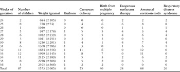
In all, 87 children were included in the study; 22 children were excluded from the study mainly because of the consequences of prematurity. None was excluded for long QT congenital syndrome. The mean gestational age was 31 weeks plus or minus 3 weeks and 2 days, with a range from 24 weeks and 2 days to 36 weeks and 6 days. The sex ratio was 1:1, that is, 47 males/40 females. The characteristics of the newborns are reported in Table 1.
Characteristics of newborns.

Weight is presented as mean plus or minus standard deviation
In all, 309 electrocardiograms were performed, most of them during the first 5 weeks of life. Regrettably, a significant number of infants could not be studied beyond 5 weeks of life because they were discharged home or transferred.
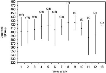
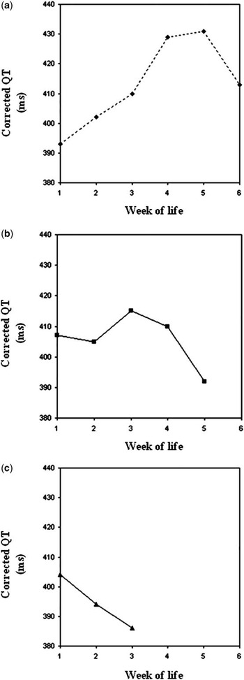
When analysed on all measurements pooled together, QT did not seem to vary significantly, nor follow a definite trend after birth (Fig 1). However, when we analysed the postnatal course of QT separately according to gestational age – 24–29 weeks of gestation, 30–32 weeks of gestation, and 33–36 weeks of gestation, we found out different trends (Fig 2). The QT of the most immature infants first increases, reaches a peak at 5 weeks of life, and then decreases (Fig 2a). The QT of more mature infants starts to decrease after 3 weeks of life (Fig 2b). The QT of even more mature infants decreases from birth (Fig 2c). The curve of postnatal evolution of QT seems to be shifting to the left as gestational age increases, suggesting that QT development follows a postmenstrual rather than a postnatal pattern.
Values of corrected QT according to the postnatal age. Results are presented as mean plus or minus standard deviation. Numbers in parentheses represent the number of children who had an electrocardiogram. Corrected QT is expressed in milliseconds (ms).

Values of corrected QT according to the term of birth and to the postnatal age. (a) For the group of pre-term infants born between 24 and 29 weeks of gestation. (b) For the group of pre-term infants born between 30 and 32 weeks of gestation. (c) For the group of pre-term infants born between 33 and 36 weeks of gestation. Corrected QT is expressed in milliseconds (ms) and is presented in/as mean.

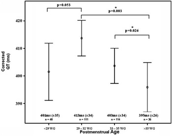
We then focused on QT evolution according to postmenstrual age and defined four groups: under 29 weeks of gestation, 29–32 weeks of gestation, 33–35 weeks of gestation, and more than 35 weeks of gestation. To do so, we pooled weekly measurements made over 3- to 4-week periods, thereby obtaining more than one measurement by infant by period. We observed a two-phase corrected QT variation: first, corrected QT tended to increase with postmenstrual age up to 32 weeks of gestation. Then, corrected QT progressively decreased from 32 weeks of gestation of postmenstrual age up to full term (Fig 3).
Evolution of corrected QT according to the postmenstrual age. Postmenstrual age is expressed in/as weeks of gestation (WG). Corrected QT is expressed in milliseconds (ms). Circles represent means and vertical bars represent confidence intervals at 95%. The difference observed between the four groups is significant: p = 0.012 (analysis of variance).

During the hospitalisation, one child died from severe neurological damages due to prematurity. Owing to the fact that they were discharged home, all children were regularly followed up, for over 3 years. Only one patient had isolated episodes of bradycardia that lasted 3 months before disappearing. For this patient, the maximum value of corrected QT was 442 milliseconds at 32 weeks of gestation. No death or syncope was reported for the other patients.
Discussion
This prospective study is, to our knowledge, the first to evaluate the evolution of the QT interval specifically in the premature infant. The electrocardiograms were obtained in a population of pre-term infants included very soon after birth, and then repeated at regular intervals during their hospitalisation, when the children's condition was stable. The QT intervals were all measured by a single observer. On the basis of these data, we demonstrated that its development depends on postmenstrual age rather than postnatal age. Our findings are very similar to those reported by in utero magnetocardiographic studies performed in the foetus: HorigomeReference Horigome, Takahashi, Asaka, Shigemitsu, Kandori and Tsukada 14 noted an increase of corrected QT in utero between 20 and 26 weeks of gestation, followed by a decrease between 27 and 37–41 weeks of gestation. We observed only a tendency of increase between the less than 29 weeks of gestation and 29–32 weeks of gestation periods, but this failure to reach significance is probably due to a lack of power. In our study, the duration of corrected QT after 35 weeks of gestation postmenstrual age is very similar to that of full-term newborns.Reference Schwartz, Montemerlo and Facchini 10 These results suggest that the evolution of corrected QT is an ontogenic process that is not modified by premature birth. Further studies are required in order to evaluate the evolution of corrected QT in premature infants after the theoretical term and to determine whether this evolution is similar to that of full-term newborns or whether, like the parasympathetic activity, it still differs for several months.Reference De Rogalski Landrot, Roche and Pichot 15
Several mechanisms may underlie these variations. The changes in QT according to postmenstrual age may be due to developmental changes in repolarising potassium currents. Wang et alReference Wang, Feng, Kondo, Sheldon and Duff 16 demonstrated that the expression of cardiac transient outward current (Ito) and inwardly rectifying potassium current (Ikr) is age dependent in mice, resulting in longer action potential and repolarisation duration in newborn compared with adult mice. Similarly, developmental changes in action potential duration have been observed in guinea pig ventricular myocytes: action potential duration decreases between foetal and neonatal periods and increases between neonatal and adult periods.Reference Kato, Masumiya, Agata, Tanaka and Shigenobu 17 These developmental changes were attributed to differences in the balance between Ikr and L-type calcium current (IcaL) densities. Generally speaking, all animal studies showed that the immature myocardium appears to have decreased repolarisation reserve, resulting in increased sensitivity to the prolonging effect of pharmacological agents.Reference Tanaka, Namekata, Nouchi, Shigenobu, Kawanishi and Takahara 18
Another possible mechanism of QT developmental changes is the maturation of the autonomic nervous system. The duration of cardiac time intervals is influenced by the autonomic nervous system.Reference Bednar, Harrigan, Anziano, Camm and Ruskin 2 , Reference Cuomo, De Caprio and Di Palma 19 Indeed, the duration of the QT interval closely depends on the autonomic balance between sympathetic and parasympathetic activities, which can be roughly estimated by the measurement of heart rate variability.Reference Pladys, Beuchée, Hernandez and Carrault 20 It was well demonstrated that premature newborns have a lower parasympathetic activity than full-term newborns because the parasympathetic system development occurs late in gestation.Reference De Rogalski Landrot, Roche and Pichot 15 , Reference Patural, Barthelemy and Pichot 21 , Reference Patural, Pichot and Jaziri 22 This depressed parasympathetic activity is associated with an increased risk of syncope and sudden death.Reference Kahn, Groswasser and Franco 23 Only one study of the autonomic cardiac control in pre-term infants sought to evaluate the variations according to postmenstrual age, but the cohort was too small to yield significant differences between the groups.Reference Patural, Pichot and Jaziri 22 In the postnatal period, the autonomic nervous system is the major factor regulating the development of cardiac ion channels involved in cardiac cell repolarisation.Reference Qu and Robinson 24 Finally, an alternative explanation for the QT evolution in our population could be prematurity-related exogenous factors such as sepsis, bronchopulmonary dysplasia, cerebral damage, or even pharmacological treatment. However, this last hypothesis seems unlikely as the occurrence of these events in premature infants is highly dependent on gestational ages, whereas the evolution of QT in our infants was mainly dependent on postmenstrual age.
There are some limitations in our study. First, we were unable to obtain a lot of electrocardiograms after 5–6 weeks of postnatal age, even in the most premature infants, because they were either discharged home or transferred to another unit closer to home. Very few infants remained in the neonatal intensive care unit after 36 weeks of postmenstrual age. Therefore, we do not have information on the QT interval in these infants when they reach full term. The technical difficulties in determining the QT interval in premature infants, due to high heart rates and QT variability, should also be acknowledged. Electrocardiogram recordings obtained from the bedside electrocardiogram monitoring were easily feasible and minimally invasive, but were composed of only three leads, compared with a 12-lead regular electrocardiogram, with a sampling rate of 500 Hertz. Despite a strict reproduction of recording conditions for each child, hysteresis is, for example, difficult to control especially when a study is conducted in a care unit.Reference Sarma, Venkataraman, Samant and Gadgil 4 , Reference Lau, Freedman, Fleming, Malik, Camm and Ward 5 In the future, a study should use an electrocardiogram Holter, with a 24-hour recording, to control physiological nycthemeral variations.
Our data suggest that if premature newborns have to be exposed to potentially QT-lengthening drugs when they reach 30–32 weeks of gestation of postmenstrual age, electrocardiogram monitoring should be reinforced. Automated continuous QT interval monitoring, which has been recently reported in the setting of neonatal intensive care units, could be beneficial in this specific situation.Reference Helfenbein, Ackerman and Rautaharju 25
We showed that corrected QT interval varies according to postmenstrual age in premature infants, increasing up to 32–33 weeks of gestation. Our results underline the need to better understand the development of the cardiac time intervals in premature newborns, to determine the patients at high risk of sudden death or serious arrhythmia. Knowledge of the physiological variations of QT interval is crucial for future neonatal pharmacological studies on the repolarisation-prolonging effect of drugs.
Acknowledgements
The authors thank Emilie Savariau for technical assistance. The authors have no conflict of interest to declare.