Introduction
The oocyte associates with somatic granulosa cells in the vertebrate ovary and becomes a structural and functional complex known as the ovarian follicle. At its earliest appearance, the oocyte enters the meiotic prophase and is wrapped by a single layer of flattened granulosa cells with little, if any, membrane associations among themselves or between the oolemma and the membrane of the granulosa cells. In mammals the earliest such fully established follicle, known as the primordial follicle, is established during fetal development or early postnatal period (Skinner, Reference Skinner2005) and in birds this occurs during hatching (Tokarz, Reference Taddei and Andreuccetti1978). With the onset of reproductive activity, a number of primordial follicles, depending on the species, develop into primary and subsequent follicular stages (Van Voorhis, Reference Van Voorhis, Knobil and Nill1999). However, in fishes, amphibians and reptiles, populations of oogonial stem cells are present in the ovary as discrete nests amidst somatic cells at locations depending upon whether the ovary is an elongated structure or formed into lobes. In the adult ovary number of oogonia produced from the stem cells, as may be required depending upon the pattern of reproduction and somatic pregranulosa cells are drawn for the development of follicles. This is referred to as follicular assembly and brings about soma–germ cell interactions that play a critical role in both the germ line and the physiological functions of the sexually mature gonad (Gilula et al., Reference Gilula, Epstein and Beers1978). Once assembled, the oogonium, which differentiates into the oocyte, is arrested in the first meiotic division (Sanchez & Villecco, Reference Sanchez, Villecco and Jamieson2003).
Amphibia are an appropriate biological model to study the interactions that take place in ovarian follicles since the follicles pass through different periods (previtellogenic, vitellogenic and maturation), when various processes related to oocyte growth and maturation occur (Sharon et al., Reference Sharon, Degani and Warburg1997; Villecco et al., Reference Villecco, Genta, Sánchez Riera and Sánchez2002). Before establishment of follicles the oocytes and pregranulosa cells exist as distinct cell populations (Wischnitzer, Reference Wischnitzer1966; Uribe, Reference Uribe, Dutta and Munshi2001, Reference Uribe, Jamieson and Sever2003; Sanchez & Villecco, 2003). Though the morphological and functional relationships between amphibian developing oocytes and their investing layer have been the subject of several investigations (Balinsky & Devis, Reference Balinsky and Devis1963; Wallace & Dumont, Reference Wallace and Dumont1968; Dumont, Reference Dumont1972; Holland & Dumont, Reference Holland and Dumont1975; Brummet & Dumont, 1976; Exbrayat & Collenot, Reference Exbrayat and Collenot1983; Exbrayat & Laurent, Reference Exbrayat and Laurent1983; Exbrayat, Reference Exbrayat1986; Villecco et al., Reference Villecco, Genta, Sánchez Riera and Sánchez2002) and the outcomes have been reviewed (Lofts, Reference Lofts1974; Saidapur, Reference Saidapur and Saidapur1989; Uribe, 2003; Sanchez & Villecco, 2003; Exbrayat, Reference Exbrayat, Jamieson and Exbrayat2006), the mechanistic details of the process of assembly of oocytes and pregranulosa cells to establish the previtellogenic follicle is only poorly understood. According to Uribe (2003), the morphological features that distinguish oogonia, the relationship of oocyte to somatic cells and the initiation of folliculogenesis require definition using the electron microscope. The ultrastructural studies of folliculogenesis in amphibians as well as the comparative morphological aspects of the process with other vertebrates are of great interest for future research.
During assembly of follicle the developing oocyte is progressively surrounded by somatic prefollicular cells (Wischnitzer, Reference Wischnitzer1966). The assembly of follicles is completed when the oocyte is surrounded by a layer of pregranulosa cells that become the granulosa cells (Dumont, Reference Dumont1972; Dumont & Brummet, Reference Dumont and Brummet1978; Wallace, Reference Wallace and Browder1985; Wallace & Selman, Reference Wallace and Selman1990; Sretarugsa et al., Reference Sretarugsa, Weerachatyanukul, Chavadej, Kruatrachue and Sobhon2001; Villecco et al., Reference Villecco, Genta, Sánchez Riera and Sánchez2002). Thus, the previtellogenic follicle consists of an oocyte, arrested in diplotene, surrounded by a single layer of follicle cells (Uribe, 2003; Sanchez & Villecco, 2003). In the salamander three stages have been identified during early folliculogenesis: stage 1, in which oogonia divide and form cell nests; stage 2 in which oogonia differentiate into oocytes; and stage 3, in which the oocyte cytoplasm increases in volume (Sharon et al., Reference Sharon, Degani and Warburg1997). Exbrayat (Reference Exbrayat, Jamieson and Exbrayat2006), in his review, discusses germinal nests, follicle cells and the previtellogenic follicles as aspects of the first (Stage A) and second (Stage B) stages in the ovary of caecilians but there is no description of assembly of follicles. Thus, a detailed description of assembly of follicles in amphibia in general and caecilians in particular, is pertinent.
Caecilians are unique among the amphibians in several aspects of reproductive anatomy, physiology and behavior. All caecilians practice internal fertilization using the eversible phallodeum as the intromittent organ and many are viviparous. Even the oviparous species are practically ovo-viviparous in the sense that the eggs at laying contain embryos. Caecilians are known to have parental care. Yet, the biology of female reproduction of caecilians has been studied for only a few species due to several reasons (Wake, Reference Wake1968, Reference Wake1970a, Reference Wakeb, Reference Wake1972, Reference Wake, Taylor and Guttman1977, Reference Wake1980; Exbrayat & Collenot, Reference Exbrayat and Collenot1983; Exbrayat & Laurent, Reference Exbrayat and Laurent1983; Exbrayat, Reference Exbrayat1986; Berois & de Sa, Reference Berois and de Sa1988; Masood-Parveez & Nadkarni, Reference Masood-Parveez and Nadkarni1993a, Reference Masood-Parveez and Nadkarnib; Anjubault & Exbrayat, Reference Anjubault and Exbrayat2004). Detailed morphological and anatomical descriptions of caecilian ovary and oogenesis are limited to three species, Chthonerpeton indistinctum (Berois & de Sa, Reference Berois and de Sa1988), Typhlonectes compressicuda (Exbrayat & Collenot, Reference Exbrayat and Collenot1983; Exbrayat & Laurent, Reference Exbrayat and Laurent1983; Exbrayat, Reference Exbrayat1986; Anjubault & Exbrayat, Reference Anjubault and Exbrayat2004) and Ichthyophis beddomei (Masood–Parveez & Nadkarni, 1993a, b). These studies have relied on conventional light microscopic histology and histochemistry. Ultrastructural descriptions of oocyte and granulosa cells during assembly of follicles and subsequent development have not yet been attempted. In this study we describe in detail the assembly of follicles in two species of caecilians of the Western Ghats of India, Ichthyophis tricolor and Gegeneophis ramaswamii.
Materials and Methods
Ichthyophis tricolor (Ichthyophiidae) and Gegeneophis ramaswamii (Caeciliidae) were collected from terraced plantations of mixed coconut and rubber from Thekkada (08°37′N, 76°57′E) in the Trivandrum district of Kerala and Maramalai (08°26′N, 77°24′E) in the Kanyakumari district of Tamil Nadu, Southern India, from June 2004 to June 2005. They are relatively abundant in the Western Ghats of Kerala and Tamil Nadu (Oommen et al., Reference Oommen, Measey, Gower and Wilkinson2000). Monthly samples (three animals from each species) were collected, anesthetized with MS-222 (tricaine methane sulphonate) and dissected to expose the female reproductive system. Slices of ovary were prepared for light microscopic histological observation (paraffin embedding; hematoxylin–eosin staining). Tissues representing different phases of ovarian structure were fixed in 2.5% glutaraldehyde prepared in cacodylate buffer, postfixed in 1% osmium tetroxide and embedded in thin viscosity resin. Semi-thin sections (1 μm thick) were stained in toluidine blue O (TBO). In addition to making observations of the status of the ovary, the diameters of oogonia, oocytes, pregranulosa cells and granulosa cells, including the nuclei, were measured in five randomly selected cells of each type using a research microscope supported with Q-Win software (Leica, Jena) and the data are presented as the range. Ultrathin sections, obtained with a Leica ultra-microtome (Jena), were stained with uranyl acetate and lead citrate and subjected to transmission electron microscopic analysis using a Philips 201C transmission electron microscope. The images were processed using Adobe Photoshop version 7.0.
Results
Gross anatomical and light microscopic observations
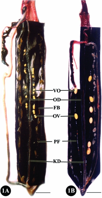
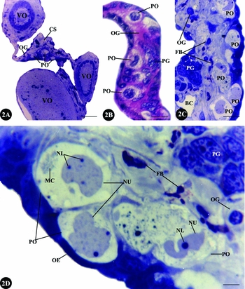
Ichthyophis tricolor and Gegeneophis ramaswamii have similar reproductive patterns, with regression of ovaries during March to June. The paired ovaries of both caecilian species are elongated sac-like structures with previtellogenic, vitellogenic or postvitellogenic follicles disposed like a string of beads. The neighbouring follicles are connected by interfollicular stromal tissue (Fig. 1A, B). (The seasonal pattern of ovarian morphology will be described elsewhere.) The nests of oogonia, premeiotic oocytes and pregranulosa cells occur as clusters throughout the year in the germinal ridges present in the stroma that connects the follicles in the string (Fig. 2A–C). The spherical oogonia are 10.2–12.6 μm diameter and have 6.7–8.3 μm diameter darkly stained nuclei lying in a slightly eosinophilic (Fig. 2B)/acidophilic (Fig. 2C) cytoplasm.
The female urinogenital system of Ichthyophis tricolor (A) and Gegeneophis ramaswamii (B). The elongated ovary (OV) on each side is a beaded structure running parallel to the oviduct (OD), kidney (KD) and fat body (FB). In both the cases there are yellowish vitellogenic (VO) and whitish previtellogenic (PF) follicles. In (B) the vitellogenic follicles are at a more advanced stage than in (A). Scale bar represents 2 cm.

The premeiotic oocytes are 40.8–51.6 μm diameter and contain spherical to irregularly shaped nuclei 10.9–31.3 μm diameter lying in a pale-stained cytoplasm. The lightly staining nuclei contain one or two prominent nucleoli. There are a few patches of slightly dense heterochromatin in the nucleus. A mitochondrial cloud, either as dense aggregate or diffuse, is present at one pole of the nucleus (Fig. 2D). The pregranulosa cells occur as a few clusters, separated from the germ cells and are highly compact. They are elongated cells 20.7–28.2 μm long and 12.1–15.6 μm diameter. The dark staining highly heterochromatic elongated nuclei are 15.4–23.2 μm long and 10.5–12.2 μm diameter. Fibroblast cells and capillaries are also present in the stroma (Fig. 2B–D).
Light micrographs of sections of ovary of Ichthyophis tricolor. (A) Toluidine blue O-stained semithin section showing the connective tissue strand (CS) connecting vitellogenic follicles (VO). There are oogonial nests (OG) and primary oocytes (PO) in the connective tissue. Scale bar represents 60 μm. (B) Magnified view of a hematoxylin- and eosin-stained section showing oogonia (OG), primary oocytes (PO) and pregranulosa cell nests (PG). Scale bar represents 30 μm. (C) A further magnified view of a TBO-stained semithin section showing oogonial nests (OG), primary oocytes (PO), fibroblast cells (FB), pregranulosa cell nests (PG) and a capillary (BC). Scale bar represents 20 μm. (D) A highly magnified view of a TBO-stained semithin section showing ovarian epithelium (OE), oogonia (OG), primary oocytes (PO), pregranulosa cell nest (PG) and fibroblast cells (FB). The primary oocyte contains an irregularly shaped nucleus (NU) with one or two prominent nucleoli (NL). The cytoplasm contains a prominent mitochondrial cloud (MC). Scale bar represents 7.5 μm.

TEM observations of oocytes
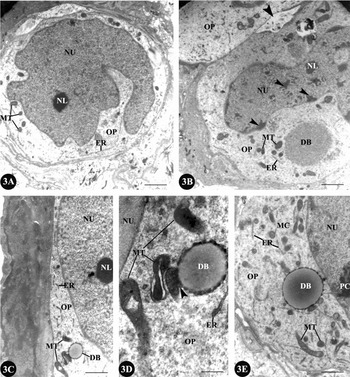
The premeiotic oocytes are spherical or elongated cells surrounded by stromal tissue (Fig. 3A, B). Neighbouring oocytes are held together by membrane juxtaposition, with an occasional intercellular space containing a heterogeneous material (Fig. 3B). The nuclei of the premeiotic oocytes are spherical to highly irregular in shape and each have a single nucleolus in a section, but there may be more than one nucleolus in an oocyte. The chromatin is fairly uniform in the premeiotic and leptotene oocytes (Fig. 3A) but forms dense aggregates in the zygotene cells (Fig. 3B). The cytoplasm stains lightly and contains mitochondria, endoplasmic reticulum, ribosomes and Golgi apparatus (not shown). The mitochondrial cloud, in view of its juxtanuclear localization, is seen only in appropriately cut sections (Fig. 3C–E).
Early meiotic prophase oocytes of Gegeneophis ramaswamii. TEM of prefollicular oocytes. (A) This leptotene oocyte has a large irregularly shaped nucleus, containing a single nucleolus. There are few mitochondria and scarce endoplasmic reticulum in the ooplasm. Scale bar represents 0.6 μm. (B) This pachytene oocyte is increased size with the nucleus becoming proportionately smaller. The nuclear chromatin exhibits synaptonemal complexes (arrows). There are many nucleoli (only one is shown) located around the periphery of the nucleus. Mitochondria have become more abundant in the ooplasm and form the juxtanuclear mitochondrial cloud. There is a dense body in the ooplasm indicating the formation of Balbiani vitelline body. The endoplasmic reticulum is still scarce. Two oocytes are held together by membrane apposition, but at places the membranes are separated resulting in a space containing an amorphous material (arrowhead). Scale bar represents 0.6 μm. (C–E) Portions of oocytes magnified, showing the gradual development of the Balbiani vitelline body. (C) Zygotene oocyte. Scale bar represents 0.2 μm. (D) A portion of (C) further magnified showing the dense body in the mitochondrial cloud. Arrowhead points to association between mitochondria and dense body. Scale bar represents 0.06 μm. (E) Pachytene oocyte showing abundant mitochondria with a dense body associating with them, forming the Balbiani vitelline body. The rough endoplasmic reticulum is also abundant. The chromatin is formed into synaptonemal complexes (PC). Scale bar represents 0.1 μm. DB, dense body of Balbiani vitelline body; ER, endoplasmic reticulum; MC, mitochondrial cloud; MT, mitochondria; NL, nucleolus; NU, nucleus; OP, ooplasm.

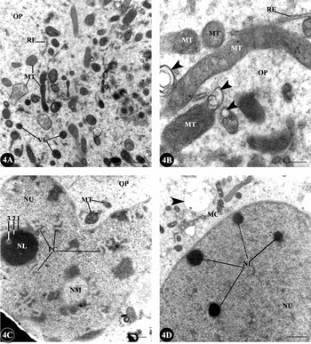
A peculiar spherical vesicle is invariably present in the cytoplasm in the vicinity of the mitochondrial cloud of meiotic oocytes (Fig. 3B, C–E). The content of these vesicles is either homogenous (Fig. 3B) or heterogeneous with a dense outer boundary and a less dense core (Fig. 3C–E). Mitochondria are physically associated with this vesicle. The mitochondrial cloud of meiotic oocytes, also known as Balbiani vitelline body (Guraya, Reference Guraya1979), contains elongated mitochondria, rough endoplasmic reticulum (RER), ribosomes and several light- to dark-stained structures, which are apparently lysosomes (Fig. 4A, B). Several mitochondria in the cloud exhibit blebs (Fig. 4B).
TEM of early meiotic prophase oocytes. (A) Gegeneophis ramaswamii. Mitochondrial cloud during early pachytene. There are dark dense bodies amongst the mitochondria, which may either be lysosomes or precursors of the large dense body. Scale bar represents 0.08 μm. (B) A portion of (A) magnified, showing blebbing of the mitochondria (arrowheads). Scale bar represents 0.02 μm. (C) Nucleus of an early pachytene oocyte of Ichthyophis tricolor showing synaptonemal complexes (PC). A large nucleolus, with differentiation as distinct zones (1–3) is also shown. There is formation of additional nucleolar material (NM). Scale bar represents 0.2 μm. (D) Gegeneophis ramaswamii. Nucleus and cytoplasm of an early diplotene oocyte. There are four nucleoli shown. There is a nuage formed (arrow) in the mitochondrial cloud. Scale bar represents 0.2 μm. MC, mitochondrial cloud; MT, mitochondria; NL, nucleoli; NM, additional nucleolar material; NU, nucleus; OP, ooplasm; RE, rough endoplasmic reticulum; VE, dark dense vesicles.

The nucleus of pachytene oocyte contains a single large nucleolus (Fig. 4C). The nucleolus has a less dense diffuse peripheral zone and a dense core. Occasionally, the dense surrounds another less-dense zone, resulting in a nucleolus that appears ring-shaped (Fig. 4C). The pairing of homologous chromosomes into synaptonemal complexes and formation of additional nucleolar material are also evident (Fig. 4C). The nucleus of diplotene oocyte contains several small nucleoli but the synaptonemal complexes are less prominent (Fig. 4D). A few nuages are evident among the mitochondria (Fig. 4C, D).
Ultrastructural organization of pregranulosa cells
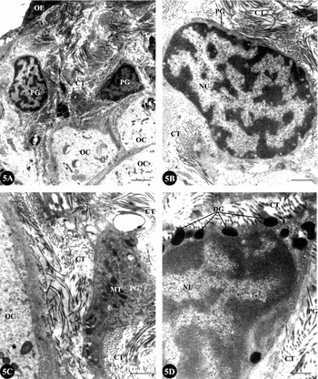
In their earliest appearance, the squamous pregranulosa cells lie embedded in the stroma in the form of discrete nests (Fig. 2B–D) but later the cells disaggregate and tend to move away from each other (Fig. 5A). The nuclei of pregranulosa cells are oval to elongated and densely heterochromatic with a continuous patch of dense heterochromatin underneath the nuclear envelope. The nucleus occupies almost the entire cell with little cytoplasm around it. The cytoplasm contains few organelles (Fig. 5A, B).
TEM showing the early events in the assembly of follicles. (A) Ichthyophis tricolor. Pregranulosa cells migrate through the stromal connective tissue and arrive at the vicinity of oocytes. Scale bar represents 1.0 μm. (B) One pregranulosa cell in (A) magnified showing the large heterochromatic nucleus and poor cytoplasmic content of the cell. Scale bar represents 0.05 μm. (C) Gegeneophis ramaswamii. On arrival closer to the oocyte, the pregranulosa cell increased in organelle content. Scale bar represents 0.02 μm. (D) Ichthyophis tricolor. Accumulation of dark dense bodies, apparently ribosomal material, in the cytoplasm of pregranulosa cell. Scale bar represents 0.02 μm. CT, connective tissue strand; DG, dense granules; MT, mitochondria; NU, nucleus; OC, oocyte; OE, ovarian epithelium; PG, pregranulosa cells.

Assembly of follicles
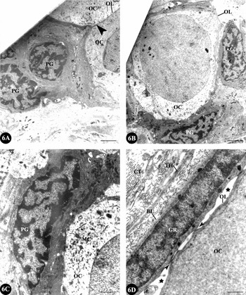
The pregranulosa cells, which are spatially separated from the premeiotic and leptotene to pachytene stage meiotic oocytes, migrate through the connective tissue fibrils of the stroma to reach the oocytes when the latter are in the early diplotene stage and stationary (Fig. 5A–D). During this migration a large number of dark dense bodies appear in the cytoplasm of pregranulosa cells (Fig. 5C, D). The migratory pregranulosa cells, on arrival at the vicinity of the oocytes, establish physical contact with the latter by displacing the connective tissue fibrils of the stroma (Fig. 6A). A few pregranulosa cells migrate between the oocytes so that the juxtaposed oocytes separate (Fig. 6A). By this time the nucleus of the oocyte has become spherical. The pregranulosa cells lodge themselves around the outer boundary of the oocytes, initially as a discontinuous layer of cuboidal pregranulosa cells (Fig. 6B). At this stage, little intercellular space is pre-sent between the oocyte and the pregranulosa cells (Fig. 6C). In the oocyte cytoplasm the mitochondrial cloud is dispersed and lysosome-like bodies appear amongst the mitochondria. During subsequent deve-lopment, the pregranulosa cells elongate and flatten, with their nuclei also transforming in shape ac-cordingly, to form the granulosa cells and the oocyte is arrested in diplotene of meiotic prophase I (Fig. 6D). The perivitelline space appears between the granulosa cells and the oocyte, with occasional junctional com-plexes, either desmosomes or gap junctions, between the two. A basal lamina appears around the follicle and sparse thecal elements associate with the follicle external to the basal lamina of the granulosa cells (Fig. 6D).
TEM showing follicular assembly. (A) Ichthyophis tricolor. Migratory pregranulosa cells are shown to establish contact with the oocyte. Still, the neighbouring oocytes are attached by juxtaposition of membranes (arrowhead). Scale bar represents 2.1 μm. (B) Gegeneophis ramaswamii. The pregranulosa cells lodge themselves around the boundary of the oocyte. Scale bar represents 2.8 μm. (C) A portion of (B) magnified showing absence of an intercellular space between oocyte and pregranulosa cell. A mitochondrial cloud is still evident in the cytoplasm of this early diplotene oocyte. Scale bar represents 1.4 μm. (D) Gegeneophis ramaswamii. The pregranulosa cell has become a flat granulosa cell. Basal lamina is seen outside the granulosa cell. The perivitelline space is under formation (asterisks) but the oolemma and granulosa cell membrane are held together by membrane junctions (arrowheads). An incipient theca is under formation. Scale bar represents 1.2 μm. BL, basal lamina; CT, connective tissue fibrils; GR, granulosa cell; MC, mitochondrial cloud; OC, oocyte; OL, oolemma; PG, pregranulosa cell; TH, theca.

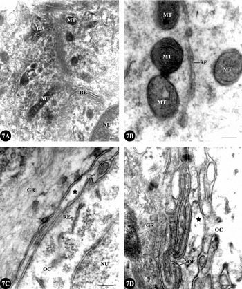
The cytoplasmic content of the granulosa cell increases and dense organelles appear in the cytoplasm (Fig. 7A, B). The organelles include mitochondria, Golgi apparatus and RER. Dense fibrils are also seen in the cytoplasm. Later, the perivitelline space becomes continuous (Fig. 7C). The granulosa cells produce highly tortuous out-pocketing, which invade the perivitelline space but these are not comparable to macrovilli that would appear later in preparation to vitellogenesis (Fig. 7D).
Gegeneophis ramaswamii. TEM. (A, B) Increase in the cytoplasmic organelle content of granulosa cell is shown. (B) is a portion of (A) magnified. Scale bar (A) represents 0.8 μm and (B) represents 0.2 μm. (C) The perivitelline space is continuous (asterisk). Scale bar represents 1.2 μm. (D) The membrane of granulosa cell forms into folds in the perivitelline space (OF). GA, Golgi apparatus; GR, granulosa cell; MT, mitochondria; NU, nucleus; OC, oocyte; RE, rough endoplasmic reticulum.

Discussion
The period of oogonial proliferation varies from species to species and may be continuous or cyclical (Tokarz, Reference Tokarz and Jones1978). In the caecilians it appears to be continuous because fully developed ovary with vitellogenic, previtellogenic and early mitotic prophase I oocytes are present through out the year in spite of regression of the ovary in size during a small part of the year (Masood-Parveez & Nadkarni, Reference Masood-Parveez and Nadkarni1993a, Reference Masood-Parveez and Nadkarnib; Exbrayat, Reference Masood-Parveez and Nadkarni2006; the present observation) as in the case of anurans (Tokarz, Reference Tokarz and Jones1978; Kanamadai & Saidapur, Reference Kanamadai and Saidapur1982; Pancharatna & Saidapur, Reference Pancharatna and Saidapur1985a, Reference Pancharatna and Saidapurb; Pramoda & Saidapur, Reference Pramoda and Saidapur1984; Saidapur, 1989; Stretarugsa et al., 2001). Following this proliferative phase, the oogonial cells must undergo a phenotypic change as they transform to oocytes (Begovac & Wallace, Reference Begovac and Wallace1988). In the caecilians, the earliest changes are increase in size with greater amounts of cellular organelles that appear as a juxtanuclear aggregate as it occurs in the toad Xenopus laevis (Al-Mukhtar & Webb, Reference Al-Mukhtar and Webb1971) and in the polyautochronic lizard Calotes versicolour (Shanbhag & Krishna Prasad, Reference Shanbhag and Krishna Prasad1993). In the latter, once a batch of follicles enters vitellogenesis, no other batch of previtellogenic follicles (stage III) is recruited to the vitellogenic stage indicating that follicle recruitment could still be continuous. This is inferred from the number of primary oocytes and primordial follicles present in the ovaries during all months of the year.
In the caecilians once the oocyte has been formed, the nuclear features with respect to synaptonemal complex formation, nucleoli fragmentation and arrest in diplotene of prophase I are similar to those in Xenopus laevis (Al-Mukhtar & Webb, Reference Al-Mukhtar and Webb1971; Coggins, Reference Coggins1973). The prominent features of the later phases of primary oocyte growth include intense transcriptional activity, formation and subsequent dispersal of the Balbiani vitelline body (mitochondrial cloud) concomitant with an increase in cytoplasmic organelles and volume (Guraya, Reference Guraya1979; Wallace & Selman, Reference Wallace and Selman1990). The several nucleoli in the pachytene and early diplotene stage oocytes and their ring-shaped structure, as observed in the caecilian early meiotic prophase oocytes, have been reported in anuran and urodelan oocytes. This is referred to as amplification of nucleoli, which is meant to produce the enormous rRNA required for protein synthesis until the mid-blastula stage (Sanchez & Villecco, 2003; Uribe, 2003). Although the amplification process in the anurans begins in premeiotic oogonia, the main period for the selective replication of rRNA is the pachytene of the meiotic prophase, followed by completion in early diplotene. The amplified nucleoli appear either as compact spheroidal bodies 2–10 μm in diameter or as ring-like structures (Sanchez & Villecco, 2003).
The mitochondrial cloud or Balbiani vitelline body has been described for many organisms (Guraya, Reference Guraya1979; Heasman et al., Reference Heasman, Quarmby and Wylie1984; Wallace & Selman, Reference Wallace and Selman1990) but is not evident in the oocyte of viviparous placentotrophic lizard Mabuya mabouya (Gόmez & Ramírez-Pinilla, 2004). The Balbiani vitelline body is the most prominent cytoplasmic structure in the premeiotic oocyte of the caecilians but it disperses through out the oocyte prior to early meiotic prophase I. During Xenopus oogenesis, the message transport organizer (METRO) pathway delivers germinal granules and localized RNAs to the vegetal cortex of the oocyte via the Balbiani vitelline body. According to the traditional model, the mitochondrial cloud is thought to break up at the onset of vitellogenesis and the germinal granules and METRO-localized RNAs are transported within the mitochondrial cloud fragments to the vegetal cortex of the oocyte. However, a study using light and electron microscopy, in situ hybridization and three-dimensional reconstruction has shown that germinal granules and METRO-localized RNAs are delivered to the oocyte cortex before the onset of mitochondrial cloud fragmentation. The delivery involves accumulation of localized RNAs and aggregation of germinal granules at the vegetal tip of the mitochondrial cloud. The subsequent internal expansion of the mitochondrial cloud is between its animal (nuclear) and vegetal tips, which drives the germinal granules and METRO-localized RNAs towards the vegetal cortex (Wilk et al., Reference Wilk, Balinski, Dougherty and Kloc2005). The situation prevailing in the caecilians in this connection is under investigation. Perhaps, the dark dense vesicles appearing within the mitochondrial cloud in the caecilian oocytes are accumulations of localized RNAs.
Follicle assembly is a critical aspect of female repro-duction, the mechanistic details of which are poorly understood. Follicle assembly requires development of oocytes from the oogonial nests and the pregranulosa cells from the somatic elements of the ovarian stroma. During follicle assembly each developing oocyte is progressively surrounded by the pregranulosa cells (Wischnitzer, Reference Wischnitzer1966). At the end of this assembly the follicle is a well established structural and functional entity but the underlying process in the amphibians has not been traced in adequate detail (Guraya, Reference Guraya1979; Uribe, 2003; Sanchez & Villecco, 2003; Exbrayat, Reference Exbrayat, Jamieson and Exbrayat2006). As of now, most descriptions of ovarian cycles in the amphibians begin with stage I, the previtellogenic follicles, which consist of oocytes wrapped by granulosa cells (Lofts, Reference Lofts1974; Rastogi et al., Reference Rastogi, Izzo-Vitiello, DiMeglio, Di Matte, Franzese, Dicostanzo, Minncci, Iela and Chieffi1983; Pancharatna & Saidapur, Reference Pancharatna and Saidapur1985a, Reference Pancharatna and Saidapurb; Saidapur, 1989; Cabada et al., Reference Cabada, Sanchez Riera, Genta, Sanchez and Barisone1996; Villecco et al., Reference Villecco, Aybar, Sánchez and Sánchez Riera1996, Reference Villecco, Aybar, Genta, Sánchez and Sánchez Riera2002; Sanchez & Villecco, 2003; Uribe, 2003; Exbrayat, Reference Exbrayat, Jamieson and Exbrayat2006). Our light microscopic and TEM description of follicle assembly in the two species of caecilians fills this gap.
Folliculogenesis in caecilians commences when oogonia differentiate into oocytes with the initiation of meiosis (Exbrayat, Reference Exbrayat, Jamieson and Exbrayat2006), as in the urodeles and anurans (Uribe, 2003; Sanchez & Villecco, 2003). In the few caecilian species so far studied in detail (Exbrayat & Collenot, Reference Exbrayat and Collenot1983; Exbrayat & Laurent, Reference Exbrayat and Laurent1983; Exbrayat, Reference Exbrayat1986; Berois & de Sa, Reference Berois and de Sa1988; Masood-Parveez & Nadkarni, Reference Masood-Parveez and Nadkarni1993a, Reference Masood-Parveez and Nadkarnib; Anjubault & Exbrayat, Reference Anjubault and Exbrayat2004), the proliferative oocytes occur as several germinal nests or proliferation areas disposed in a segmental fashion along the entire length of the ovary. The ovarian follicles lie between the islets of germinal elements (Masood-Parveez & Nadkarni, Reference Masood-Parveez and Nadkarni1993b), as is the case in the caecilians in this study. The earliest stage of follicles so far described for a caecilian is the primary follicle, which contains the oocyte with the central nucleus possessing one to several nucleoli, surrounded by a discontinuous single layer of flattened epithelial cells (Exbrayat, Reference Exbrayat1986; Exbryat & Collenot, 1983; Masood-Perveez & Nadkarni, 1993a, b). Exbrayat (Reference Exbrayat, Jamieson and Exbrayat2006) describes isolated oogonia in the caecilian ovary as the first stage or Stage A and when the oogonia transform into primary oocytes, which are either naked or surrounded by a small layer of flattened follicle cells, it is the second stage or Stage B. The sequential light microscopic and ultra structural changes taking place during these stages are described in this study.
After the assembly of follicles the granulosa cells either remain as a simple layer consisting of a single cell type as in most anuran and urodelan amphibians (Dumont & Brummet, Reference Dumont and Brummet1978; Saidapur, 1989; De Oliviera & Santos, 2004), chelonian and crocodilian reptiles (Sarkar, et al., Reference Sarkar, Sarkar, Das and Maiti1996; Uribe & Guillette, Reference Uribe and Guillette2000; Calderon et al., Reference Calderon, De Perez and Ramirez-Pinilla2004) and domestic goose (Kovacs et al., Reference Kovacs, Forgo and Peczely1992), or a simple layer differentiating into dark cells and clear cells as in the anuran Ceratophrys cranwelli (Sanchez & Villecco, 2003) and the Japanese quail (Callebault, Reference Callebault1991) or differentiate into a hetero-geneous and multilayered structure consisting of three types of cells as in the case of the spotted rays Torpedo marmorata (Marina, et al., Reference Marina, Loredana and Piero2002, Reference Marina, Salvatore, Maurizio, Loredana, Annamaria, Vincenza, Ermelinda and Piero2004) and Urolophus jamaicensis (Hamlett et al., Reference Hamlett, Jezior and Spieler1999), the hylid anuran Scinax fuscovarius (De Oliviera & Santos, 2004), the dove (Zarnescu, Reference Zarnescu2004) and the squamate reptiles (Andreuccetti et al., Reference Andreuccetti, Taddei and Filosa1978; Filosa et al., Reference Filosa, Taddei and Andreuccetti1979; Klosterman, Reference Klosterman1987; Sarkar & Shivanandappa, Reference Sarkar, Shivanandappa and Saidapur1989; Ibrahim & Wilson, Reference Ibrahim and Wilson1989; Taddei & Andreuccetti, Reference Taddei and Andreuccetti1990; Andreuccetti, Reference Andreuccetti1992; Gόmez & Ramírez-Pinilla, Reference Gόmez and Ramírez-Pinilla2004; Ricchiari et al., Reference Ricchiari, Carmela, Marina, Rosa, Annamaria and Piero2004; Hernandez-Franyutti et al., Reference Hernandez-Franyutti, Uribe-Aranzabal and Guillette2005). Whether caecilian follicular granulosa cells continue to remain as a single-layered homogenous structure or become a single-layered heterogeneous structure or a multilayered heterogeneous structure during subsequent development is under investigation.
The structural changes in the granulosa cells during folliculogenesis in the caecilians indicate that the granulosa cells are the primary cell type in the ovary that provide the physical support and the microenvironment required for the developing oocytes. In the anurans and urodeles the follicle cells form the interface between the blood capillaries and the oocyte and play critical roles in the sequestration of vitellogenin, the precursor of yolk protein, from the blood to the oocyte (Wallace & Dumont, Reference Wallace and Dumont1968; Wallace & Bergink, Reference Wallace and Bergink1974; Wallace & Jared, Reference Wallace and Jared1976; Brummet & Dumont, 1977; Dumont, Reference Dumont1978; Polzonetti-Magni, Reference Polzonetti-Magni, Knobil and Nill1998; Wallace and Selman, Reference Wallace and Selman1990; Hamlett et al., Reference Hamlett, Jezior and Spieler1999; Stretarugsa et al., 2001; Villecco et al., Reference Villecco, Genta, Sánchez Riera and Sánchez2002; Uribe, 2003; Sanchez & Villecco, 2003). In addition to this passive role, the follicle cells in other amphibians play an active role in the formation of vitelline envelope (Dumont & Brummet, Reference Dumont and Brummet1978), steroid biosynthesis (Redshaw, Reference Redshaw1972; Guraya, Reference Guraya1979; Wallace, 1985; Mahmoud et al., Reference Mahmoud, Ba-Omar and Alkindi2006), providing cAMP, which would be a regulatory molecule in the meiotic arrest of the oocyte at diplotene (Villecco et al., Reference Villecco, Aybar, Sánchez and Sánchez Riera1996, Reference Villecco, Aybar, Genta, Sánchez and Sánchez Riera2000, Reference Villecco, Genta, Sánchez Riera and Sánchez2002). The production of a maturation inducing hormone (MIH) brings about oocyte maturational competence (OMC) so as to respond to MIH (Villecco et al., Reference Villecco, Aybar, Sánchez and Sánchez Riera1996; Patino et al., Reference Patino, Yoshizaki, Thomas and Kagawa2001), synthesis and secretion of the components of the vitelline envelope (Cabada et al., Reference Cabada, Sanchez Riera, Genta, Sanchez and Barisone1996). The possible role of the follicle cells in these aspects in the caecilians is under investigation.
During previtellogenesis in the anuran follicles, the granulosa cells increase in the content of RER, free ribosomes and glycogen. They become metabolically very active and are involved in the synthesis of nucleic acids. It is conceivable that the follicle cells surrounding the oocytes of caecilians are concerned with these processes. A possible involvement of granulosa cells in the transfer of ribosomes to the oocyte in lizards has been proposed (Taddei, Reference Taddei1972; Andreuccetti et al., Reference Andreuccetti, Taddei and Filosa1978; Klosterman, Reference Klosterman1987). Cytoplasmic dense-cored granules accumulate in the granulosa cells in close association with fenestrated cisternae and networks of tubule derived from the RER of domestic goose. These granules consist of spheres and strands of an amorphous substance of unknown origin (Kovacs et al., Reference Kovacs, Forgo and Peczely1992). The dark dense inclusions that appear in the pregranulosa cells during their migration towards the oocyte and later present in these cells less prominently after the assembly of follicle, in the caecilians are comparable with these granules. Perhaps, these granules are packaged ribosomes for export to the oocyte, an aspect worthy of further investigation. Intermediate filament bundles containing keratin have been shown in the follicle cells of the lizard Podarcis sicula (Maurizii et al., Reference Maurizii, Alibardi and Taddei2000). The filaments in the pregranulosa cells of the caecilian early follicles may correspond to these fibrils and provide a cytoskeletal framework. The junction between granulosa cells and oocyte in the caecilians are either tight or gap junctions. In Xenopus laevis oocytes these junctions were identified as gap junctions based on the passage of microinjected fluorescent dye from oocytes to follicle cells (Browne et al., Reference Browne, Wiley and Dumont1979). Before beginning of vitellogenesis in the anurans, the membranes of granulosa cells are apposed to the oocyte membrane, without an interface. Soon the granulosa cell plasma membrane happens to separate from the oolemma, forming an interface made up of small spaces with a homogenous material. Only at certain points are both membranes in close apposition (Sanchez & Villecco, 2003). This matches with the observation made in the caecilians in this study.
The establishment of primordial follicles in the mammalian ovary requires individual oocytes to segregate and associate with pregranulosa cells. To provide for this, nests of associated oocytes undergo random apoptosis of individual oocytes to derive isolated oocytes, which then associate with precursor squamous granulosa cells (Skinner, Reference Skinner2005). During the establishment of follicles in the caecilian ovary the pregranulosa cells physically separate the juxtaposed oocytes thereby not requiring apoptotic death of oocytes for purpose of isolating them from one another. On the contrary in the common snook, Centropomus undecimali, cytoplasmic processes of epithelial cells and not the entire cells, encompass meiotic oocytes and transform into prefollicle cells, which become follicle cells at the completion of folliculogenesis (Grier, Reference Grier2000).
Thus, this study describes the sequential changes in the transformation of premeiotic oocyte into diplotene oocyte. When it is arrested in the meiotic prophase, the assembly of diplotene oocyte and pregranulosa cells occur. The latter becomes granulosa cells in the two species of caecilians.
Acknowledgements
The TEM facility of Welcome Trust Research Laboratory, Christian Medical College and Hospital, Vellore, is heartily acknowledged. The research was supported with funds from the Kerala State Council for Science, Technology and Environment (KSCSTE), through the Science and Research Development (SARD) facility. Support under the FIST scheme of Department of Science and Technology, Government of India, New Delhi, to the Department of Zoology, University of Kerala, Thiruvananthapuram and the Department of Animal Science, Bharathidasan University, Tiruchirappalli, is also acknowledged. We thank Dr B. Kadalmani for help in the image processing.