Introduction
Much attention has been paid in recent years to the medicinal applications of clay minerals due to their abundance in nature and unlimited potential (Reichle, Reference Reichle1986; Choy, Reference Choy2004). Clay minerals have been used especially for different biological applications including biocompatible materials, biosensors, pharmaceuticals, cosmetics, and nanomedicine (Choy et al. Reference Choy, Kwak, Park, Jeong and Portier1999; Choy et al. Reference Choy, Kwak, Jeong and Park2000; Choi et al. Reference Choi, Eom, Vinu and Choy2018). This is surely due to their excellent physicochemical and biological properties, such as large surface area, large ion exchange capacity, intercalative swelling behavior, excellent biocompatibility, and low toxicity (Baek et al. Reference Baek, Lee and Choi2012; Gaharwar et al., Reference Gaharwar, Mihaila, Swami, Patel, Sant, Reis, Marques, Gomes and Khademhosseini2013; Xavier et al., Reference Xavier, Thakur, Desai, Jaiswal, Sears, Cosgriff-Hernandez, Kaunas and Gaharwar2015).
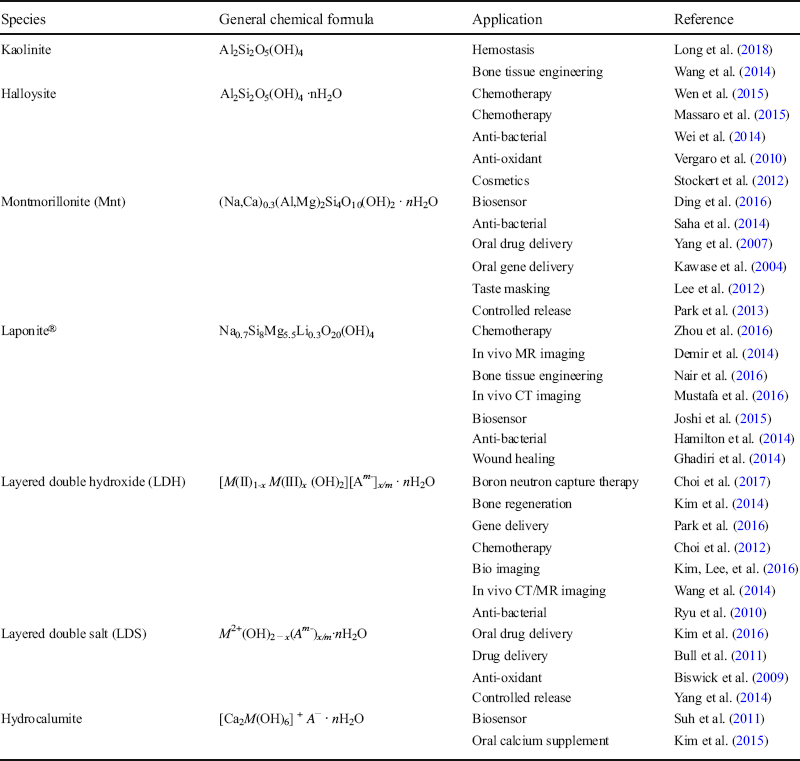
Clay minerals can be classified into neutral, cationic, and anionic clays with respect to their layer charge (Table 1). A wide variety of clay minerals such as halloysite, montmorillonite (Mnt), Laponite®, layered double hydroxide (LDH), layered double salt (LDS), and others have been studied extensively as vectorized clays, namely, advanced drug delivery vehicles for their potential biomedical applications.
Bio-applications of clay minerals
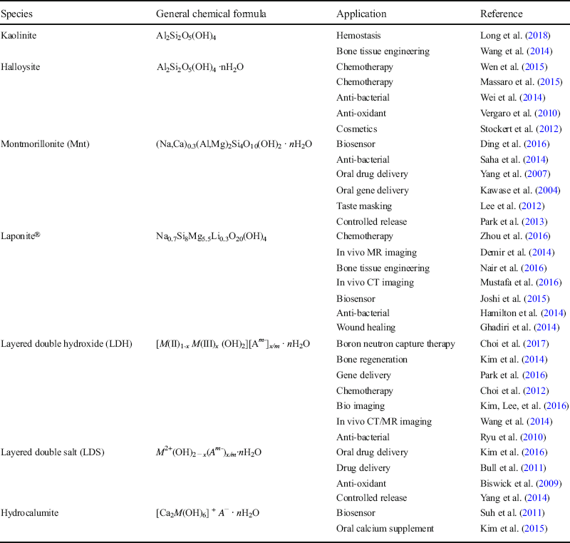
In this review, the key aspects of clay chemistry will be discussed with a specific focus on the nanomedical applications of cationic and anionic clay minerals, and their nanohybrids formed with drugs or bioactive molecules.
Cationic clay
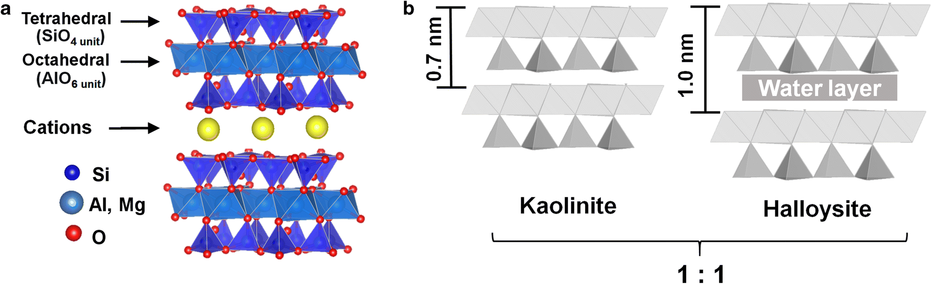
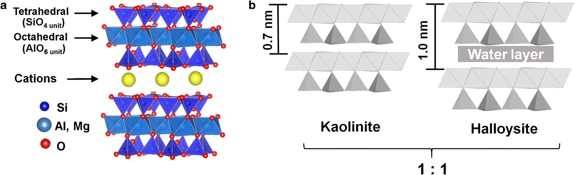
Cationic clays are phyllosilicates comprising tetrahedral silicate sheets and octahedral Mg or Al oxyhydroxide sheets (Fig. 1A). They are divided into two main classes: 1:1 and 2:1 types (Fig. 1A,B), depending on their arrangements of octahedral and tetrahedral sheets along the crystallographic c axis. As for the 1:1 type clay (e.g. kaolinite and halloysite) with the unit-cell formula of Al2Si2O5(OH)4, each layer is electrically neutral, and its interlayer interaction is of a molecular nature such as hydrogen bonding or van der Waals force (Bergaya & Lagaly, Reference Bergaya, Lagaly, Bergaya, Theng and Lagaly2006). One thing to note here is that the 1:1 type clay minerals, in general, have very small cation exchange capacities (CEC) compared with 2:1 type clay minerals. Unlike kaolinite, halloysite layers are hydrated and rolled up into nanotubes giving rise to larger specific surface areas (SSA) with different internal and external surface properties (Yuan et al. Reference Yuan, Tan and Annabi-Bergaya2015). Such an unusual porous structure enables halloysite nanotubes to be drug delivery vectors in nanomedicine (Lvov et al. Reference Lvov, Devilliers and Fakhrullin2016). For the 2:1 type clays with the unit-cell formula of M x Si4O10(OH)2 (e.g. mica, chlorite, vermiculite, and montmorillonite), each layer consists of an octahedral sheet sandwiched between two tetrahedral ones, where M x is Al2 3+ for the dioctahedral clay, and M x is Mg3 2+ for the trioctahedral sheet. If the octahedral and/or tetrahedral metal ions were substituted by cations with smaller valencies, a negative layer charge could be developed. In order to balance this layer charge and its distribution, cations are stabilized in the interlayer space of a clay lattice and in general solvated, which can, however, be replaced by other inorganic or organic cations by a simple ion-exchange reaction depending on the CEC values of the 2:1 type clays. Negatively charged drugs or bioactive molecules, therefore, have been encapsulated in the interlayer space of the clay for exploring advanced drug delivery systems based on clays.
Schematic crystal structures for a 2:1 type, and b 1:1 type clay minerals along the c axis
Anionic clay
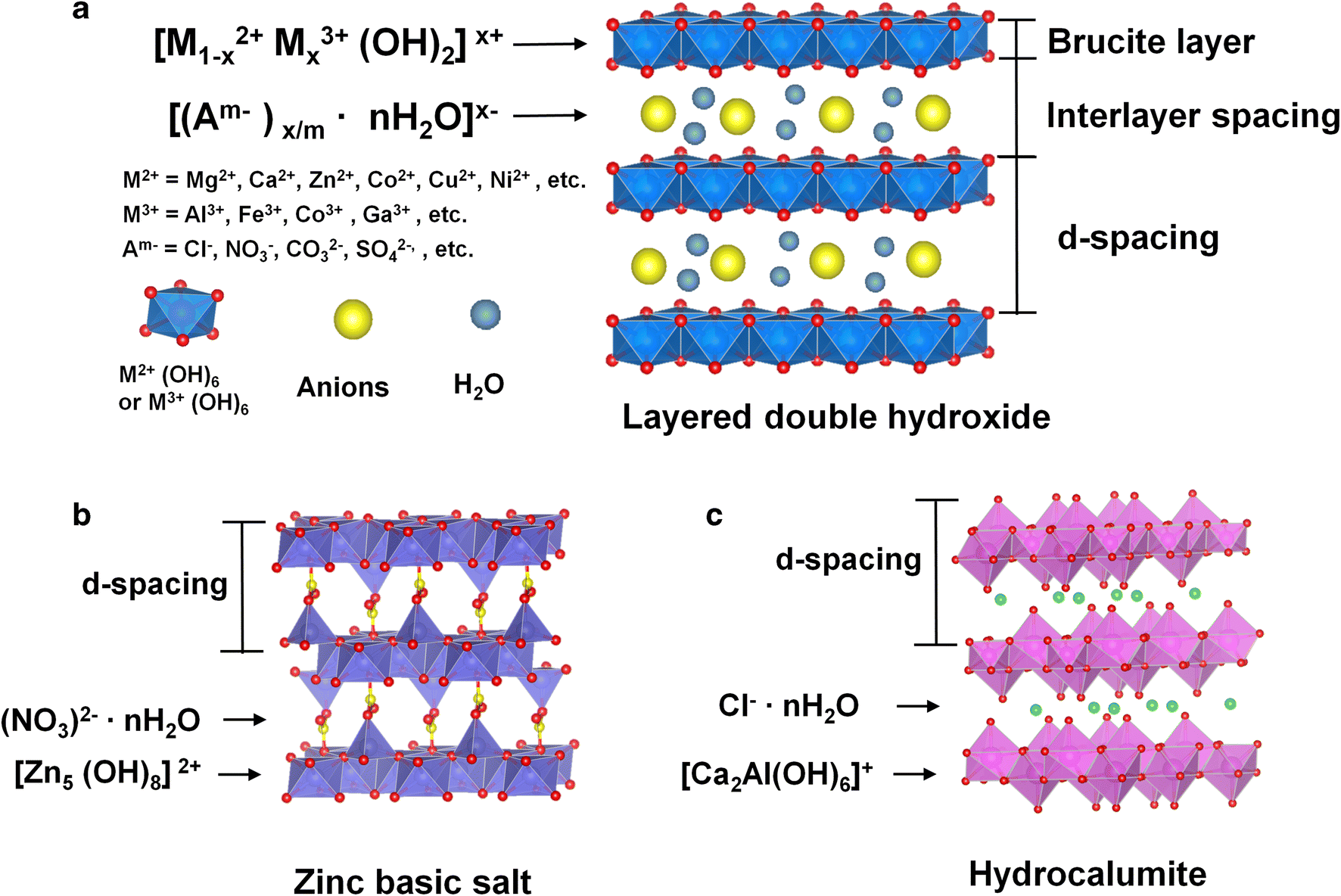
Layered double hydroxides (LDH) belong to a class of anionic clays. Unlike cationic clays, individual cationic brucite-like layers are stacked on top of each other with exchangeable anions in the interlayer space (along with water molecules) (Fig. 2a). The general formula of LDH can be expressed as [M(II)1–x M(III) x (OH)2][A m–] x/m ·nH2O, where M(II) is a divalent cation (Mg2+, Ca2+, Zn2+, Co2+, Cu2+, Ni2+, etc.), M(III) is an isomorphously substituted trivalent cation (Al3+, Fe3+, Co3+, Ga3+, etc.), and A m– is an interlayer anion with charge m (Cl−, NO3 −, CO3 −, SO4 2−, etc.). Various kinds of inorganic or organic anions have been introduced between the hydroxide layers by simple anion exchange reaction or co-precipitation reaction (Choy et al., Reference Choy, Kwak, Park, Jeong and Portier1999). Layered double salt (LDS) is a family of LDH, and very similar in terms of crystal structure, which consists of positively charged layers and charge-compensating anions along the crystallographic c axis, but different in its layer composition (Kaassis et al., Reference Kaassis, Xu, Guan, Evans, Wei and Williams2016; Bull et al. Reference Bull, Marklan, Williams and O'Hare2011). In the LDS structure, however, only divalent metal cations are octahedrally coordinated with hydroxo ligands to form hydroxide layers, and its general formula can be described as M 2+(OH)2 − x (A m– x/m ·nH2O, where M is a divalent cation (Zn2+, Co2+, Cu2+, Ni2+, etc.), and A m– is an anion with charge m (Cl−, NO3 −, CO3 2−, SO4 2−,etc.). For example, Zn-containing LDS is known as the zinc basic salt (ZBS) with a chemical formula [Zn5(OH)8](NO3)2·nH2O (Fig. 2B). The crystal structure of ZBS comprises edge-sharing Zn(OH)6 octahedra, and tetrahedrally coordinated Zn(OH)4 units, which are formed on the upper and lower sides of vacant octahedral sites. Those units, therefore, are exposed to the internal surface of ZBS layers and further ligated with water molecules (Yang et al., Reference Yang, Han, Park, Park, Hwang and Choy2007; Kim et al., Reference Kim, Yang, Lee, Choi, Park, Jo, Choi and Choy2015). Such a ZBS has been known as a family of 2D compounds structurally well defined and chemically stable, and, therefore, studied extensively as biomolecular reservoirs or advanced drug delivery systems (Oh et al., Reference Oh, Biswick and Choy2009).
Schematic crystal structures of a layered double hydroxide (LDH), b zinc basic salt (ZBS), and c hydrocalumite
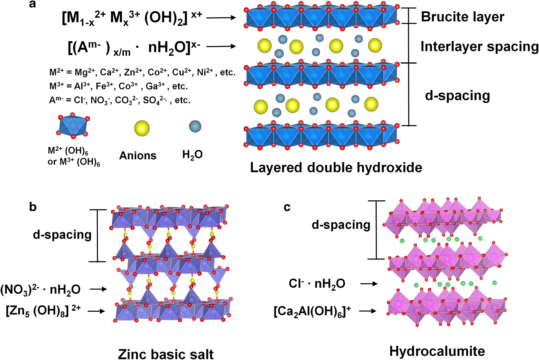
Hydrocalumite belongs to LDHs in a broad sense, and its general formula can be expressed as [Ca2 M(OH)6]+ A −∙nH2O. From the crystal structure point of view, hydrocalumite is similar to LDHs, as it also consists of self-assembled (Ca(OH)2) layers where Ca2+ ions are replaced partially by M 3+ (Al3+ or Fe3+) ions and in such a way a positive layer charge can be generated. As a result, incorporation of interlayer anions, such as CO3 2−, Cl−, and OH−, are required to balance the resulting positive layer charge (Wen et al. Reference Wen, Yang, Yan and Xie2015) (Fig. 2c). Obviously, hydrocalumites have some common characteristics with LDHs (Kim et al. Reference Kim, Lee, Choi and Oh2014).
Toxicity and Biocompatibility
In vitro
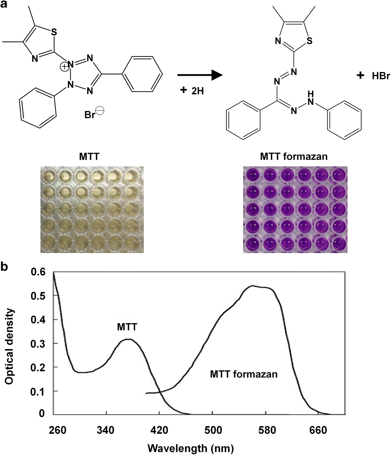
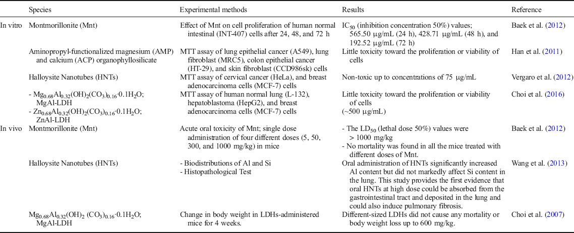
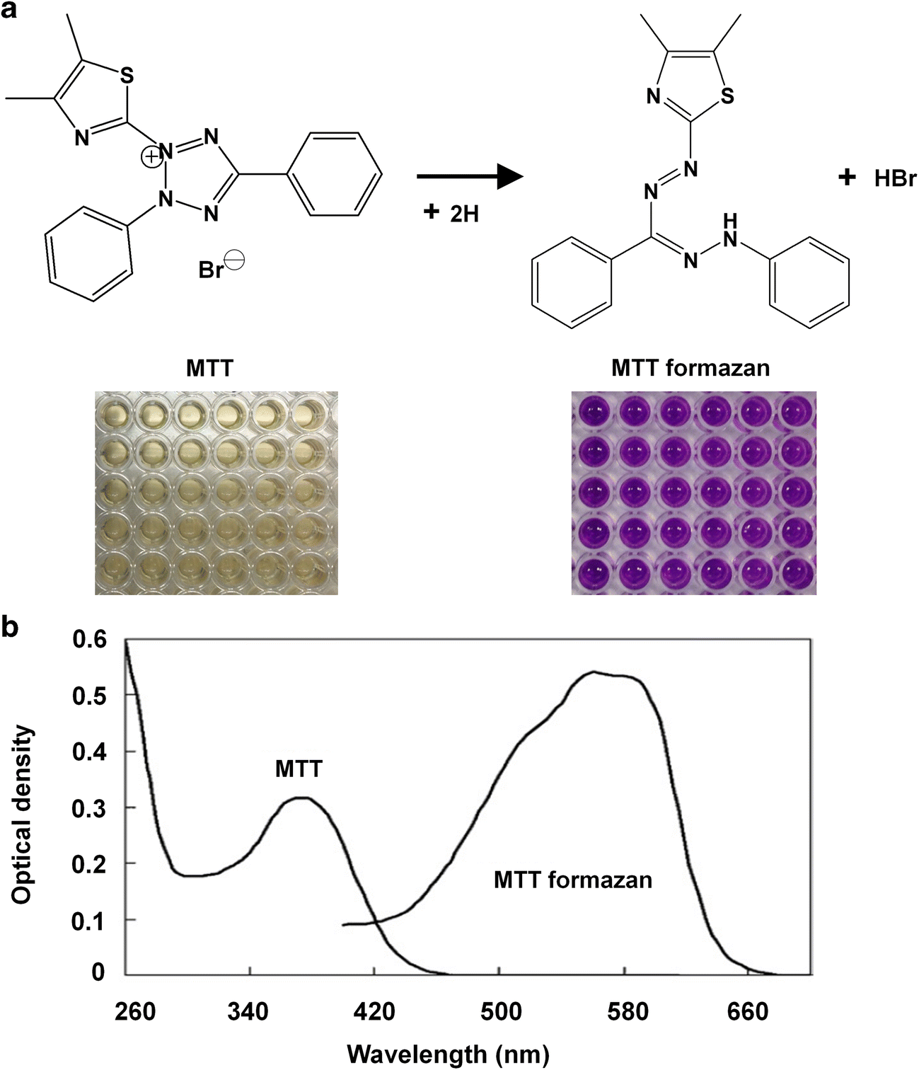
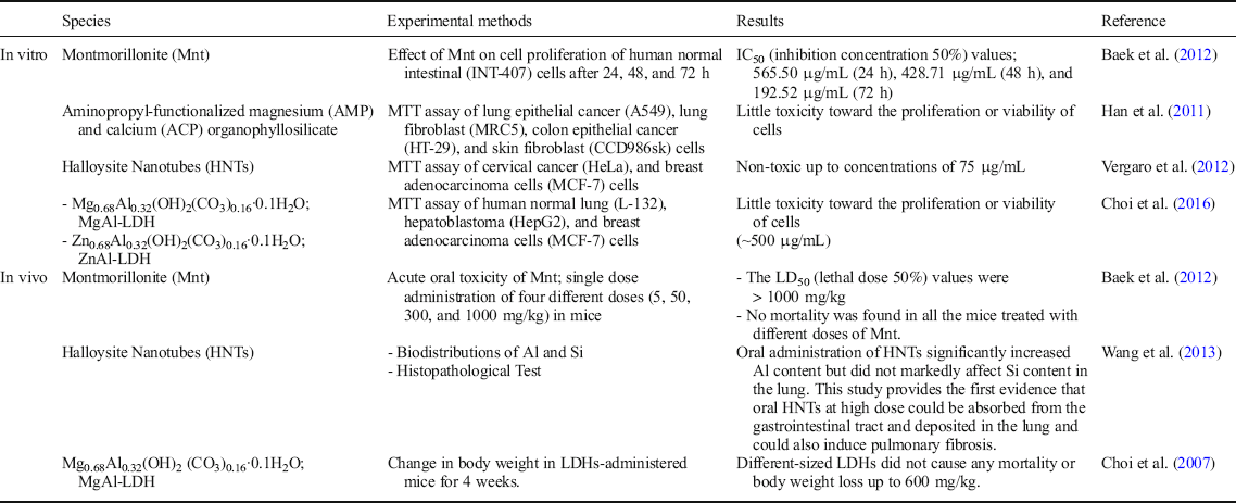
Various cell lines have been used to study the toxicities induced by clays and clay-containing nanocomposites (Maisanaba et al., Reference Maisanaba, Pichardo, Puerto, Gutiérrez-Praena, Cameán and Jos2015). In the case of in vitro toxicological evaluation based on a colorimetric assay using the tetrazolium salt thiazolyl blue, the MTT assay after methyl-thiazolyl-tetrazolium (Mosmann, Reference Mosmann1983) has been applied widely for estimating cytotoxicity, cell viability, and proliferation. The MTT agent yields a yellowish solution, but results in violet-blue formazan crystals upon reduction by dehydrogenases present in metabolically active cells (Fig. 3). The lipid soluble formazan product can be extracted using organic solvents and evaluated by spectrophotometry (Stockert, Blázquez-Castro, Cañete, Horobin, & Villanueva, Reference Stockert, Blázquez-Castro, Cañete, Horobin and Villanueva2012). A 2:1 type clay such as montmorillonite (Mnt) inhibits significantly cell proliferation after 24–72 h incubation time and at concentration levels >100 μg/mL (Table 2; Baek et al., Reference Baek, Lee and Choi2012). MgAl-LDH and ZnAl-LDH, the most frequently studied drug delivery carriers, have been determined to be very low in toxicity at the concentration level of practical application as drug delivery systems. LDH clay at the nano-scale, therefore, has been suggested as an advanced drug delivery vector (Choi, Oh, Park, & Choy, Reference Choi, Oh, Park and Choy2007).
(a) Chemical structures and colors of MTT and its reduced formazan product, and (b) absorption spectra of MTT and MTT formazan (after Stockert et al. Reference Stockert, Blázquez-Castro, Cañete, Horobin and Villanueva2012, reproduced with permission from Elsevier)
In vitro and in vivo toxicities of clay minerals and their clay hybrids
In vivo
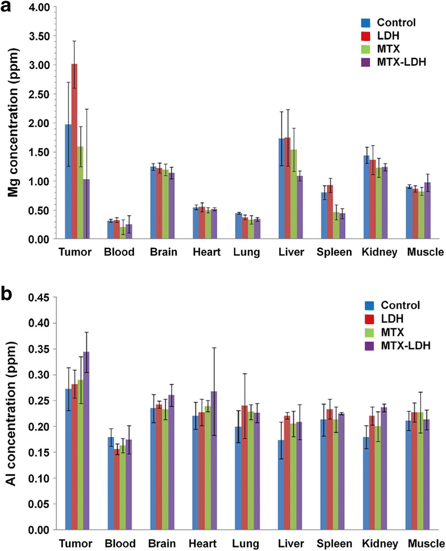
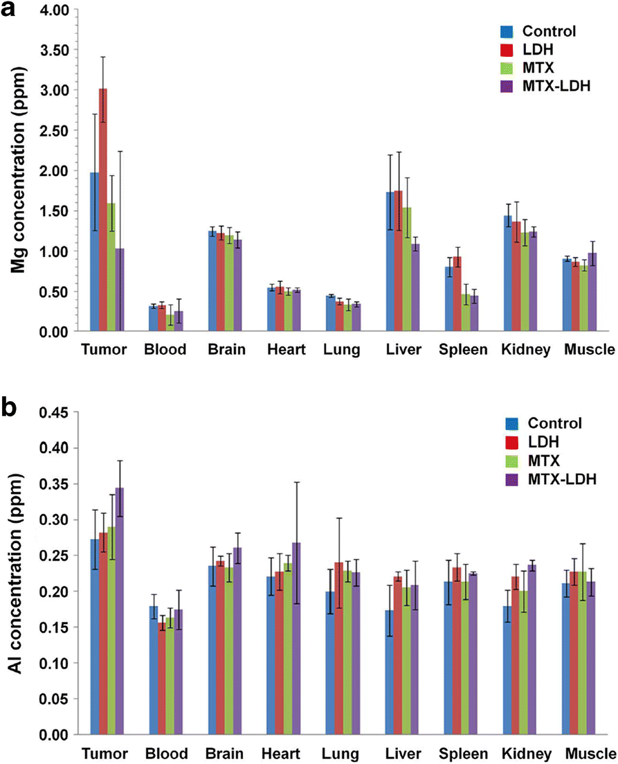
According to bio-distribution studies of LDH nanoparticles in various organs (Fig. 4), the ionic concentrations of Mg and Al in LDH can be determined by inductively coupled plasma-atomic emission spectrometry (ICP-AES). In in vivo studies, tumors and various organs were harvested from each treatment group 1 day after the last injection, and then dissolved completely in an aqueous solution of HCl and H2O2. The Mg and Al ion concentrations were similar among the groups within the error limit for all the organs evaluated (Fig. 4). Surprisingly, LDH clay nanoparticles were not accumulated in tissue upon treatment with the MCF7/mot orthotropic breast-cancer mouse model (Choi, Kwon, Oh, Yun, & Choy, Reference Choi, Kwon, Oh, Yun and Choy2014).
(a) and (b) accumulation of LDH nanoparticles in organs of tumor-bearing mice treated with PBS (control), LDH, MTX, or MTX-LDH for 5 weeks (n = 3 for each group). On the first day after the final treatment, the mice were sacrificed and organs, blood, and tumors were collected for measurement of Mg and Al contents, the major components of LDH (after Choi et al. Reference Choi, Kwon, Oh, Yun and Choy2014, reproduced with the permission of Nature Research)
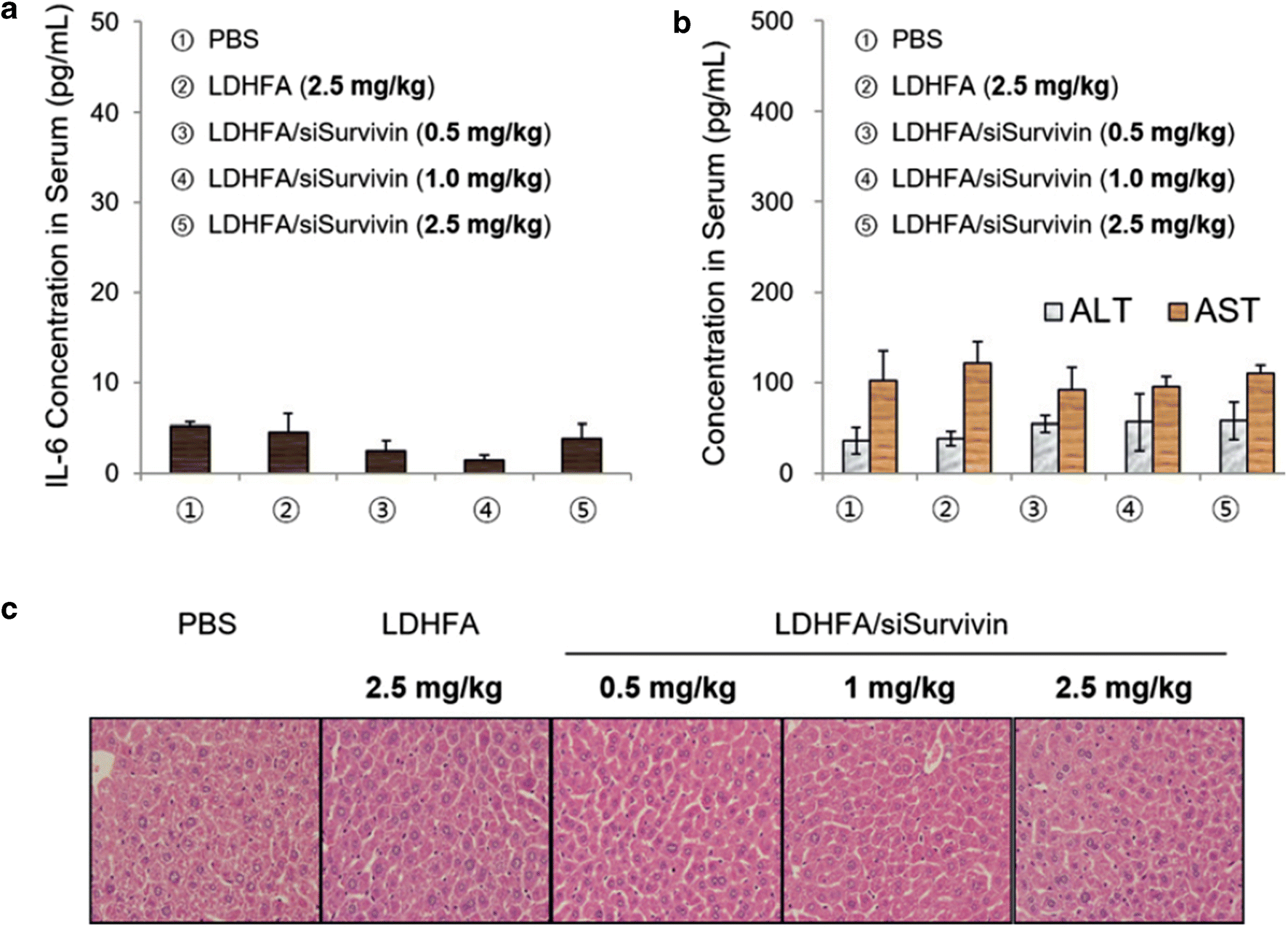
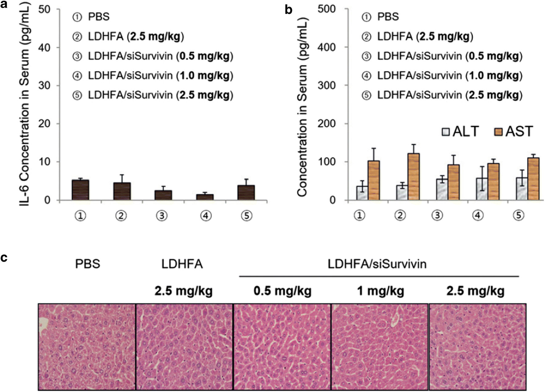
Park et al. (Reference Park, Cho, Kwon, Yun and Choy2016) reported recently on in vivo safety of LDH as an anionic clay nanovehicle. No liver toxicity was observed up to a dosage of 125 mg/kg of LDH-FA(Folate)/siSurvivin corresponding to 2.5 mg/kg of siSurvivin (Fig. 5); the levels of inflammatory cytokine interleukin (IL)-6 (3.8 pg/mL), alanine transaminase (ALT) (57.7 pg/mL), and aspartate transaminase (AST) (109.6 pg/mL) were found to be in the normal ranges. Liver tissues were also studied using the hematoxylin and eosin (H&E) staining method, and found to be undamaged. The in vivo toxicity of FA-conjugated LDH (LDH-FA) nanocarrier was also tested in order to evaluate the therapeutic dosages and the toxicity of LDH-FA/siSurvivin. The LD50 (lethal dose 50%) value for LDH-FA was found to be >600 mg/kg, placing it in the non-toxic class.
Innate immune responses and liver toxicity of the LDH-FA/siSurvivin treatment (n = 4 per group). (a) Inflammatory cytokine level (IL-6 level) analyzed at 6 h after LDH-FA/siSurvivin treatment, (b) ALT and AST levels, and (c) H&E staining of liver tissues performed on day 3 after treatment (original magnification: × 100) (after Park et al., Reference Park, Cho, Kwon, Yun and Choy2016, reproduced with the permission of Wiley)
Design of Clay Hybrid Systems
Clay minerals have been designed as hybrid materials in order to optimize their characteristics for specific biological and medical applications. Because clay minerals have large surface areas, adsorption capacity, and ion exchange capacity, they can be hybridized easily with drugs, genes, bioactive molecules, biopolymers, and other functional organic molecules.
Modification of clay hybrid materials
Many studies of hybridization of clay minerals have been focused on the surface modification by surfactants and polymers. Prepared hybrid-clays can thus be utilized as the drug delivery systems with designed functions such as controlled drug release, targeted drug delivery, enhanced solubility, improved chemical stability, etc.
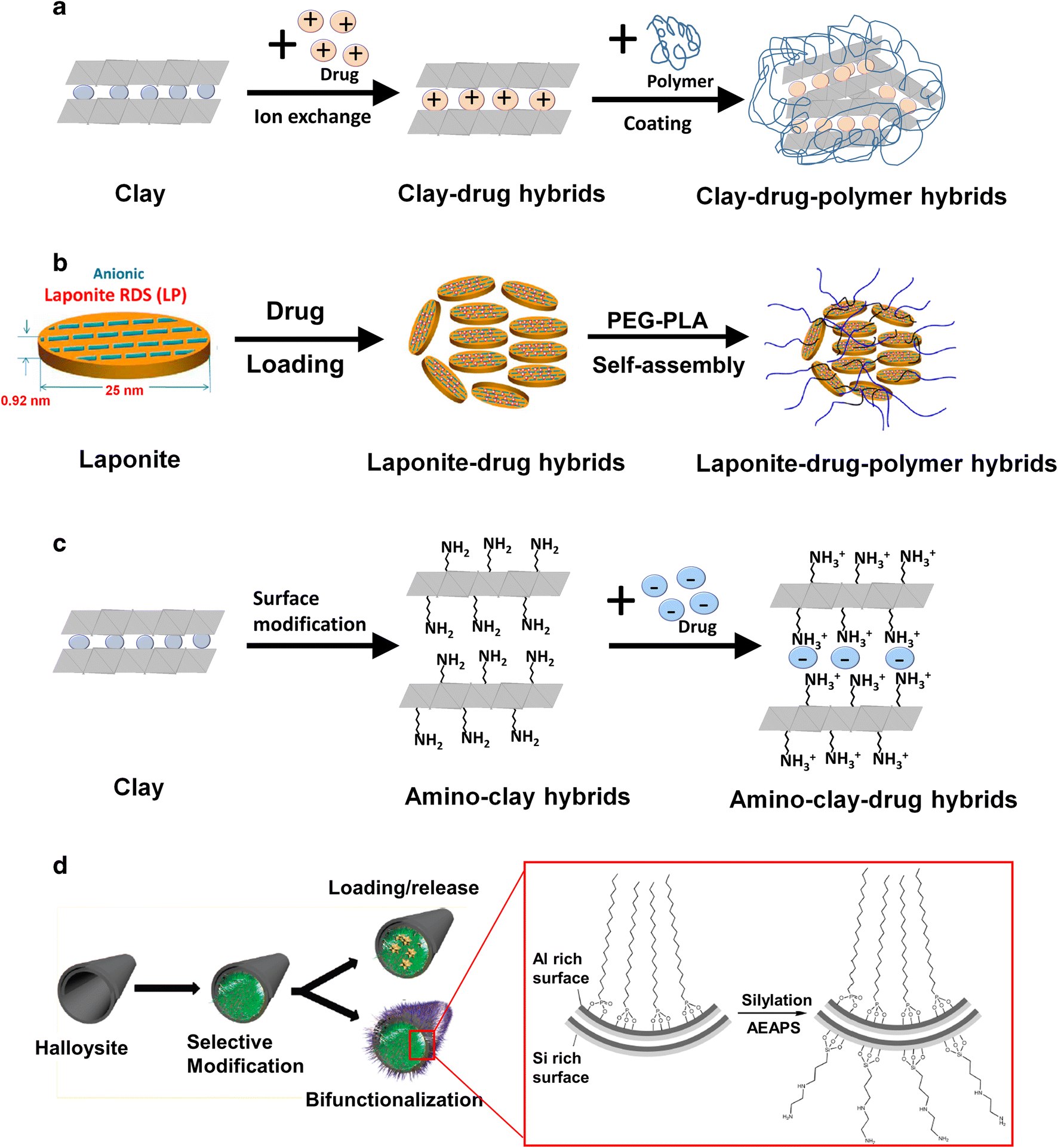
Lee et al. (Reference Lee, Choi, Oh, Park, Choy, Park, Yoon, Lee, Chang and Choy2012), for example, intercalated a cationic drug (sildenafil) into the negatively charged Mnt layers through an ion-exchange reaction. To improve the taste-masking efficiency and enhance the rate of drug-release, the sildenafil-Mnt hybrids were further modified with AEA (polyvinylacetal diethylaminoacetate) (Fig. 6a). For oral medication, AEA is insoluble in the buccal cavity, but its release rate can be enhanced in gastric juice, due to the high solubility of AEA in acidic conditions.
Chemical modifications of clay hybrid materials. Schematic procedures for (a) polymer coating on clay-drug nanohybrid, (b) self-assembling of Laponite®-drug hybrid with polymer (after Wang et al., Reference Wang, Gong, Rong, Gui, Hu and Xu2018), (c) immobilizing drug molecules in aminoclay nanohybrid, and (d) bifunctionalizing internal and external surfaces of halloysite nanotubes (after Yah et al., Reference Yah, Takahara and Lvov2012, reproduced with the permission of the American Chemical Society)
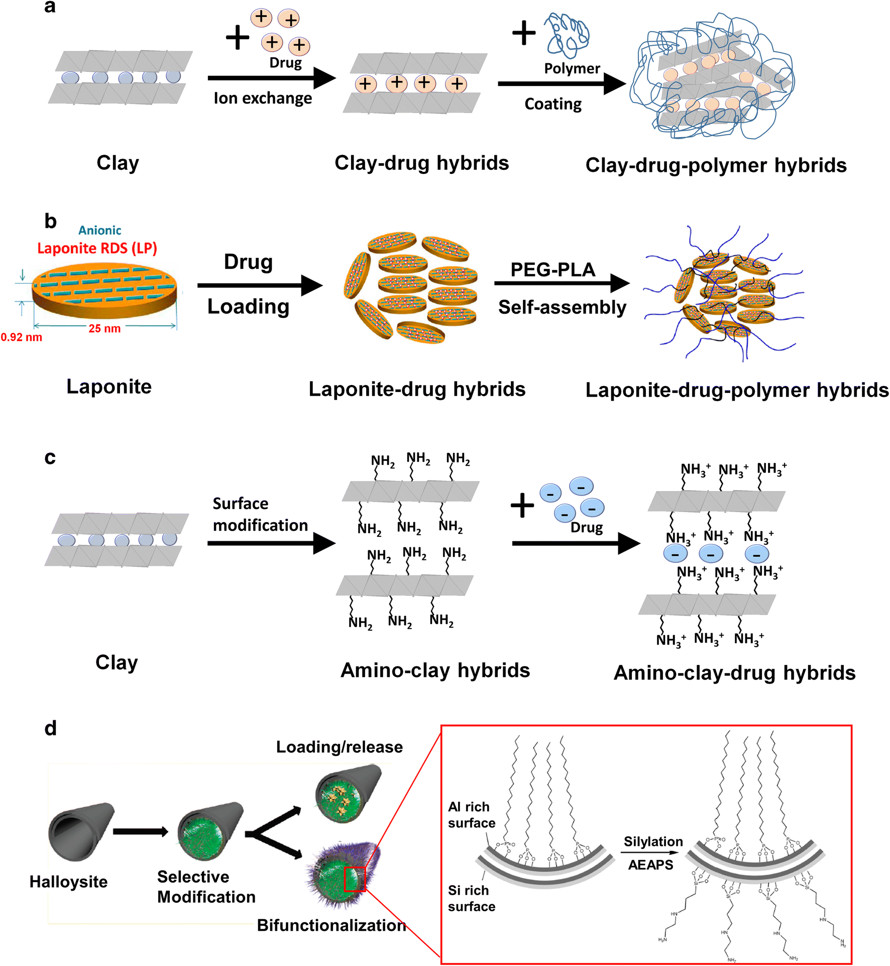
Using a different approach, Wang et al. (Reference Wang, Maciel, Wu, Rodrigues, Shi, Yuan, Liu, Tomas and Li2014) reported Laponite®-drug-polymer nanohybrids with enhanced cytocompatibility, pH-stimulative controlled release property, and physiological stability. In this case, a cationic drug (doxorubicin) was intercalated into Laponite® and then further modified with PEG-PLA (polyethylene glycol-polylacic acid) copolymer by a self-assembling method (Fig. 6b), but a bio-distribution study is required to understand accumulation of the drug in various organs in the body.
Yang et al. (Reference Yang, Shao and Han2014) also tried to intercalate telmisartan, an antihypertensive drug (poor water solubility), into 3-aminopropyl functionalized magnesium phyllosilicate (aminoclay) in order to improve the bioavailability of the drug for oral administration, because the surface charge of an aminoclay becomes positive due to the amine groups functionalized on clay. A positively charged aminoclay, therefore, can intercalate or adsorb negatively charged telmisartan via an electrostatic interaction resulting in the formation of a weakly coupled telmisartan-aminoclay complex, which showed an enhanced bioavailability of a poorly soluble drug (Fig. 6c).
Yah et al. (Reference Yah, Takahara and Lvov2012) studied functional halloysite by modifying its surface with various molecules, taking advantage of the inner-surface chemistry of halloysite nanotubes which is different from the outer-surface chemistry. Selective functionalization of the halloysite clay lumen resulted in a hydrophobic aliphatic chain core with micelle-like architecture and a hydrophilic silicate shell. The internal surface of halloysite was first modified with octadecylphosphonic acid (Fig. 6d), and then the external surface with silylating agent, N-(2-aminoethyl)-3-aminopropyltriethoxysilane, giving rise to a difference in chemical reactivity between the inner and outer surfaces of the clay nanotubes, and, therefore, the functionalized halloysite can be used for drug reservoir and delivery.
Synthetic method of clay hybrid materials
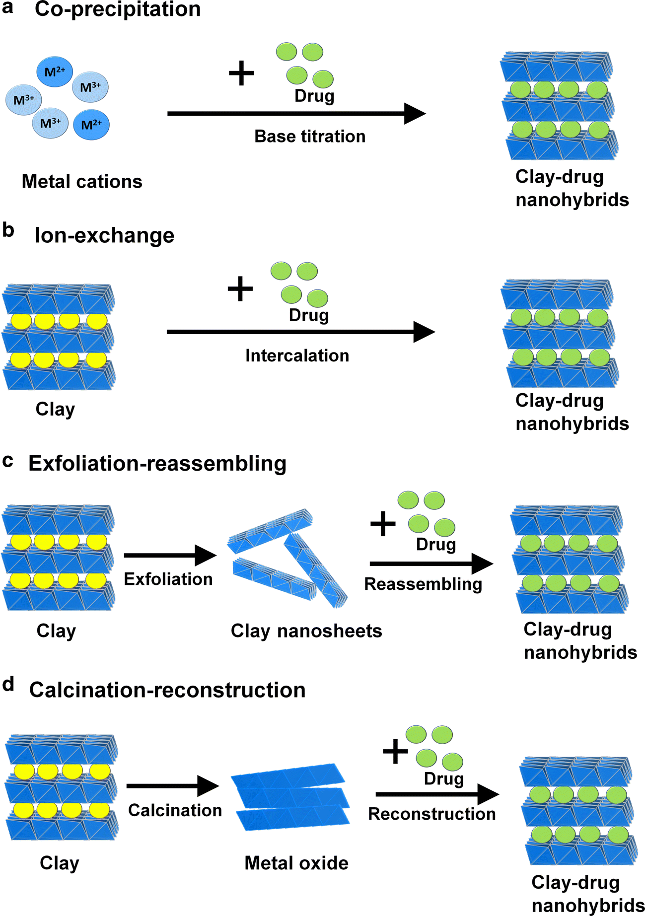
Despite the variety of clay minerals in nature, clay hybrid systems can be explored on the basis of a synthetic clay for medical applications. Several synthetic routes to anionic clay nanohybrid materials, such as co-precipitation, ion-exchange, exfoliation-reassembling, and calcination-reconstruction (Margarita et al. Reference Margarita, López-Blanco, Aranda, Leroux and Ruiz-Hitzky2005; Park et al., Reference Park, Hwang, Oh, Yang and Choy2013; Choi et al., Reference Choi, Eom, Vinu and Choy2018), have been studied extensively (Fig. 7).
Reaction routes for clay hybrid materials; (a) co-precipitation, (b) ion-exchange, (c) exfoliation-reassembling, and (d) calcination-reconstruction
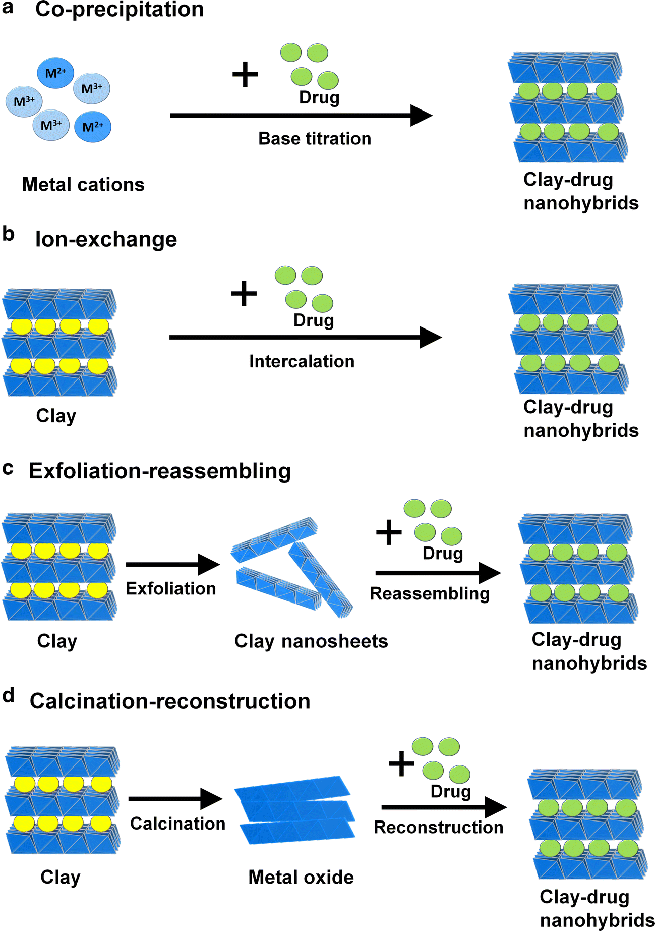
The co-precipitation method is a simple and economic technique for the preparation of LDH hybrid materials, which can be achieved by base titration of a metal salt [M 2+ and M 3+ (or mixtures)] precursor solution in the presence of anionic drug or biofunctional molecules. In this way, anionic drug or guest molecules can be incorporated simultaneously into the LDH lattice to form LDH hybrid. To obtain LDH hybrid as a precipitate, the solution pH must be adjusted carefully to a coprecipitation range of corresponding metal hydroxides by considering the pCi [log(ionic concentration)] values with respect to pH and the pKa values of functional groups in biomolecules. Based on this solution chemistry, a wide variety of biomolecules can be intercalated directly into an anionic clay lattice. To control the particle size and enhance the crystallinity of the clay, the hydrothermal method can be used selectively.
The ion exchange process is a classical way of preparing clay hybrid materials, where the negatively charged guest molecules in the solution can be exchanged with the anions present in the interlayer species of clays. The ion exchange reaction can, in general, be affected by some factors, such as the ion exchange capacity of LDH, the solution pH, and the thermodynamic stability between host LDH and guest molecules.
The exfoliation-reassembling process is also suggested as a useful way of encapsulating or intercalating bulky and large-sized biomolecules into lamellar clays, whether they are cationic or anionic. If the layer charge were not that big, the multilayers consisting of cationic or anionic layers could be exfoliated into a single layer by intercalating polar solvent molecules (Mnt: e.g. ammonium salts with long alkyl chains, polymer natrix; LDH: e.g. formamide) (James et al. Reference James, Groen and Coveney2015; Ma et al. Reference Ma, Liu, Li, Lyi and Sasaki2006). Once exfoliated, they are then allowed to reassemble in the presence of guest molecules in a solution. In this way guest molecules can be intercalated into the clay lattice to build the clay hybrid with 1:1 heterostructure.
The final route to clay hybrids is the calcination-reconstruction process. Thanks to the structural memory effect of LDHs, the pristine Mg-Al LDH is, at first, calcined at ~400–500°C in such a way that layered metal hydroxide is thermally decomposed into amorphous metal oxides along with a weakly developed periclase MgO phase (JCPDS 43–1022), followed by dehydration and subsequent dehydroxylation reactions. An amorphous oxide phase prepared in this manner can be recrystallized into LDH upon rehydration. If the rehydration reaction occurs in the presence of guest molecules in a solution, the clay hybrid can simply be regenerated.
Clay Minerals in Medicinal Applications
Oral medicine
Various drugs such as antimalaria (Kim et al., Reference Kim, Yang, Lee, Choi, Park, Jo, Choi and Choy2015), anti-psychotic (Oh et al., Reference Oh, Choi, Choy, Park, Park, Lee, Yoon, Chang and Choy2013), antibiotic (Jung et al. Reference Jung, Kim, Choy, Hwang and Choy2008; Yang et al. Reference Yang, Jung, Kim, Yo and Choy2013; Ghadiri et al. Reference Ghadiri, Chrzanowski and Rohanizadeh2014), and anticancer (Kevadiya et al., Reference Kevadiya, Thumbar, Rajput, Rajkumar, Brambhatt, Joshi, Dangi, Mody, Gadhia and Bajaj2012; Iliescu et al., Reference Iliescu, Andronescu, Ghitulica, Voicu, Ficai and Hoteteu2014; Massaro et al., Reference Massaro, Colletti, Noto, Riela, Poma, Guernelli, Parisi, Milioto and Lazzara2015) have been explored extensively for oral administration, but some were found to have drawbacks such as low bioavailability, poor water solubility, unpleasant taste, etc. In order to overcome such issues, advanced drug delivery systems with suitable delivery carriers are required urgently. Amongst various drug delivery carriers, clay minerals were considered as promising because the drug molecules can be incorporated into the interlayer of montmorillonite by an intercalative ion-exchange reaction, or into the nanotube of halloysite by capillary condensation, and also released from the drug-clay hybrid into a body fluid. Clay minerals, moreover, were found to be biocompatible with low toxicity, large drug-loading capacity, and sustained-release property.
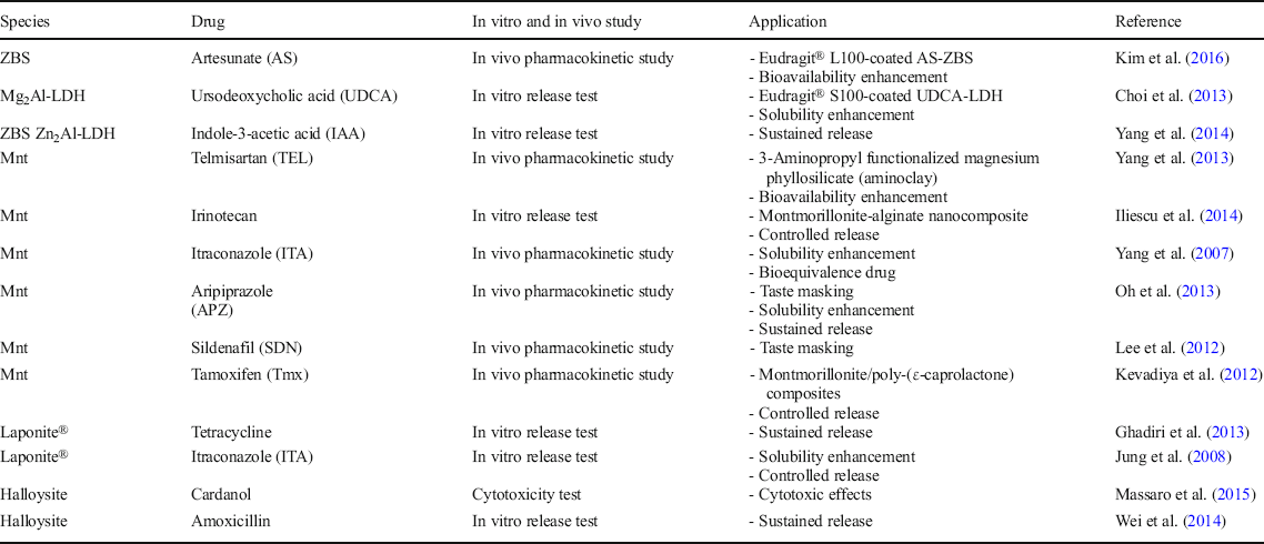
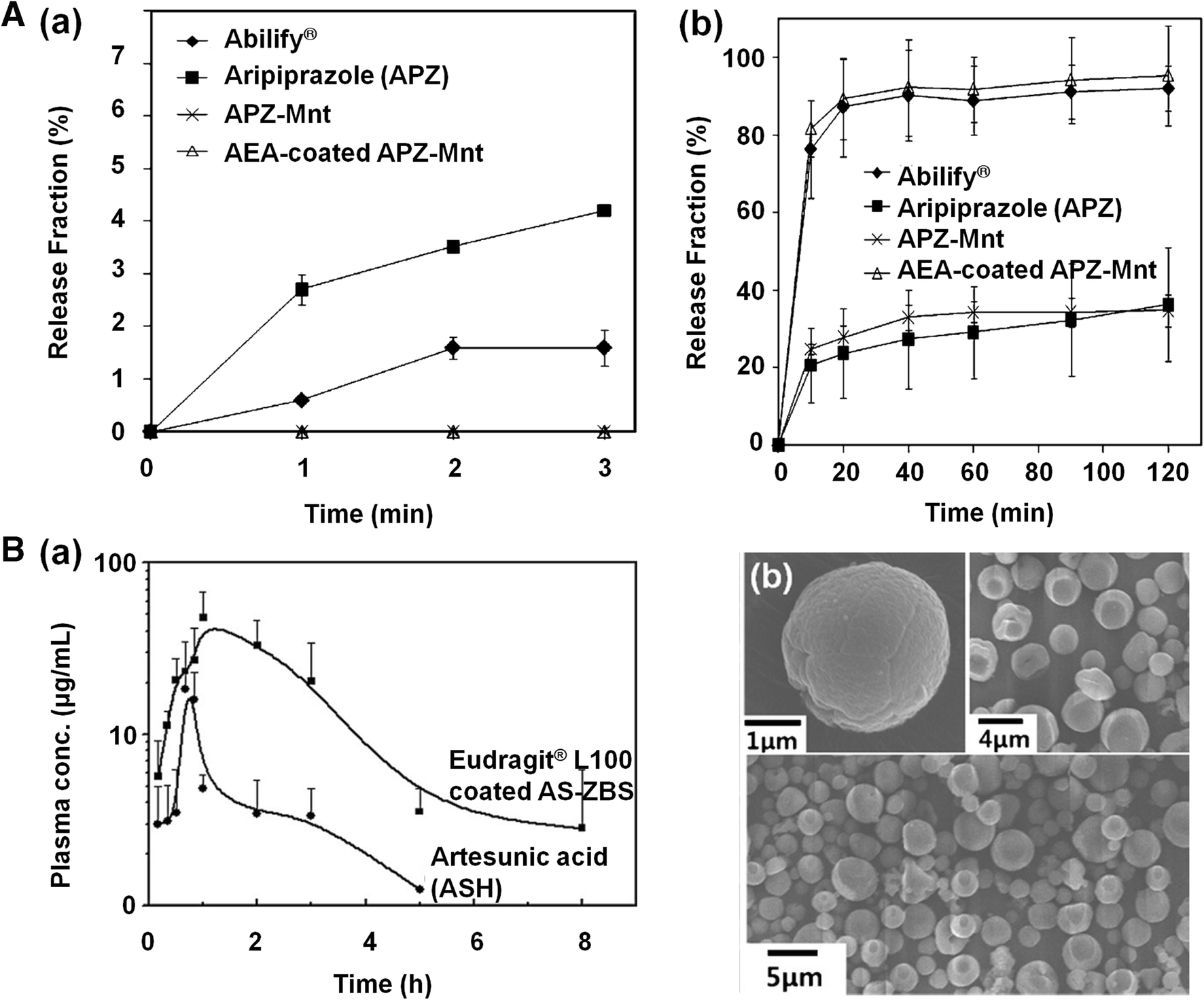
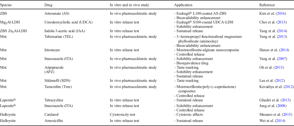
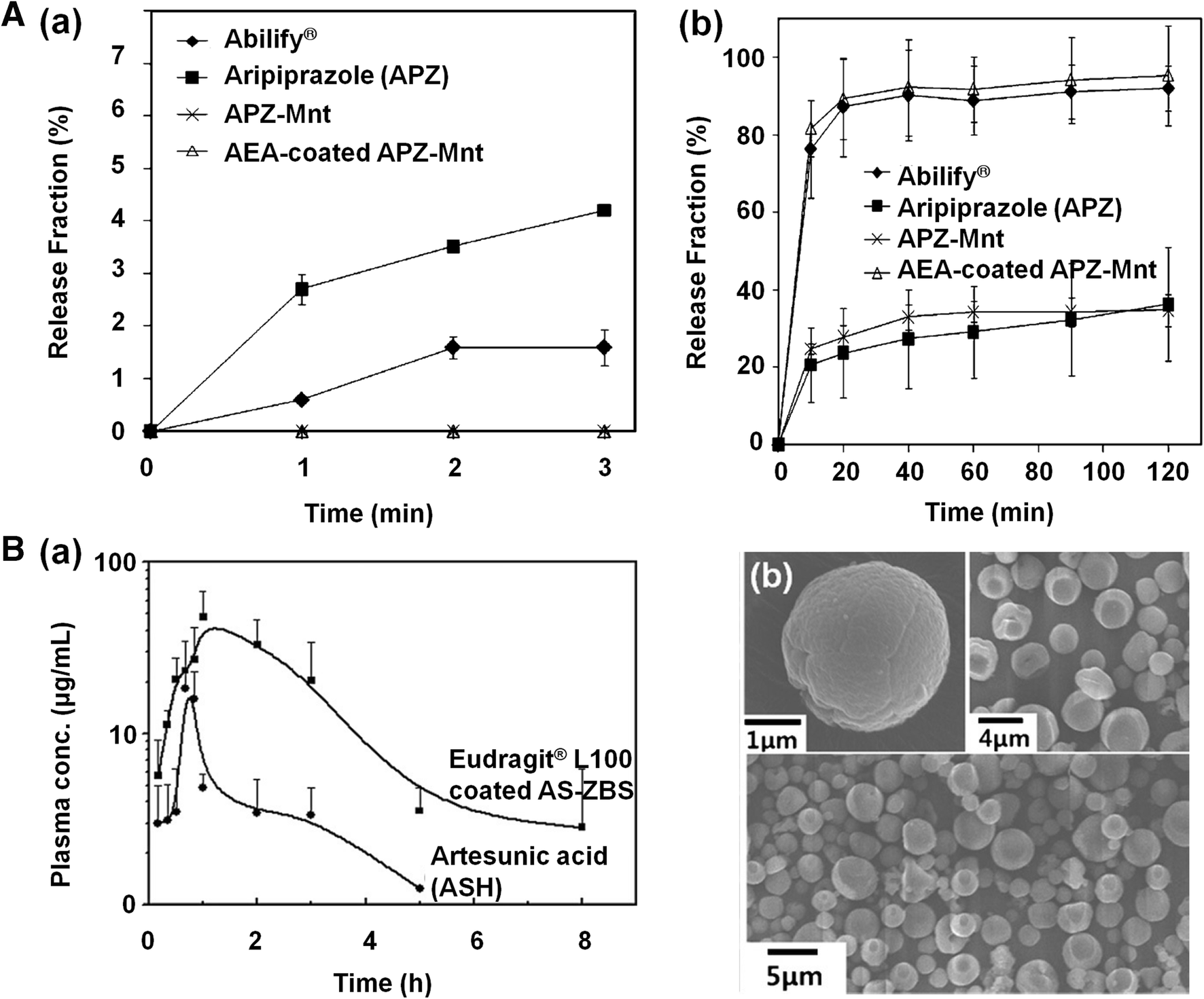
Several approaches have been made to develop drug-clay hybrids for oral medications (Table 3). Oh et al. (Reference Oh, Choi, Choy, Park, Park, Lee, Yoon, Chang and Choy2013), for example, reported an aripiprazole (APZ)-montmorillonite (Mnt) hybrid, which was further encapsulated with polyvinylacetal diethyl-amino acetate (AEA) in order to overcome the poor aqueous solubility and unpleasant taste of aripiprazole (APZ: 7-{4-[4-(2,3-dichlorophenyl)piperazin-1-yl]butoxy}-3,4-dihydro quinolin-2(1H)-one). APZ has been used widely for treating the negative and positive symptoms of schizophrenia with reduced extrapyramidal side effects compared to the control drug, the commercially used tablet for oral delivery (Abilify®, Otsuka Pharmaceutical) (Oh et al., Reference Oh, Choi, Choy, Park, Park, Lee, Yoon, Chang and Choy2013; Harrison & Perry, Reference Harrison and Perry2004; Green, Reference Green2004) According to in vitro dissolution tests at neutral pH (Fig. 8A(a)), APZ drug release from AEA-coated APZ-Mnt was greatly reduced (<1%) for up to 3 min, thus masking the taste of the APZ. In a simulated gastric solution at pH 1.2, however, the total percentage of APZ released over a 2 h period was up to 95% in the case of AEA-coated APZ-Mnt (Fig. 8A(b)). The release profile of APZ in vitro was the same as from the Abilify®, indicating that the present formulation of AEA-coated APZ-Mnt could be developed further as a new generic drug or an incrementally modified new drug (IMD). Kim et al. (Reference Kim, Yang, Lee, Choi, Park, Jo, Choi and Choy2015) studied an antimalarial drug (artesunic acid; ASH), which orally, has poor bioavailability as the aqueous solubility of ASH is extremely low. To overcome this problem, artesunate (AS) was immobilized in the ZBS interlayer space by the co-precipitation method, and then further coated with Eudragit® L100. According to the in vivo pharmacokinetic results (Fig. 8B(a)), the AS level was considerably higher in the rat plasma administered with Eudragit® L100-coated AS-ZBS than with pure ASH. When the Eudragit® L100-coated AS-ZBS nanohybrid was administered orally, the AUC parameter of Eudragit® L100-coated AS-ZBS was determined to be 5.5 times larger than that of intact ASH. Eudragit® L100-coated AS-ZBS can, therefore, be recommended as a hybrid drug-delivery system for enhancing drug solubility and bioavailability (Kim et al., Reference Kim, Yang, Lee, Choi, Park, Jo, Choi and Choy2015). Many previous reports including the two mentioned above have confirmed that cationic and anionic clays can play a role as drug delivery carriers and their nanohybrids can provide a novel drug delivery platform of oral medication for taste masking, solubility enhancement, bioavailability enhancement, and sustained and controlled release.
Summary of oral medicine using clay minerals
(A) Release profiles of APZ (a) in deionized water with 1% Tween 80, and (b) in simulated gastric fluid (pH 1.2) with 1% Tween 80 (modified from Oh et al., Reference Oh, Choi, Choy, Park, Park, Lee, Yoon, Chang and Choy2013). (B) Pharmacokinetic curves of AS after oral administration (a), and scanning electron microscopy (SEM) images of Eudragit® L100-coated AS-ZBS (b) (after Kim et al., Reference Kim, Hur, Choi, Min, Choy and Choy2016, reproduced with the permission of Wiley)
Injectable medicine
As already suggested by Choy’s group (Oh et al. Reference Oh, Choi, Kim and Choy2006, Reference Oh, Choi, Lee, Kim and Choy2009), the ~100 nm-sized LDH nanoparticles are, in general, biocompatible and targetable to tumor tissues and cells, not only due to their particle-size-dependent tissue-targeting function, ascribed to their enhanced permeability and retention (EPR) effects, but also due to their intrinsic cell targeting function known as the intercellular pathway based on the clathrin-mediated endocytosis mechanism.
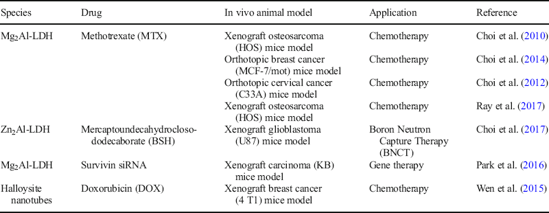
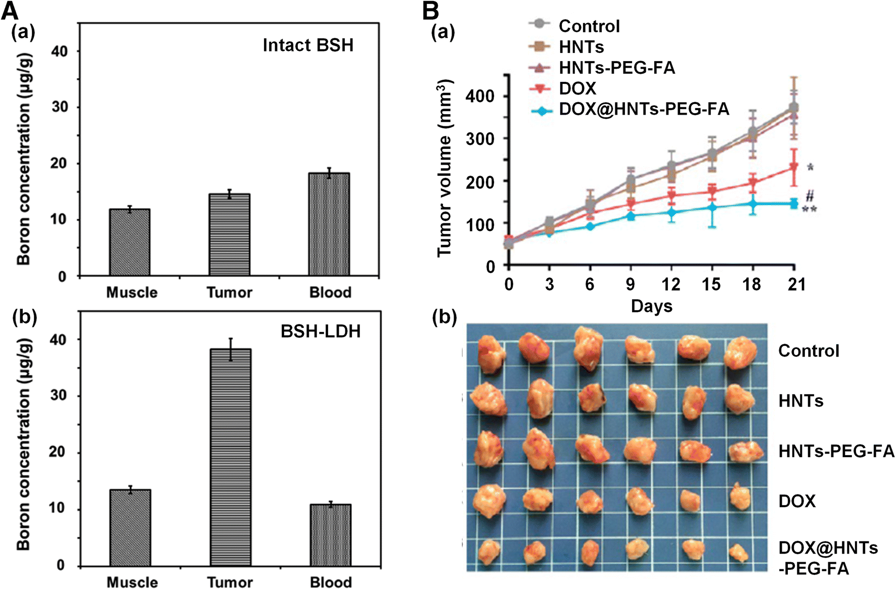
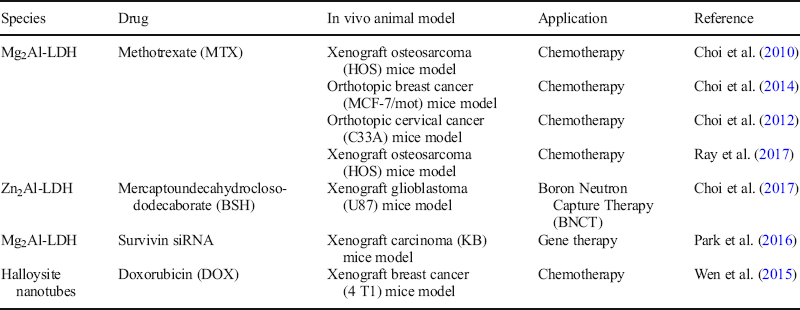
Many attempts have been made to develop LDH nanoparticles using injectable medicine for chemo-, gene-, and radiation-therapy (Table 4). Choy, Oh, and co-workers, for example, were successful in encapsulating methotrexate (MTX), an anticancer drug, in an LDH delivery vehicle to prepare a potential DDS as an MTX-LDH nanohybrid with a particle size of ~100 nm (Choi et al. Reference Choi, Kim, Oh and Choy2012, Reference Choi, Oh, Chung, Hong, Kim and Choy2013, Reference Choi, Kwon, Oh, Yun and Choy2014, Reference Choi, Piao, Alothman, Vinu, Yun and Choy2016; Oh et al., Reference Oh, Choi, Kim and Choy2006). According to the Trypan Blue assay, the cell viability with MTX-LDH was more significantly reduced than that with intact MTX. The IC50 value for intact MTX in osteosarcoma MNNG/HOS cells was determined to be ~2.5 times greater than that for MTX-LDH, suggesting that the latter could permeate through the cell membrane more efficiently than the former, resulting in improved drug delivery, and eventually higher drug efficacy. The cell viability, furtheremore, was not significantly influenced by the concentrations of LDH nanoparticles up to concentrations of 500 μg/mL (Oh et al., Reference Oh, Choi, Kim and Choy2006), indicating its low cytotoxicity. Choi et al. (Reference Choi, Kwon, Oh, Yun and Choy2014) also prepared an injectable MTX-LDH nanohybrid system by the co-precipitation route, and examined drug efficacy for the first time in an orthotopic breast cancer model. According to the biodistribution studies, the mice injected with MTX-LDH showed that six times more MTX was delivered to the tumor than those treated with intact MTX, indicating the tumor targeting function of the drug delivery vehicle, LDH. Furthermore, an MTX-LDH nanohybrid showed better drug efficacy than intact MTX in a tumor growth inhibition test with the same orthotopic mice model (Choi et al., Reference Choi, Kwon, Oh, Yun and Choy2014). More recently, Choi et al. (Reference Choi, Jeon, Piao and Choy2017) studied an injectable LDH nanovehicle hybridized with mercaptoundecahydrocloso-dodecaborate (BSH) for boron neutron capture therapy (BNCT). According to the biodistribution studies in the xenograft glioblastoma (U87) mice model (Fig. 9a), the tumour MTX-to-blood MTX ratio of BSH in the BSH-LDH-treated-group was found to be 4.4 times greater than that in the intact BSH-treated group 2 h after drug treatment (Choi et al., Reference Choi, Jeon, Piao and Choy2017). Such a targeting phenomenon in vivo can be explained not only by the enhanced permeability and retention (EPR) effect of the nanosized BSH-LDH particles (~100 nm), which deliver BSH molecules preferentially to tumor tissue and eventually to tumor cells, but also by their excellent cellular uptake effect due to clathrin-mediated endocytosis (Oh et al., Reference Oh, Choi, Kim and Choy2006).
Summary of injectable medicine using clay minerals
A Biodistribution studies of boron in each tissue of U87 xenograft tumor-bearing mice treated with (a) BSH and (b) BSH-LDH for 2 h after administration (after Choi et al., Reference Choi, Jeon, Piao and Choy2017). B Antitumor effects of DOX@HNTs-PEG-FA in four T1-bearing mice; (a) tumor growth curves of mice treated with saline, HNTs, HNTs-PEG-FA, DOX, and DOX@HNTs-PEG-FA (5 mg DOX equiv./kg) via intravenous injection, and (b) photographs of excised 4 T1 solid tumor from different groups on the 22nd day. The values are represented as mean ± SD (n = 6). *P < 0.05, **P < 0.01, vs. control; # P < 0.05 vs. DOX (after Wen et al. Reference Wen, Yang, Yan and Xie2015, reproduced with the permission of the Royal Society of Chemistry)
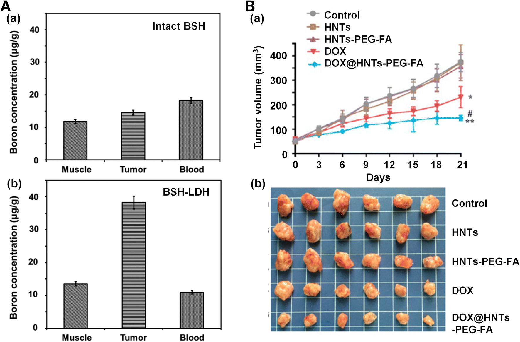
In the case of cationic clays, however, only a few studies have been done on injectable drug-clay systems, no doubt because of the difficulties in controlling particle sizes at <100 nm, and the insolubility in body fluid resulting in problems of organ accumulation after injection. However, Wu et al. (Reference Wu, Yang, Gao, Shen, Jiang, Zhou, Li, He and Liu2018) reported recently the use of folate-conjugated halloysite nanotubes (HNTs) for doxorubicin (DOX) delivery. The particle size of HNTs was controlled at 200 nm by ultrasonic scission, and thus prepared, nanoparticles were functionalized with amide groups to induce chemical bonding with N-hydroxylsuccinimide-polyethylene glycol carboxylic acid (NHS-PEG-COOH) and folate (FA). DOX@HNTs-PEG-FA was then synthesized by adding DOX on HNTs-PEG-FA via simple physical adsorption. According to the in vivo studies, an antitumor effect of DOX@HNTs-PEG-FA was clearly confirmed in a breast cancer (4 T1 cells) mice model through intravenous injection. As expected, the tumor volume and tumor weight in 4 T1-bearing mice for both cases, DOX (P < 0.05) and DOX@HNTs-PEG-FA (P < 0.01) treatments, were strongly reduced with respect to the control group, while no significant change could be observed for the HNTs and HNTs-PEG-FA treatment groups (Fig. 9B) (Wu et al., Reference Wu, Yang, Gao, Shen, Jiang, Zhou, Li, He and Liu2018). Even though HNT is non-degradable, it was used as a drug delivery carrier due to its low toxicity in vivo (Wu et al., Reference Wu, Yang, Gao, Shen, Jiang, Zhou, Li, He and Liu2018). However, a long-term biodistribution study is required to follow its trafficking pathways and to understand its fate after injection.
Diagnostics
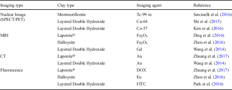
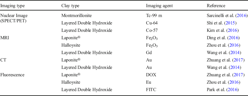
The diagnosis of cancer can be made using a variety of molecular imaging techniques such as magnetic resonance imaging (MRI), computerized tomography (CT), nuclear imaging including positron emission tomography (PET), single photon emission computed tomography (SPECT), and optical imaging, etc. For precise cancer diagnosis, various inorganic and/or organic materials, including liposome, dendrimer, and clay minerals immobilized with imaging agents have been developed (Xing et al. Reference Xing, Hwang and Lu2016; Cheng et al. Reference Cheng, Zhao, Li and Xu2011; Choi et al., Reference Choi, Eom, Vinu and Choy2018). Various clay minerals such as montmorillonite (Mnt), Laponite®, halloysite, and layered double hydroxide have already been explored as carriers of molecular imaging agents for diagnostics (Table 5).
Clay minerals as carriers of molecular imaging agents for nanomedicine
Nuclear molecular imaging
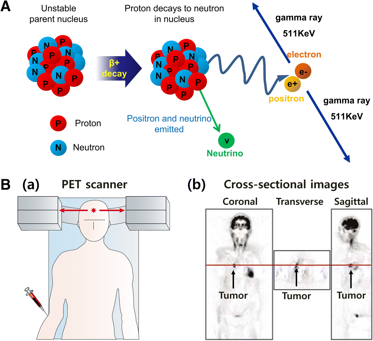
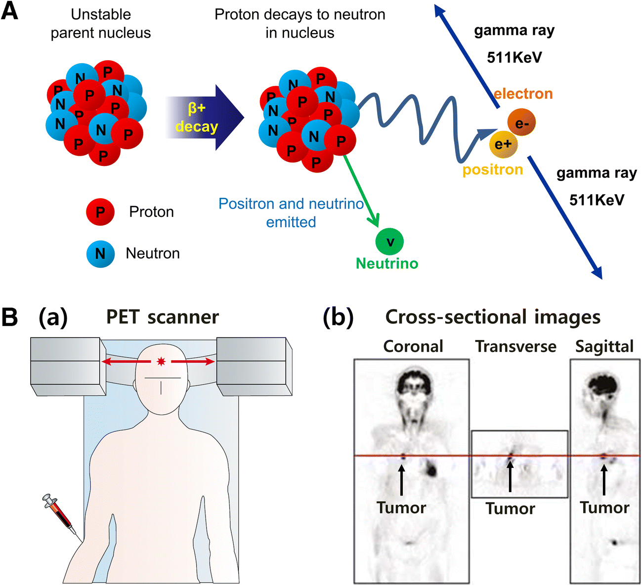
Radiology was started after the discovery of X-rays by Roentgen in 1895, and nuclear medicine began after the discovery of radium and its radioactivity by Becquerel and the Curies in 1896. In nuclear medicine, the gamma camera was developed by Anger in 1958 for tomographic imaging, and today it is used in the form of SPECT and PET (McRobbie et al. Reference McRobbie, Moore and Graves2017). The detection sensitivity of SPECT and PET is very high, even at very low concentrations with a picomolar or a nanomolar range (Khalil et al. Reference Khalil, Tremoleda, Bayomy and Gsell2011). The imaging agents for SPECT are, in general, doped with radioisotopes, such as 99mTc, 111In, 123I, 57Co, and 67Ga, which can emit gamma rays. On the other hand, radioisotopes emitting positrons are employed for PET, and the positron emitters known so far are: 15O, 13N, 11C, 18F, 64Cu, and 68Ga (Gambhir, Reference Gambhir2002). PET images can be made from high-energy gamma-rays emitted by annihilation between electrons and positrons upon decay of the radioactive isotope (Fig. 10). For example, decay of 11C can be represented by the following equation: 116C → 115B + e+ (positron) + ve (neutrino). The positron thus emitted happens to meet an electron from the surrounding environment after decaying, and then these two particles, e+ and e−, combine and ‘annihilate’ each other generating two 0.511 MeV gamma-rays in opposite directions (Fig. 10a). A PET scan can detect these two gamma-rays, and then images can be represented by showing the positions and concentration of the imaging agent (Fig. 10B(a)). Finally, cross-sectional PET images for coronal, transverse, and sagittal planes can be obtained (Fig. 10B(b)). Radioisotopes themselves have an imaging function, however, not a targeting function to malignant tissues and cells. Exploring new carriers with a targeting function to immobilize gamma ray or positron emitters is, therefore, necessary.
A Positron emission from radioisotope. B (a) Positron emission tomography (PET) scanner and (b) cross-sectional PET image after injection with 2-[18F]fluoro-2-deoxy-D-glucose (FDG) (after Gambhir (Reference Gambhir2002, reproduced with the permission of Nature Reviews))
As described above, clay minerals doped with a radioisotope were explored for in vitro and in vivo diagnostics for tumors (Table 5). Sarcinelli et al. (Reference Sarcinelli, de Souza Albernaz, Szwed, Iscaife, Leite, Junqueira, Bernardes, Silva, Tavares and Santos-Oliveira2016), for example, investigated detection of breast cancer using polylactic acid (PLA)/polyvinyl alcohol (PVA)/montmorillonite (Mnt)/trastuzumab nanoparticles labeled with 99mTc. The targeting property of LDH with 57Co incorporated through isomorphous substitution was shown in an animal experiment with the CT-26 (colon carcinoma cell) xenografted mice model (Kim et al. Reference Kim, Lee, Kim, Park and Oh2016). According to the in vivo biodistribution study, the radioisotope could clearly be delivered selectively to the tumor, thanks to the LDH carrier. Such experimental results indicate that the radioisotope-labeled clay product can be suitable as a cancer imaging agent.
Magnetic Resonance Imaging (MRI)
Magnetic resonance imaging (MRI) is a medical imaging technique employed to obtain information about the anatomy and the physiological processes of the patient body. Unlike CT scanning, MRI is not related to any strong ionizing radiation, and its scanners use only strong magnetic fields, electric field gradients, and radio waves to produce images of the organs in the body (Bushong et al., Reference Bushong and Clarke2014). MRI has significantly higher resolution than SPECT and PET, while its sensitivity is very low in comparison (Table 6) (Khalil et al., Reference Khalil, Tremoleda, Bayomy and Gsell2011).
Important parameters for different imaging technologies (after Dufort et al. Reference Dufort, Sancey, Wenk, Josserand and Coll2010, reproduced with the permission of Elsevier; Data from Korea Institute of Radiological & Medical Sciences)

In the MR image, the differences in proton density result in MRI contrast in soft tissue; the two modes are: T 1 (spin-lattice relaxation time) and T 2 (spin-spin relaxation time) of protons. The former is also called longitudinal relaxation, which is related to how rapidly the magnetization is recovered parallel to the magnetic field after a RF pulse. Protons that relax rapidly (short T 1) recover magnetization completely along the longitudinal axis and generate large signal intensities. For protons which relax more slowly (long T 1), magnetization cannot be recovered fully before subsequent RF pulses and, therefore, inherently produce less signal. T 1-weighted images are well demonstrated in anatomy, and are the preferred option when a clear image of a structure is needed (Stephen, Kievit, & Zhang, Reference Stephen, Kievit and Zhang2011). T 2 relates to how fast the in-plane magnetization vertical to the static magnetic field loses coherence, as represented by transverse relaxation. Upon a RF pulse, proton nuclei spin in-phase. After the pulse, however, the magnetic fields of all the nuclei interact with each other, and, as a consequence, energy is exchanged between them. The nuclei lose their phase coherence and eventually tend to spin in a random fashion (Estelrich, Sánchez-Martín, & Busquets, Reference Estelrich, Sánchez-Martín and Busquets2015).
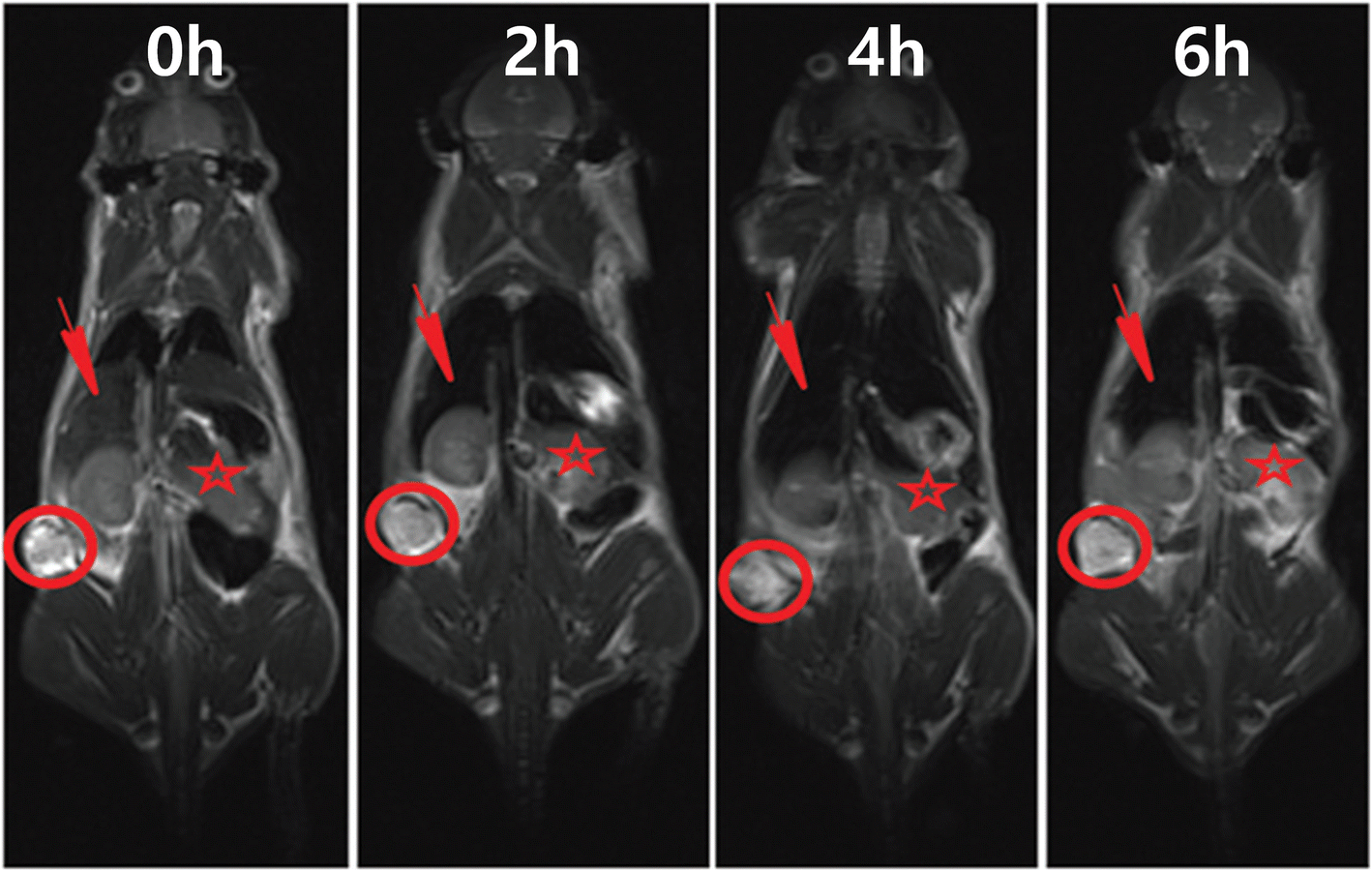
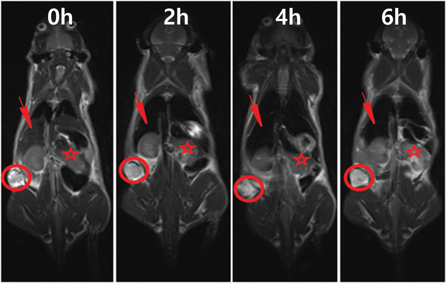
High-spin paramagnetic metals such as gadolinium (Gd3+), manganese (Mn2+), or iron (Fe3+) are used commonly as T 1 contrast agents, while ferro- and ferrimagnetic or superparamagnetic materials such as iron oxide (Fe3O4) have been used as T 2 contrast agents. These T 1 and T 2 contrast agents themselves do not show any targeting effect, and they are also excreted quickly by short blood circulation time in the body due to small particle sizes (<50 nm). As a result, clay minerals (Laponite®, halloysite, and LDH) immobilized with MR contrast agents have been studied for MR imaging in order to improve the passive targeting effect and blood circulation time (Table 5). After hybridizing commercially available Laponite® clay with superparamagnetic Fe3O4 nanoparticles having large T 2 relaxivity, the Laponite®-Fe3O4 hybrid was injected into the HeLa (human cervical carcinoma cell) xenograft mice model. Surprisingly, bright and dark MR images were observed, respectively, in tumor and liver tissues after 2 h, 4 h, and 6 h (Fig. 11) (Ding et al., Reference Ding, Hu, Luo, Zhu, Wu, Cao, Peng, Shi and Guo2016). LDH clay was also doped with Gd3+ ions (LDH-Gd), well known as a MR contrast agent, and demonstrated on 4 T1 murine breast tumor-bearing mice for T 1-weighted MR images related to the spin-lattice relaxation mode. MR signal intensity of LDH-Gd was increased gradually from 4688.7 to 4904.5 and 5166.7, respectively, 1 h and 4 h after injection, indicating that LDH-Gd could be an effective MR imaging system for tumors (Wang et al., Reference Wang, Xing, Zhang, Ren, Pan, Zhang, Bu, Zheng, Zhou, Peng, Hua and Shi2013). In conclusion, clay minerals, whether they are cationic or anionic, are potential carriers for MR contrasting agents due to their biocompatibility and passive targeting effect.
In vivo T 2-weighted MR images of tumor (red circle), liver (red arrow), and kidney (red star) after intravenous injection of Laponite®-Fe3O4 nanoparticles for 0 h, 2 h, 4 h, and 6 h (after Ding et al. Reference Ding, Hu, Luo, Zhu, Wu, Cao, Peng, Shi and Guo2016, reproduced with the permission of the Royal Society of Chemistry)
Transdermal medicine
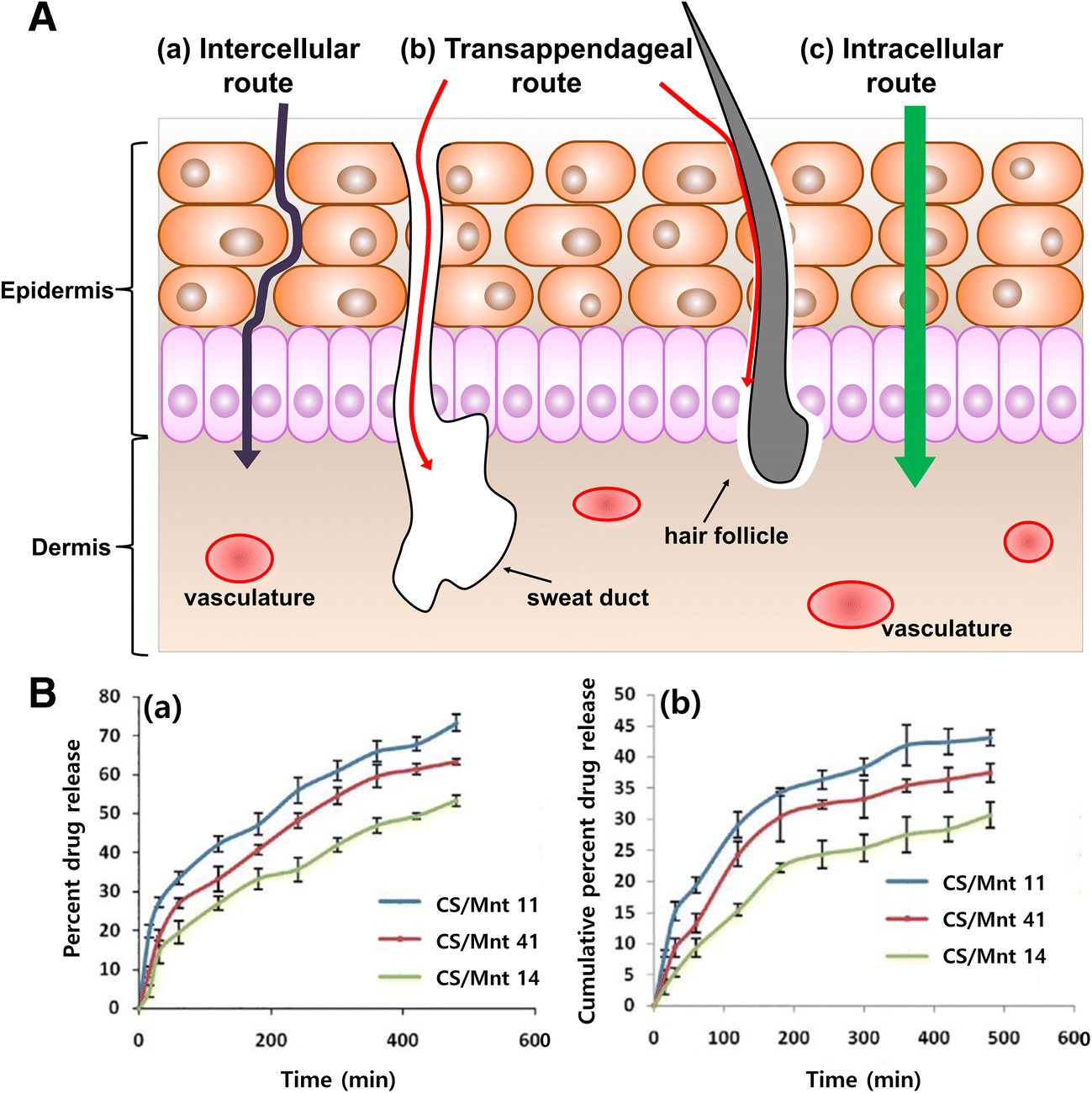
Transdermal delivery refers to delivery of drug molecules across the skin into the blood circulation at a fixed rate (Marwah, Garg, Goyal, & Rath, Reference Marwah, Garg, Goyal and Rath2016; Lee et al., 2017). Three routes are possible for drug delivery across intact skin, namely the intercellular, intracellular, and trans-appendiceal pathways (Fig. 12a). The intercellular and intracellular pathways involve passage through the stratum corneum, an architecturally diverse, multi-layered, and multi-cellular barrier. The intercellular route is the common pathway, allowing diffusion of lipophilic or non-polar solutes across the continuous lipid matrix. The intracellular route through corneocytes, terminally differentiated keratinocytes, however, allows the transportation of hydrophilic or polar solutes. The transappendageal route is related to passage through sweat glands and across hair follicles, which provides a continuous channel for drug permeation but is blocked easily due to the presence of hair follicles and sweat ducts (Alkilani et al. Reference Alkilani, McCrudden and Donnelly2015; Marwah et al., Reference Marwah, Garg, Goyal and Rath2016). In practice, transdermal patches have been developed as adhesive ones that can be attached to the skin. The polymer matrix is one of the main components of the patch due to its flexibility and drug reservoir property (Prausnitz et al., Reference Prausnitz and Langer2008). To give an effective drug delivery property for transdermal medicine, the design of the drug delivery systems not only to have large drug-loading capacity but also to have sustained drug release behavior is very important. For this reason, clay minerals have been in the spotlight due to their large drug-loading capacity and controlled release behavior in the body fluid systems.
(A) Different transdermal pathways of clay nanoparticles. (B) (a) In vitro drug release, and (b) permeation behaviors of the chitosan/montmorillonite K transdermal films (after Thakur et al., Reference Thakur, Singh and Singh2016, reproduced with the permission of Wolters Kluwer Medknow Publications)
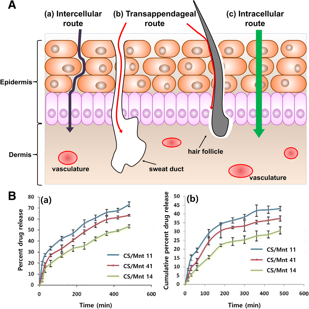
Chitosan/montmorillonite K 10 (CS/Mnt) clay minerals were prepared as a transdermal film type for curcumin delivery (Thakur et al. Reference Thakur, Singh and Singh2016). According to the release study of curcumin from the CS/Mnt films (Fig. 12B(a), the sustained release could be observed in three cases: 53.34 ± 1.26%, 63.23 ± 1.56%, and 73.27 ± 1.71% for CS/Mnt 14, 41, and 11, respectively (where 14, 41, and 11 indicate the ratios between CS and Mnt (1:4, 4:1, and 1:1)). For in vitro drug permeation studies of CS/Mnt films, CS/Mnt 11, CS/Mnt 41, and CS/Mnt 14 were used on rat skin with a size of 1 cm2 for 24 h. The drug releases of CS/Mnt 11, CS/Mnt 41, and CS/Mnt 14 films were 30.63 ± 2.04%, 37.45 ± 1.49%, and 43.03 ± 1.09%, respectively, after 8 h of permeation (Fig. 12B(b)). As a result, a permeation efficacy of 59% was found for the CS/Mnt 14 film, indicating that drug molecules indeed penetrated the skin by their controlled-release properties. In conclusion, a patch (or film) containing a drug-clay hybrid could be suggested as a promising transdermal material for biomedicine.
Summary and Perspectives
Clay minerals, including a variety of cationic and anionic clays, have been used as delivery carriers of therapeutic and diagnostic agents for nanomedicine including oral, injectable, and transdermal medications, etc. Depending on the nanomedical applications, clay minerals could be designed and functionalized by various synthetic routes and their special parameters eventually optimized, such as size, dispersibility, drug-release property, and chemical- and biostability, etc. Based on much experimental evidence from in vitro and in vivo studies as described in this tutorial review, such a proof of concept of clay delivery carriers has great implications for actual future application in nanomedicine. Many challenges remain, however, because the long-term in vivo evaluations of therapeutic efficacy and diagnostic properties, including bio-distributions and excretion mechanisms, of clay nanoparticles should be done under clinical-trial conditions, and their long-term toxicology studies are also needed.
Acknowledgments
This work was supported by the National Research Foundation of Korea (NRF) Grants funded by the Korean Government (MSIP) (No. 2017R1A6A3A11034149, No. 2016R1D1A1A02937308, and No. 2017K2A9A2A10013104).