Introduction
Thyroid haemorrhage is usually due to a recognised cause. Patients commonly present with an acute, painful neck swelling. Very rarely, there is significant neck swelling and airway compromise.
Using the key words ‘thyroid gland’, ‘thyroid cyst’, ‘haemorrhage’, ‘haematoma’ and ‘swelling’, we searched PubMed Medical Subject Headings up to September 2011. We found only one case report of spontaneous rupture of a nodular goitre secondary to acute haemorrhage, presenting as a large neck mass with dyspnoea.Reference Chang, Chou, Tiu, Chiou, Wang and Chiou1
Case report
A 93-year-old woman presented to the accident and emergency department with a 3-hour history of acute external neck swelling, dysphagia, odynophagia and a non-productive cough. She was unable to swallow her own saliva. There was no obvious precipitant. She had no prior difficulty in deglutition. She was otherwise systemically well, with no fever or shortness of breath. She had no known history of thyroid or neck disease. She was not taking anti-coagulants.
On examination, the patient was able to speak in full sentences and maintain her own airway. Her blood oxygen saturation was 99 per cent on air and her respiratory rate was 16 breaths per minute. There was no stridor. She had a 5 cm swelling extending from the midline to the left side of the neck. The lump was well demarcated, firm, tender and moved with deglutition. It was non-pulsatile, with no bruit audible on auscultation. The oropharyngeal examination was normal.
Flexible nasolaryngoscopy revealed a significant left hypopharyngeal wall swelling with medialisation.
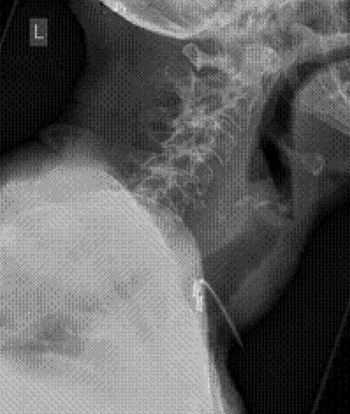
The patient was investigated initially with a lateral soft tissue neck X-ray taken in the accident and emergency department (Figure 1). This showed a soft tissue mass in the root of the neck, displacing the trachea anteriorly. Calcification within the mass suggested it was long-standing and probably of thyroid origin, given the location.
Lateral neck X-ray showing a soft tissue mass in the root of the neck displacing the trachea anteriorly. Calcification within the mass suggests it is long-standing and probably of thyroid origin given the location. L = lateral

An urgent ultrasound suggested a long-standing thyroid cyst.
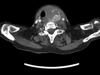
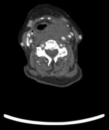
An urgent computed tomography scan of the neck and chest showed acute bleeding into a thyroid cyst, with secondary tissue swelling causing a considerable mass effect and tracheal deviation anteriorly and to the right (Figures 2 and 3).
Axial computed tomography neck scan with contrast, showing a mass effect with tracheal deviation to the right and loss of the normal fat planes.

Axial computed tomography scan at the supraglottic level, showing blood tracking round into the retropharyngeal space, compressing and effacing the pharynx and paralaryngeal fat and displacing the carotid sheath posterolaterally.

A 21 Ch gauge needle was used to aspirate the swelling. Fourteen millilitres of old blood was aspirated, with significant swelling reduction.
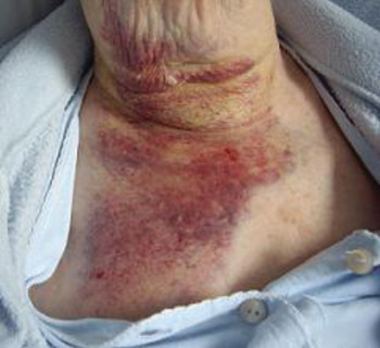
The patient was admitted for observation and further treatment consisting of intravenous hydrocortisone 200 mg 8-hourly and intravenous fluid maintenance. Within 2 days, the swelling had completely subsided, with residual bruising of the anterior neck and chest (Figure 4).
Clinical photograph showing post-aspiration anterior chest wall bruising within the subplatysmal plane.

The patient was fit for discharge 3 days later, and was referred to an ENT consultant with a thyroid interest for definitive management. Upon review, it was decided to manage the cyst conservatively.
Discussion
Thyroid cysts are common: up to 50 per cent of the adult population have palpable cysts.Reference Buzduga, Mogos, Galesanu, Vulpoi, Ungureanu and Cristea2 Paleri et al. have described a case of spontaneous haemorrhage into the retropharyngeal and parapharyngeal spaces secondary to bleeding from a thyroid cyst.Reference Paleri, Maroju, Ali and Ruckley3 Forty cases of non-traumatic retropharyngeal and parapharyngeal haematomas have been reported. However, there have been no reports describing patients presenting with hypopharyngeal swelling secondary to a spontaneous thyroid cyst haemorrhage.
• Spontaneous thyroid cyst haemorrhage can cause hypopharyngeal swelling and potential airway compromise
• Prompt diagnosis and treatment can prevent fatal airway obstruction
Our case is unusual because of the risk of airway compromise at the level of the hypopharynx secondary to a thyroid cyst haemorrhage. Management, as in this case, is straightforward if the correct diagnosis is made quickly.