Introduction
The Atlantic Forest biome is distributed along the eastern coast of South America, predominantly in Brazil, extending from Rio Grande do Norte to Rio Grande do Sul states, reaching the central region of the country in the states of Minas Gerais, São Paulo, Paraná, Bahia, Espírito Santo, and Santa Catarina. It comprises coastal plains, the Serra do Mar and Serra da Mantiqueira mountains, and highland plateaus, ranging in altitude from sea level to over 2,000 m. The region has a tropical humid to subtropical climate with high levels of rainfall. Owing to its wide latitudinal and altitudinal range, the biome encompasses several ecosystems, including dense ombrophilous forests, semideciduous seasonal forests, mixed ombrophilous forests (Araucaria Forest), restinga, mangroves, and high-altitude grasslands. Despite its exuberance and recognition as a global hotspot of biodiversity, the biome is threatened by deforestation, urbanization, habitat fragmentation, and agricultural expansion.
Moreover, deforestation has reduced the Atlantic Forest to only approximately 12% of its original extent (Sobral-Souza et al. Reference Sobral-Souza, Stropp, Santos, Prasniewski, Szinwelski, Vilela, Freitas, Ribeiro and Hortal2021), and habitat fragmentation has led to a decline in endemic species and, consequently, their parasites (Myers et al. Reference Myers, Mittermeler, Mittermeler, Da Fonseca and Kent2000). Habitat fragmentation in the Atlantic Forest significantly alters the distribution of mammalian species and the composition of helminth communities, thereby constraining the accurate assessment of parasitological patterns and host–parasite dynamics within these ecosystems (Lúcio et al. Reference Lucio, Gentile, Cardoso, Santos, Teixeira, Maldonado Júnior and D’Andrea2019).
The genus Akodon Meyen, 1833 (Rodentia: Cricetidae: Sigmodontinae) comprises 42 species and is widely distributed across the Neotropical region of South America (Brandão et al. Reference Brandão, Carmignotto, Percequillo, Christoff, Mendes-Oliveira and Geise2022; Labaroni et al. Reference Labaroni, Paez Coll Mairhofer, Ojeda, Novillo, Teta, Jayat, Ojeda, Buschiazzo, Cálcena, Bolzan and Lanzone2023; MDD, 2026). In Brazil, 11 species of Akodon are distributed (Abreu et al. Reference Abreu, Casali, Garbino, Loss, Moras, Nascimento, Oliveira, Pavan, Percequillo and Nascimento2025; Paglia et al. Reference Paglia, Fonseca, Rylands, Herrmann, Aguiar and Chiarello2012), including the cursorial grass mouse Akodon cursor (Winge, 1888), the most prevalent rodent in the coastal areas of the Atlantic Forest, occupying both preserved habitats and areas subjected to anthropogenic disturbance (Simões et al. Reference Simões, Souza, Maldonado Júnior and Luque2011).
The trematodes of the family Dicrocoeliidae Looss, 1899, comprise 48 genera and over 400 species (Pojmańska Reference Pojmańska, Bray, Gibson and Jones2008). The genus Platynosomoides Yamaguti, Reference Yamaguti1971, is known to parasitize the bile ducts and liver of placental mammals (Guerreiro Martins et al. Reference Guerreiro Martins, Panisse, Robles, Diaz and Navone2022). The life cycle of Platynosomoides spp. is indirect, with invertebrates, usually snails and arthropods, acting as primary and secondary hosts (Pojmańska Reference Pojmańska, Bray, Gibson and Jones2008; Yamaguti Reference Yamaguti1971). The diagnosis of the genus is based on a combination of key morphological features, including the orientation of the testis, location of the genital pore, arrangement of the vitelline bands, and the relative size of the ventral sucker (Pojmańska Reference Pojmańska, Bray, Gibson and Jones2008). Currently, three species of Platynosomoides are recognized: P. muris (Stcherbakova, Reference Stcherbakova1942) infecting the coxing’s white-bellied rat Niviventer coninga (Swinhoe, 1864), endemic to Taiwan; P. verschureni (Baer, Reference Baer1959) described as parasitizing shrews (Soricidae) in the Belgian Congo, Africa; and P. lunaschiae Guerreiro Martins, Reference Guerreiro Martins, Panisse, Robles, Diaz and Navone2022 parasitizing the sigmodontine rodent Akodon montensis Thomas, 1913 in Argentina (Baer Reference Baer1959; Guerreiro Martins et al. Reference Guerreiro Martins, Panisse, Robles, Diaz and Navone2022; Stcherbakova Reference Stcherbakova1942).
During a survey to assess the diversity of small mammals and their parasites in Itatiaia National Park (PNI), several trematode flatworms were recovered from the bile ducts of cursorial grass mice, Akodon cursor. The flukes were examined using light and scanning electron microscopy, and molecular phylogenetic analyses were performed to determine the phylogenetic position of the genus within Dicrocoeliidae.
Material and methods
This study was conducted as part of a broader study on the small mammal fauna of Itatiaia National Park (PNI), situated between the states of Minas Gerais and Rio de Janeiro in Brazil. As a crucial area for Atlantic Forest protection, PNI is included in the strategic conservation initiative and is recognized as a part of the Atlantic Forest Biosphere Reserve (RBMA 2023). Field surveys were conducted from May 24 to June 4, 2022, in Montane Ombrophilous Dense Forest areas located at elevations between 500 and 1,500 m in PNI (22°14′33″–22°27′54″S; 44°34′04″–44°46′11″W). The climate of PNI is strongly influenced by altitude and orographic effects. Annual rainfall averages 1,500–1,800 mm at approximately 1,000 m reaching 2,000–3,000 mm in high-altitude grasslands around 2,000 m (Aximoff and Rodrigues Reference Aximoff and Rodrigues2011). Mean annual temperatures at the plateau reach 11.4°C, with extremes from 21.4°C in summer to –15.4°C in winter (Santos Reference Santos2000).
Akodon cursor specimens were captured using Sherman (7.62 cm × 9.53 cm × 30.48 cm) live-traps, suitable for capturing small mammals. Mammals were anesthetized, sexed, and biometric data were recorded before euthanasia under veterinary supervision. Necropsies were performed for helminth recovery, and voucher specimens were taxidermized and deposited in the Integrated Collection of Wild Reservoir Mammals (COLMASTO, formerly LBCE n° 23053) of the Oswaldo Cruz Institute, Oswaldo Cruz Foundation (IOC/Fiocruz). This study was approved by the Ethics Committee on the Use of Animals of the Oswaldo Cruz Institute (CEUA/IOC, protocol no. L-038/2018), and by the Chico Mendes Institute for Biodiversity Conservation (ICMBio), under authorization N° 74498-1 in accordance with Brazilian legislation regulating the ethical use of mammals in scientific research.
The helminths were fixed in 70% ethanol for morphological analyses. For taxonomic identification, the trematodes were stained with hydrochloric carmine, dehydrated using an alcohol series, cleared in eugenol, and examined under a light microscope (Olympus BX51). Morphological identification was performed following the keys of Travassos (Reference Travassos, Freitas and Kohn1969), Yamaguti (Reference Yamaguti1971), Bray et al. (Reference Bray, Gibson and Jones2008), and other relevant articles. The prevalence (P), mean intensity (MI), and mean abundance (MA) were calculated according to Bush et al. (Reference Bush, Lafferty, Lotz and Shostak1997).
Optical microscopy images were obtained using Olympus cellSens Standard software. All morphological characters were measured. Measurements are given in millimetres unless otherwise stated, with the mean followed by the range in parentheses. The scales in the figures are expressed in micrometres (μm). The measurements were based on eleven individuals. The relative size of the testes was expressed as the ratio of testicular width to total body width, and the proportion of suckers was calculated using the ventral/oral sucker maximum diameter ratio.
For SEM analysis, five trematode individuals were dehydrated through a graded ethanol series (30–100%) for 2 h prior to critical point drying using CO2 (CPD 030, Balzers, Switzerland). The trematodes were placed on aluminium stubs, coated with a 20-nm gold layer, and observed with a AURIGA 600 Compact Zeiss (Zeiss, Germany) at the Unidade de Pesquisa Urogenital, Universidade do Estado do Rio de Janeiro, and Jeol JSM 6390LV SEM (operating at 15 kV) (JEOL, Akishima, Tokyo, Japan) at the Rudolf Barth Electron Microscopy Platform, Instituto Oswaldo Cruz, Rio de Janeiro.
For molecular genetic analyses, total genomic DNA was isolated from a pool of five trematodes using the QIAamp DNA Mini Kit, following the manufacturer’s protocol (QIAGEN, Hilden, Germany). DNA concentration and purity were measured using a NanoDrop Lite Plus Spectrophotometer (Thermo Fisher Scientific, Wilmington, DE, USA). Amplification of the 18S rRNA gene was performed using four distinct primers: the forward primer 5′-CATTAAATCAGCTATGTTCCTTAG-3′ (primer A) and the reverse primer 5′-TGGATCGCGCCAGACAAACTACAG-3′ (primer C); and the reverse primers 5′-CGGTATTCTGATCGTCTTCGAACCTC-3′ (primer B) and 5′-AATGATCAAGTTTGGTCGTCTTCTC-3′ (primer D). Additionally, we used the forward primer 5′-CATTAAATCAGCTATGTTCCTTAG-3′ (primer A) and the reverse primer 5′-AATGATCAAGTTTGGTCGTCTTCTC-3′ (primer D) to obtain a larger region of the gene. These primers were specifically designed based on conserved regions of the 18S rRNA gene of the trematode family Dicrocoeliidae by Cai et al. (Reference Cai, Zhang and Ye2012).
Amplicons were cycle sequenced using the Big Dye Terminator v3.1 Cycle Sequencing Kit (Applied Biosystems, USA) on both strands using only the PCR primers C and D, resulting in bidirectional sequencing with improved data accuracy. Sequencing was performed using an ABI3730 DNA Analyzer. Both procedures and cycle-sequenced products precipitation were conducted at the Platform of the Oswaldo Cruz Foundation for DNA sequencing by capillary electrophoresis (P01-001-RPT/FIOCRUZ).
Each PCR reaction had a final volume of 25 μL, comprising 3 μL (~30 ng/μL) of total genomic DNA, 12.5 μL of PCR Master Mix (Promega Corporation, Madison, USA), 8.5 μL of DNA-free water, and 0.5 μL of each forward and reverse primer (10 μM). The thermocycling conditions included initial denaturation at 95°C for 4 min, followed by 35 cycles of denaturation at 94°C for 1 min, annealing at 58°C for 40 s, and extension at 72°C for 1 min, with a final elongation step at 72°C for 7 min. PCR products were visualized by electrophoresis on a 1.5% agarose gel stained with GelRed nucleic acid gel stains (Biotium, Hayward, California, USA) and examined under UV transiluminator. Amplified DNA fragments were purified using the direct purification with the Illustra GFX PCR DNA and Gel Band Purification Kit according to the manufacturer’s protocol (GE Healthcare, Little Chalfont, UK). Amplicons were cycle-sequenced using the Big Dye Terminator v3.1 Cycle Sequencing Kit (Applied Biosystems, USA) on both strands using the PCR primers mentioned above, resulting in bidirectional sequencing for improved data accuracy. Sequencing was performed using an ABI3730 DNA Analyzer. Both procedures and cycle-sequenced products of precipitation were conducted at the Platform of the Oswaldo Cruz Foundation for DNA sequencing by capillary electrophoresis (P01-001-RPT/FIOCRUZ). The resulting sequences were validated using the NCBI BLAST tool to confirm the target specificity. The nucleotide sequence obtained in this study was deposited in the GenBank database under accession number PX233969.
Phylogenetic analyses of the 18S rRNA gene sequences were performed using MEGA version 11 (Tamura et al. Reference Tamura, Peterson, Peterson, Stecher, Nei and Kumar2011). Multiple sequence alignments were conducted using the ClustalW algorithm implemented in MEGA software. Phylogenetic reconstruction of the 18S rRNA gene matrix, including sequences of Platinosomoides macrotestis sp. nov. (accession number PX233969), was carried out based on reference sequences of species of the family Dicrocoeliidae retrieved from GenBank. The best-fitting nucleotide substitution model was selected in MEGA using the “Find Best DNA/Protein Models (ML)” tool and subsequently applied in Maximum Likelihood (ML) analyses, with branch support values estimated. Bayesian inference (BI) was performed using MrBayes 3.2.6 (Ronquist et al. Reference Ronquist, Teslenko, van der Mark, Ayres, Darling, Höhna, Larget, Liu, Suchard and Huelsenbeck2012), implemented as a plugin in Geneious Prime 2025.2 (Kearse et al. Reference Kearse, Moir, Wilson, Stones-Havas, Cheung, Sturrock, Buxton, Cooper, Markowitz, Duran, Thierer, Ashton, Meintjes and Drummond2012), with nodal support values also estimated. Markov chain Monte Carlo (MCMC) samplings were performed for 10,000,000 generations, with four simultaneous chains, in two runs. Branch supports were assessed by Bayesian posterior probabilities (BPP), calculated from trees sampled every 1,000 generations, after a 25% fraction burn-in removal. For the phylogenetic tree rooting, a sequence of Fasciola hepatica (Linnaeus, 1758), belonging to the suborder Echinostomata (Looss, 1902), was used as an outgroup.
The nomenclatural acts have been registered in ZooBank (LSID: urn:lsid:zoobank.org:act:50A21362-A42F-44E0-AE8E-336CC6D3E8C0)
Results
Order Plagiorchiida La Rue, 1957
Suborder Xiphidiata Olson, Cribb, Tkach, Bray & Littlewood, 2003
Superfamily Gorgoderoidea, Looss, 1901
Family Dicrocoeliidae Looss, 1899
Genus Platynosomoides Yamaguti, Reference Yamaguti1971
Platynosomoides macrotestis sp. nov.
Platynosomoides macrotestis sp. nov. Description based on 11 individuals; measurements are listed in Table 1. Body elongated, fusiform, posteriorly tapered, 2.05 (1.70–2.54) long, 0.76 (0.58–0.94) wide (Figs. 1a, 2a). The muscular suckers were well-developed (Fig. 2b). Oral sucker subterminal, rounded, 0.19 (0.14–0.23) × 0.095 (0.05–0.16), margins poorly defined (Fig. 2c). Ventral sucker rounded, 0.33 (0.30–0.38) × 0.20 (0.16–0.24), ~60% larger than oral sucker, located in proximal third of body, edges prominent (Figs. 2b, 2d). The anterior region had inconspicuous cuticular papillae (Fig. 2c). Pharynx well developed, strongly muscular, 0.12 (0.08–0.19) × 0.12 (0.09–0.15), followed by short oesophagus (Figs. 1b, 1c). Genital pore at the pharyngeal level (Figs. 1c, 2c). Cecal bifurcation in forebody, anterior to ventral sucker. Ceca were long and extended near the posterior extremity (Figs. 1a, 2a).
Morphological measurements of Platynosomoides sp. in rodents

Platynosomoides macrotestis sp. nov. Light microscope image: a) complete adult flatworm, ventral view showing oral sucker (Os), pharynx (Ph), right testis (Rt), left testis (Lt), ovary (Ov), seminal receptacle (Sr), uterus (U), cecum (Ce), and excretory pore (Ep). Extracellular vitelline bands (Vb) are indicated (arrowheads); b) detail of oral (Os) and ventral (Vs) suckers, pharynx (Ph), cirrus (c), cirrus sac (Cs), and internal seminal vesicle (isv); c) detail of the cirrus sac (Cs) with the internal seminal vesicle (isv), external seminal vessel (esv), cirrus (c), muscle pharynx (Ph), and genital pore (gp); d) middle third region showing the ovary (Ov), seminal receptacle (Sr), ootype (oo), oviduct (ovd), and uterus (U). e) Posterior region detailing the excretory pore (Ep).

Platynosomoides macrotestis sp. nov. Scanning electron microscopy (SEM) image: a) ventral view showing the full body; b) anterior region detailing the oral sucker (Os) and ventral sucker (Vs); c) anterior end highlighting the cephalic papillae (arrows) and cirrus (c) protruding from the genital pore (arrowhead); d) detail of ventral sucker (Vs).

The testes were rounded and immediately posterolateral, overlapping the distal half of the ventral sucker (Fig. 1a). Right testis 0.26 (0.22–0.30) × 0.24 (0.15–0.30); left testis slightly more posterior, 0.26 (0.23–0.31) × 0.25 (0.17–0.32) (Fig. 1d). Cirrus sac 0.13 (0.11–0.14) × 0.10 (0.09–0.11), containing internal seminal vesicle (Figs. 1c, 3b); external seminal vesicle slightly curved (Figs. 1c, 3b). Cirrus claviform (Figs. 1c, 2b). Ovary rounded, 0.14 (0.08–0.19) × 0.12 (0.08–0.16), immediately posterior to testes (Figs. 1a, 1d). Seminal receptacle well developed, posterior and slightly lateral to ovary (Fig. 1d). The uterus diffuse and occupying the intercecal space (Fig. 1a). The extracecal, asymmetrical vitelline glands began at the ventral sucker level; the left vitellarium extended to the mid-region of the second body third, and the right vitellarium reached the anterior margin of the posterior third (Fig. 1a). Excretory pore subterminal (Fig. 1e). Vitelline bands not visible in Fig. 1a but extend to the mid-body, as confirmed by microscopy; the extent is shown in Fig. 3a.
Platynosomoides macrotestis sp. nov. Line drawing; a) ventral view of the adult specimen; b) details of cirrus sac (Cs) and cirrus (c) with the internal (isv) and external (esv) seminal vesicles; c) details of the ovarian complex, showing the ovary (ov), oviduct (ovd), seminal receptacle (sr), and ootype (oo).

The SEM images (Fig. 2) provide insights into the morphology of the oral and ventral suckers of Platynosomoides macrotestis sp. nov. The oral sucker displayed poorly defined margins, with its lobes clearly invaginating into the structure and a transverse opening (Fig. 2b). In contrast, the ventral sucker was characterized by elevated, well-defined circumferential edges and two lobes that joined vertically at the centre (Fig. 2d). Additionally, the cirrus was identifiable, exhibiting a claviform (club-shaped), slightly flattened, and swollen structure with a smooth surface (Fig. 2c). However, limitations in image resolution and specimen preservation hindered a more precise morphological descriptions.
Taxonomic summary
Species: Platynosomoides macrotestis sp. nov.
Type host: Akodon cursor (Winge, 1888); voucher n° LBCE 23053
Locality: Itatiaia National Park, Atlantic Forest of Rio de Janeiro, Brazil
Site of infection: Bile duct
Prevalence: 4% (1 rodent infected/ 25 rodents collected)
Mean intensity: 41 (41 trematodes in one infected rodent)
Mean abundance: 1.64 (41 trematodes in the 25 rodents collected)
Etymology: The specific epithet refers to the prominent anatomical features that distinguish this species.
Deposited specimens: Holotype (CHIOC No. 39817a) and paratype (CHIOC No. 39817b).
Molecular genetic characterization
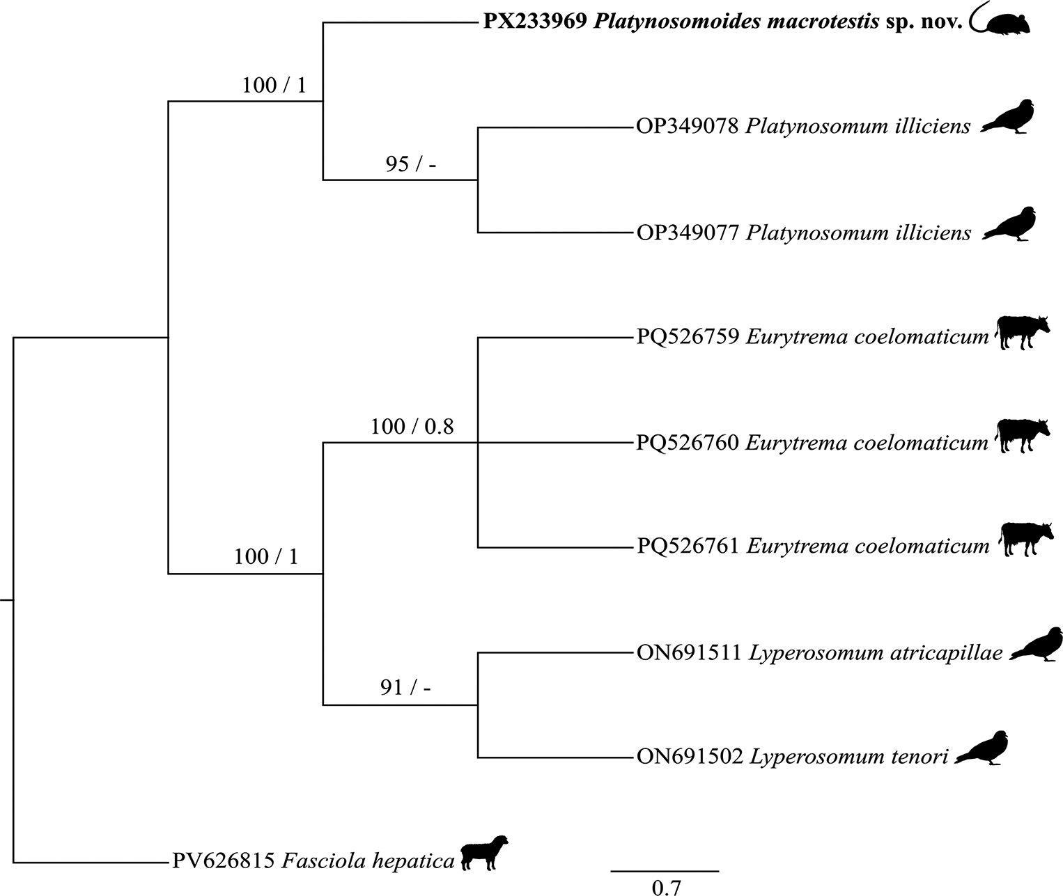
A partial consensus sequence of the 18S rRNA gene of Platynosomoides macrotestis sp. nov., generated in the present study, comprised 751 bp and was deposited in the GenBank database (http://www.ncbi.nlm.nih.gov) under accession number PX233969 (Table 2). Alignment of this sequence with other taxa, after trimming terminal regions, resulted in a data matrix including nine taxa. The final alignment consisted of 434 bp, containing 97 conserved sites, 312 variable sites, and 209 parsimony-informative sites. The Kimura two-parameter model (K2) was selected by MEGA as the best-fitting nucleotide substitution model for this dataset. Bayesian Inference was conducted in Geneious Prime using the MrBayes plugin, yielding a mean marginal likelihood estimate of −1708.546 and a median of −1708.212. The effective sample size (ESS = 3493.309) indicated high sampling efficiency and adequate chain convergence, supporting the robustness of all parameter estimates. The sequenced specimen of Platynosomoides macrotestis sp. nov. clustered within a well-supported monophyletic clade comprising Platynosomum illiciens (OP349077; OP349078) (MLBP = 100%, BPP = 1.0) (Fig. 4). The internal node connecting the two Platynosomum sequences received strong ML support (MLBP = 95%, BPP = 0.6). Similarly, sequences of Eurytrema coelomaticum (PQ526759, PQ526760, and PQ526761) formed a well-supported monophyletic group (MLBP = 100%, BPP = 0.8). The genus Lyperosomum also formed a distinct monophyletic clade composed of L. atricapillae (ON691511) and L. tenori (ON691502), with moderate support (MLBP = 91%, BPP = 0.6).
List of species, genetic markers, and corresponding GenBank accession numbers used in the phylogenetic analysis

Phylogenetic relationships among 18S rRNA gene sequences of selected species of the family Dicrocoeliidae were inferred, including the sequence of Platynosomoides macrotestis sp. nov. Phylogenetic reconstruction was performed using Maximum Likelihood (ML) and Bayesian Inference (BI) methods, and the topology obtained from BI is presented. Nodal support values are indicated as bootstrap percentages for ML (MLBP), shown to the left of the nodes, and posterior probabilities for BI (BPP), shown to the right. Only support values greater than 70% (MLBP) and 0.7 (BPP) are displayed. The symbol “–” indicates nodal support values below these thresholds.

The clade comprising Platynosomoides macrotestis sp. nov. and Platynosomum was recovered as the sister group to the cluster formed by Eurytrema and Lyperosomum, indicating that these lineages share a relatively recent common ancestor within the Dicrocoeliidae family.
Discussion
Currently, three species of Platynosomoides are recognized: P. muris (Stcherbakova, Reference Stcherbakova1942), which parasitizes Apodemus sylvaticus (Linnaeus, 1758) and Leopoldamys siporanus (Thomas, 1895) in Asia and Western Europe; P. verschureni (Baer, Reference Baer1959), found infecting Crocidura olivieri (Lesson, 1827) in the Congo; and P. lunaschiae (Guerreiro Martins et al., Reference Guerreiro Martins, Panisse, Robles, Diaz and Navone2022), identified in A. montensis in Argentina.
The taxonomy of the family Dicrocoeliidae remains complex, particularly concerning species classification within the genus Platynosomoides (Guerreiro Martins et al., Reference Guerreiro Martins, Panisse, Robles, Diaz and Navone2022). For example, P. muris was originally described as Skrjabinus muris by Stcherbakova in 1942, later reassigned to the genus Platynosomum by Skrjabin in 1952, and subsequently transferred to Platynosomoides by Yamaguti in 1971. However, Nguyen and Pham (Reference Nguyen and Pham2004) proposed a new taxonomic combination, reclassifying this species as Dictyonograptus muris comb. nov., based on morphological traits deemed characteristic of the genus Dictyonograptus, such as ovary morphology, the position of the genital pore anterior to the cecal bifurcation, and testis placement posterior to the ventral sucker. We did not adopt that reclassification, as the species exhibits diagnostic features consistent with Platynosomoides, in agreement with Yamaguti (Reference Yamaguti1971), including vitelline bands that extend from the level of the ventral sucker, well beyond the ovaries.
Similarly, P. verschureni was initially assigned to Platynosomum by Baer (Reference Baer1959), but Yamaguti (Reference Yamaguti1971) reclassified it to Platynosomoides based on characteristics such as elongated and narrow ceca extending well beyond the mid-body, testes located posteriorly and laterally to the ventral sucker, an ovary positioned posterior to the testes, and vitelline glands forming lateral bands beginning at the ventral sucker and extending beyond the ovary. These classifications were later supported by Pojmańska (Reference Pojmańska, Bray, Gibson and Jones2008), who provided diagnostic morphological keys for species identification within the genus Platynosomoides.
The morphological and morphometric characteristics of Platynosomoides macrotestis sp. nov., as observed through light microscopy and SEM, align well with the established features of the genus Platynosomoides in the family Dicrocoeliidae. These morphometric traits were compared with those of the three previously described Platynosomoides species (Table 1), further reinforcing their placement in the genus.
Consistent with these established characteristics, Platynosomoides species exhibit well-developed testes situated anterior to the ovary, a bifurcated cecum extending along the fusiform body of the parasite, a diffuse uterus, vitelline bands spanning approximately two-thirds of the lateral margins of the body, and a notably large and prominent ventral sucker (Pojmańska Reference Pojmańska, Bray, Gibson and Jones2008; Yamaguti Reference Yamaguti1971).
Platynosomoides macrotestis sp. nov. is distinguished by its shorter body length (1.7–2.54 mm) compared to P. muris (3.27–3.41 mm) and P. verschureni (3–4.5 mm). In Platynosomoides macrotestis sp. nov., the relative size of the testes in relation to the body width is noteworthy. The ratio of testis width to total body width was 0.32, whereas in P. lunaschiae and P. muris, this proportion was considerably lower, corresponding to 0.12 and 0.21, respectively. In addition, the testes of Platynosomoides macrotestis sp. nov. are conspicuously enlarged and partially overlap the posterior region of the ventral sucker, whereas those of P. lunaschiae are comparatively smaller and positioned strictly posterolateral to the ventral sucker. These proportional differences are corroborated by morphometric data, which indicate that the testis width in Platynosomoides macrotestis sp. nov. accounts for approximately one-third of the body width, in contrast to approximately one-eighth in P. lunaschiae.
Another consistent and diagnostic morphological feature is the relative sucker size. In Platynosomoides macrotestis sp. nov., the ventral sucker was markedly larger than the oral sucker, with a ventral sucker–to–oral sucker ratio of 1.73, and had well-defined, elevated circumferential margins. In contrast, in P. muris and P. lunaschiae, the ventral sucker is only slightly larger than the oral sucker, with ratios of 1.12 and 1.22, respectively, and lacks a pronounced marginal definition.
The ovary of Platynosomoides macrotestis sp. nov. (0.08–0.16 mm) was smaller than that of P. muris (0.23–0.24 mm). Moreover, a new species is characterized by long ceca extending to the posterior end of the body and asymmetrical extracecal vitelline bands originating at the ventral sucker level, occupying the middle third of the body, and extending posteriorly to the anterior limit of the final third (Fig. 1a). This contrasts with P. lunaschiae, where ceca and vitelline bands extend to three-quarters of the body length; P. muris, which has vitelline bands reaching full two-thirds of the body; and P. verschureni, in which both ceca and vitelline bands are limited to the middle of the body. The cirrus sac was significantly smaller in Platynosomoides macrotestis sp. nov. than in all other described species (Table 1). Compared to P. lunaschiae (Guerreiro Martins et al. Reference Guerreiro Martins, Panisse, Robles, Diaz and Navone2022), Platynosomoides macrotestis sp. nov. has a more developed seminal receptacle, with dimensions similar to those of the ovary and located posterior to it. Thus, based on the presented data, the trematodes were all identified as a new species within the genus Platynosomoides.
Platynosomoides macrotestis sp. nov. was found to parasitize the bile duct of A. cursor in the Atlantic Forest in the state of Rio de Janeiro, Brazil. In total, six Dicrocoeliidae species have been documented to parasitize rodents of the Akodontini tribe in South America: Canaania obesa Travassos, Reference Travassos1944 in A. cursor and A. montensis in Brazil (Maldonado et al. Reference Maldonado, Pinheiro, Simões and Lanfredi2010; Simões et al. Reference Simões, Souza, Maldonado Júnior and Luque2011), Platynosomoides sp. in A. montensis in Argentina (Guerreiro Martins et al. Reference Guerreiro Martins, Robles, Diaz, Panisse and Navone2019, Reference Guerreiro Martins, Panisse, Robles, Diaz and Navone2022; Panisse et al. Reference Panisse, Robles, Digiani, Notarnicola, Galliari and Navone2017), Yungasicola travassosi Gardner & Pérez-Ponce, Reference Gardner and Pérez-Ponce de León2002 described from Akodon fumeus (Thomas, 1902) and Akodon mimus (Thomas, 1901) from Bolivia (Gardner and Pérez-Ponce de León Reference Gardner and Pérez-Ponce de León2002), Conspicuum pulchrum Travassos, 1919 in Akodon spp. in Brazil (Travassos Reference Travassos1944), Conspicuum minor Mane Garzon & Holcman-Spector, Reference Mañé-Garzón and Holcman-Spector1975 parasitizing Scapteromys aquaticus (Thomas, 1920) in Argentina (Sutton Reference Sutton1998), and Skrjabinus oxymycterae Sutton, 1983 in Akodon azarae (Fischer, 1829) also in Argentina (Sutton Reference Sutton1983).
Canaania obesa has previously been described in A. cursor in the Atlantic Forest, where it infects the bile duct (Cardoso et al. Reference Cardoso, Costa-Neto, Braga, Weksler, Simões, Maldonado Júnior, Luque and Gentile2020). Subsequently, dicrocoeliids were reported in other sigmodontine rodents, such as A. montensis, Oligoryzomys nigripes Olfers, 1818, and Nectomys squamipes Brants, 1827, suggesting that rodents of the genus Akodon are among the primary hosts (Cardoso et al. Reference Cardoso, Costa-Neto, Braga, Weksler, Simões, Maldonado Júnior, Luque and Gentile2020; Maldonado et al. Reference Maldonado, Pinheiro, Simões and Lanfredi2010). This fluke is characterized by a leaf-shaped body with pointed ends and a central ventral bulge, which is the widest at the acetabulum, located mid-body, and is two to three times larger than the subterminal oral sucker. It has long ceca that do not reach the end of the body, testes positioned posterior to the acetabulum, and a single lateral ovary opposite the testes. The vitellaria extend from the pre-acetabular region to approximately two-thirds of the body length, without overlapping the ceca ends. The excretory pore is subterminal, and the eggs are elliptical, operculated, and have knobs at the opposite end. These features are essential for species identification and distinguishing it from Platynosomoides macrotestis sp. nov., which has an elongated fusiform body shape, proportionally larger testes relative to the total body size, ovary positioned posterior to the testis and aligned along the median line of the body, and distinct size and distribution of the vitelline bands.
Despite the remarkable diversity of dicrocoeliid species infecting rodents in the Americas, no molecular data for this group were available in GenBank. In this context, the present study constitutes the first molecular record of dicrocoeliid trematodes infecting rodents in this region. Despite the limitations of the obtained sequences, this study provides novel genetic information for a taxonomic group that previously lacked molecular characterization, thereby contributing to the baseline for future phylogenetic and systematic investigations.
In conclusion, this study provides a morphological description by light and scanning electron microscopy of a new species of Platynosomoides, a parasite of the bile ducts in the cricetid rodent A. cursor from the Atlantic Forest. Our results strongly support the validity of Platynosomoides macrotestis sp. nov. and indicate the existence of a recent common ancestor with the genus Platynosomum, thereby expanding the current knowledge of the genus Platynosomoides and the Dicrocoeliidae family. These findings highlight the importance of preservation programs in the Atlantic Forest for maintaining helminth biodiversity and host–parasite ecological network.
Acknowledgements
Thanks to Fundação Carlos Chagas Filho de Amparo à Pesquisa do Estado do Rio de Janeiro (FAPERJ) and Conselho Nacional de Desenvolvimento Científico e Tecnológico (CNPq) for research support. We also would like to thank the Laboratório de Biologia e Parasitologia de Mamíferos Silvestres Reservatórios da Fundação Oswaldo Cruz (IOC/FIOCRUZ), Unidade de Pesquisa Urogenital (Universidade do Estado do Rio de Janeiro), and the Plataforma de Microscopia Eletrônica Rudolf Barth (IOC/FIOCRUZ).