Implications
Several studies have shown the existence of numerous dissimilarities in metabolic activity between Iberian and modern pig breeds that might be explained by genotypic differences. Primary hepatocyte cultures are a valuable tool to study specific metabolic liver functions, without the interference of changing levels of metabolites and hormones found in vivo. These cultures give more clear-cut results than freshly isolated cells, perfused liver, liver slices or in vivo experiments. Furthermore, primary cultures, unlike immortalized cell lines, retain the specific liver functions and characteristics of the donor animal.
Introduction
It is well established that genetic background of piglets have a deep impact on their growth and development, which may be mediated by differences in metabolism and nutrient partitioning between fat and protein accretion among pig breeds. Considering the vascular route followed by ingested nutrients, the liver, for its anatomic position and metabolic functions, plays an essential role in nutrient distribution to peripheral tissues. In terms of whole-body metabolism, the liver has a disproportionate influence with respect to its mass and under certain circumstances its high metabolic rate may compromise nutrient availability to the periphery.
Primary hepatocytes in monolayer culture is a good technique to study functional aspects of the liver where metabolic events can be studied in a defined, easily manipulated, population of cells over a period of days or even weeks, enabling long-term tests. Moreover, in animal science, hepatocytes are a valuable tool to elucidate regulation of metabolism by hormones or mechanism of action of nutrients or additives after digestive absorption by the animal. Nevertheless, there is a lack of information about differences in metabolic activity in hepatocytes from breeds with divergent metabolic profile. The Iberian pig is an obese slow growing Mediterranean breed with higher whole-body fat content than lean-type pigs and distinct hormone and metabolite profile (Fernández-Fígares et al., Reference Fernández-Fígares, Lachica, Nieto, Rivera-Ferre and Aguilera2007). When comparing breeds with disparate metabolic profiles it is presumable that hepatocytes may behave differently. In addition, porcine hepatocytes more often serve as a non-rodent model in pharmacology and toxicology research and they have not been extensively characterized. Therefore, the aim of the present experiment was to determine differences in metabolic activity in primary hepatocytes isolated from pure Iberian and Landrace pigs that may help to explain the divergent metabolic profile found in vivo.
Material and methods
Isolation of liver cells and cell culture
Care and treatment of all pigs were approved in advance by the Institutional Bioethical Committee of CSIC. Except where noted, reagents were purchased from Sigma Aldrich, Tres Cantos, Madrid, Spain. Hepatocytes were isolated from pure Iberian (n=10, 9 to 45 kg BW) and Landrace (n=8, 23 to 41 kg BW) pigs. Piglets were restrictively fed (85% ad libitum) a standard commercial diet. The day before slaughter they had free access to food. After stunning by electric shock they were exsanguinated and livers were immediately excised, the left lateral lobe removed and transported from the abattoir to the cell culture laboratory in ice cold sterile transport media (Minimum Essential Medium Eagle with Hanks’ salts Sigma M9288, 4.46 mM NaCO3H, 0.2 mM NaOH) as quick as possible (5 min). Hepatocytes were isolated essentially as published by Conde-Aguilera et al. (Reference Conde-Aguilera, Lachica, Nieto and Fernández-Fígares2012). Viability of isolated hepatocytes from animals was 81.4±4.7% (trypan blue dye exclusion) and cell yield from each liver preparation was 4.9±0.5×10⁸ viable hepatocytes (n=18).
Cell culture was described elsewhere (Conde-Aguilera et al., Reference Conde-Aguilera, Lachica, Nieto and Fernández-Fígares2012). Insulin (0.173 and 17.3 nM) and glucagon (0.287, 2.87 and 28.7 nM) concentrations are indicated for each assay. Media glucose concentration was 11.1 mM except for gluconeogenesis (GNG) assay. Duplicate flasks were used for each experimental condition.
GNG
Cells were cultured on day 1 for 24 h in serum-free basal medium with low insulin (0.173 nM)/glucagon (28.7 nM) ratio, mimicking fasting state, to deplete glycogen stores and washed twice with warm glucose-free 4-(2-hydroxyethyl)-1-piperazineethanesulfonic acid (HEPES)-buffered saline and subsequently cultured for 2 h in glucose free Dulbecco’s Modified Eagle’s medium (DMEM; Sigma D5030) containing amino acids (5.17 mM), lactate and pyruvate (0.5 mM) and a low insulin (0.173 nM)/glucagon (28.7 nM) ratio. GNG was estimated as glucose released to the media assuming negligible glycogenolysis (Conde-Aguilera et al., Reference Conde-Aguilera, Lachica, Nieto and Fernández-Fígares2012). Free glucose in the media was measured using an enzymatic method, Glucose Trinder GOD-POD (Labkit, Chemelex, S.A., Barcelona, Spain).
Glycogen degradation
Cells were cultured on day 1 for 24 h in culture medium with high insulin (17.3 nM)/glucagon (0.287 nM) ratio to achieve maximal glycogen content and two flasks harvested to set the starting point for glycogen degradation estimation. The rest of the flasks were washed twice with warm glucose free HEPES-buffered saline and cultured for 3 h in glucose-free DMEM with glucagon (2.87 nM) to ease glycogen degradation and insulin (0.173 nM). Cells were harvested and frozen at −80°C. Glycogen degradation was quantified as the difference in glycogen content of cells with full glycogen load (after 24 h culture with 17.3 nM insulin) and cells where degradation was facilitated (2.87 nM glucagon). Glycogen in cell homogenates was determined as described by Fernández-Fígares et al. (Reference Fernández-Fígares, Shannon, Wray-Cahen and Caperna2004), except that the enzymatic method used was Glucose Trinder.
Triglycerides (TG) content and esterification from fatty acids
Cells were cultured for 24 h in basal medium with high insulin (17.3 nM)/glucagon (0.287 nM) ratio and washed twice with warm HEPES-buffered saline. To study the effect of type of free fatty acid supplementation on TG esterification, cells were subsequently cultured for 96 h in the same media supplemented or not with CLA or LA (0.2 mM) as TG precursors. Lipids were extracted and dissolved in isopropanol. Intracellular TG content was determined using the glycerol determination procedure (Labkit, Chemelex, S.A.). Fatty acid esterification was estimated (Conde-Aguilera et al., Reference Conde-Aguilera, Lachica, Nieto and Fernández-Fígares2012) as the difference in TG content of cells cultured in media supplemented with CLA or LA (de novo lipogenesis plus esterification) and cells cultured in media without free fatty acids (de novo lipogenesis; basal).
Urea synthesis
On day 1, cells were cultured for 24 h in basal medium with high insulin (17.3 nM)/glucagon (0.287 nM) ratio, mimicking postprandial state, washed twice with warm HEPES-buffered saline and cultured in the same medium added with different concentrations of NH4 (0, 2.5, 5 or 10 mM) for 3 h. As a source of ammonia, NH4Cl was used. Rates of urea production (µg urea in the media/mg protein per hour) were estimated as the difference in urea concentration in the media between plates with 0 and 2.5, 5 or 10 mM ammonium. Urea was determined by UV/VIS spectrophotometry using an enzymatic procedure in which the urease reaction is coupled to the concurrent amination of α-oxoglutarate and the oxidation of NADH by glutamate. Disappearance of NADH is proportional to urea concentration in the media. The commercial kit used was Urease-GLDH Kinetic UV (Labkit, Chemelex, S.A.).
Albumin synthesis
Cells were cultured from day 1 for 24 h in bovine serum albumin (BSA) free basal medium with high insulin (17.3 nM)/glucagon (0.287 nM) ratio. Subsequently, flasks were washed twice with warm HEPES-buffered saline and cultured in BSA-free basal medium supplemented either with low insulin (0.173 nM)/glucagon (28.7 nM) or high insulin (17.3 nM)/glucagon (0.287 nM) ratio, mimicking fasting or postprandial state, respectively, and media collected after 24 h. Albumin concentration was measured in the culture media through an ELISA technique using a commercial kit (Pig albumin ELISA quantitation kit; Bethyl, Montgomery, AL, USA) according to the manufacturer protocol.
ß-hydroxybutyrate (BHB) synthesis
Cells were cultured from day 1 for 48 h in basal medium with high insulin (17.3 nM)/glucagon (0.287 nM) ratio, mimicking postprandial state, or low insulin (0.173 nM)/glucagon (28.7 nM) ratio, mimicking fasting state. Media was changed every 24 h and collected at 48 h. As ketone production was expected to be low in media from pig hepatocytes, a sensitive micro-method in 96-well plate format (Fernández-Fígares et al., Reference Fernández-Fígares, Shannon, Wray-Cahen and Caperna2004) was used to analyze BHB using a fluorescence microtitre plate reader (Victor X5 2030 Multilabel Reader; Perkin-Elmer, Turku, Finland). Data were expressed as nmol BHB/mg cell protein per 24 h.
IGF-1 synthesis
Cells were cultured in basal medium with high insulin (17.3 nM)/glucagon (0.287 nM) ratio for 48 h and media changed every 24 h following two washes with warm HEPES-buffered saline. After 48 h, media and cells were harvested and frozen at −80°C. Before analysis of IGF-1, media was concentrated 12× by means of a rotary evaporator (SpeedVac SPD131DDA-230; Thermo, Milford, MA, USA). A previously validated (Fernández-Fígares et al., Reference Fernández-Fígares, Lachica, Nieto, Rivera-Ferre and Aguilera2007) two-site immunoradiometric assay (cat. no. DSL-5600, Diagnostic Systems Laboratories Inc., Webster, TX, USA) was used to analyze IGF-1. The analytical procedure included an extraction step (acid-ethanol), in which IGF-1 was separated from its binding proteins, for detection of total IGF-1 in culture media. The assay used antibodies against human IGF-1 and human IGF-1 as the standard. The intra and inter-assay CV were 2.5% and 6.7%, respectively.
Protein determination
Protein in cell homogenates was determined by a modified Lowry procedure as described by Conde-Aguilera et al. (Reference Conde-Aguilera, Lachica, Nieto and Fernández-Fígares2012). All data were normalized to protein content.
Statistical analysis
In all, 10 Iberian and 8 Landrace cell preparations from individual pigs constituted an experiment, and treatments within each preparation were replicated in two flasks. All data from individual flasks were used in the ANOVA and analyzed using a mixed model analysis (SAS Institute 2004, version 9.1.3.). The factors introduced in the model were breed (Iberian or Landrace) for all traits analyzed; in addition, insulin/glucagon ratio was included for albumin and BHB synthesis data; concentration of ammonia in the media (0, 2.5, 5 and 10 mM) was included in the model for urea synthesis. All possible treatment combinations were tested. Results were expressed as least square means±s.e.m. of duplicate flasks and the number of individual preparations indicated. Statistical significance was assessed using Bonferroni multiple comparison test to determine differences between treatment groups. The level of significance was set to 5%. In addition, regression analysis was performed of ammonia concentration in the media on urea production using the REG procedure of SAS. Linear and exponential models were used. R 2 was used to evaluate the fit of the models.
Results and discussion
Some considerations were taken into account when designing the present experiment. It was a concern to be able to use piglets of comparable physiological characteristics for both breeds in order to compare their metabolic characteristics at the hepatic level. As there were no available breeders that weaned the piglets of Iberian and Landrace at the same time, it was decided to perform all the isolation from Landrace pigs and continue with Iberian pig isolations, although we would have preferred alternate isolation of hepatocytes of each breed. In addition, the developmental age of the animals of different breed may differ, and a decision must be made whether to use pigs of the same BW or age. The study was made on pigs of 10 to 50 kg BW because developmental age difference at this early state was relatively small. We designed the experiments so that aspects of glucose (GNG and glycogen degradation), protein (albumin synthesis and urea production) and fat (fatty acid esterification and BHB production) metabolism as well as hormone secretion (IGF-1) were evaluated. L-carnitine was routinely added to all culture media to ensure that fatty acid entry into mitochondria was not limited by carnitine availability and all fatty acids were complexed to albumin at concentrations that simulated physiological conditions (Stangl et al., Reference Stangl, Müller and Kirchgessner1999).
GNG and glycogen degradation
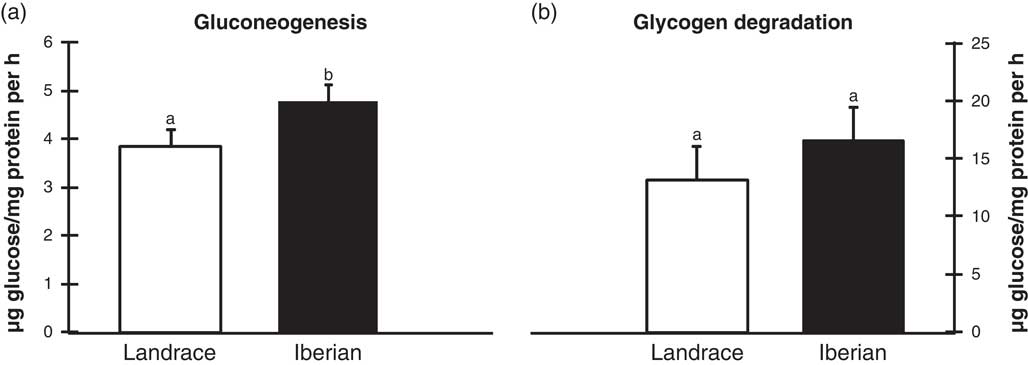
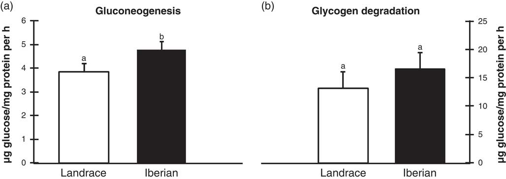
It was previously shown that differences related to genotype for glycemia and plasma insulin levels existed when Iberian and Landrace pigs were compared (Fernández-Fígares et al., Reference Fernández-Fígares, Lachica, Nieto, Rivera-Ferre and Aguilera2007). In the present study, incubating hepatocytes with glucagon (28.7 nM) for 24 h virtually depleted glycogen stores (data not shown). Although some glycogenolysis cannot be discarded, because of depletion of glycogen stores after pre-culture with glucagon (28.7 nM) for 24 h, media glucose was a consequence, essentially of GNG. We designed our experiment using a low insulin/glucagon ratio to detect differences between breeds in conditions that stimulate GNG. Hepatocytes from Iberian had greater glucose production from gluconeogenic precursors (24%, P<0.05; Figure 1a) than from Landrace pigs although no difference in glycogen degradation between breeds was found (P>0.10; Figure 1b). In line with our results, greater potential for GNG in Ossabaw compared with Yorkshire pigs has been reported (Kasser et al., Reference Kasser, Martin, Gahagan and Wangsness1981). As in the transition from fasted to fed liver glycogen deposition is predominantly a gluconeogenic process in rats (Katz et al., Reference Katz, Kuwajima, Foster and McGarry1986), we could speculate that hepatocytes from Iberian pigs would have greater glycogen synthesis as a result of their increased gluconeogenic capacity compared with Landrace. Unfortunately, we did not measure glycogen synthetic capacity in our experiments. In vivo, however, Iberian had lower fasting serum glucose and greater insulin concentration than Landrace growing gilts (Fernández-Fígares et al., Reference Fernández-Fígares, Lachica, Nieto, Rivera-Ferre and Aguilera2007). Similarly, serum glucose concentrations were generally lower in obese compared with lean pigs (Pond et al., Reference Pond, Yen, Lindvall and Hill1980). It has been suggested that seasonal variations in food availability, and therefore nutrient intake, may be related to the development of obesity in native pig breeds (McCusker et al., Reference McCusker, Wangsness, Griel and Kavanaugh1985). Iberian pigs have a high potential for fat accumulation (Nieto et al., Reference Nieto, Lara, Barea, García-Valverde, Aguinaga, Conde-Aguilera and Aguilera2012) and the ability to store excess fat during feed abundance enables the Iberian pigs survival during periods of scarcity. Low fasting glycemia of Iberian pigs compared with Landrace (Fernández-Fígares et al., Reference Fernández-Fígares, Lachica, Nieto, Rivera-Ferre and Aguilera2007) could be related to an increased basal and insulin-stimulated glucose transport early in the development of obesity, a common pattern in many animal models of obesity.
Effect of breed (Landrace or Iberian) on gluconeogenesis (a) and glycogen degradation (b) in porcine hepatocytes. For gluconeogenesis, cells depleted of glycogen were cultured in the presence of amino acids, lactate and pyruvate and a low insulin (0.173 nM)/glucagon (28.7 nM) ratio for 2 h and glucose measured in the media. For glycogen degradation, hepatocytes loaded with glycogen were cultured for 3 h in glucose-free media supplemented with glucagon (2.87 nM) and cells harvested for glycogen determination. Data are least square means and s.e.m. for duplicate cultures from 8 (Landrace) or 10 (Iberian) pigs. Means with different letters differ (P<0.05).
TG content and fatty acids esterification
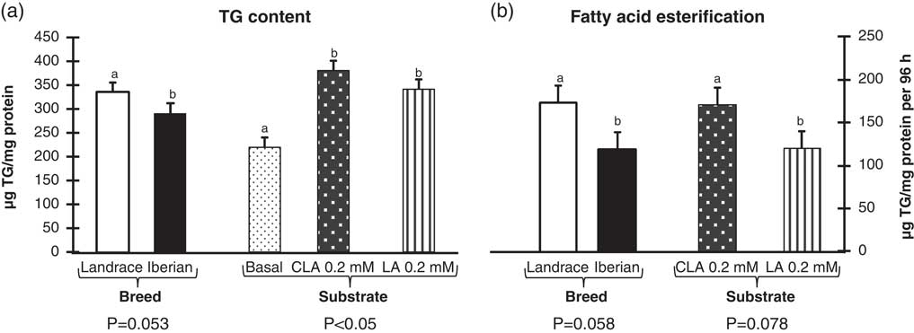
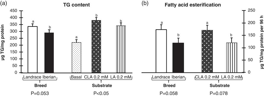
Although pig liver is a minor site of lipogenesis in mature pigs (Gondret et al., Reference Gondret, Ferré and Dugail2001), its importance is greater than in adipose tissue in pre-weaned piglets (Fenton et al., Reference Fenton, Roehrig, Mahan and Corley1985). Nonetheless, little attention has been paid to the study of liver lipogenesis in young pigs. In our study, difference in TG content of basal flasks was due essentially to lipogenesis from glucose or gluconeogenic substrates as they had no added free fatty acid in the media. On the other hand, flasks supplemented with free fatty acids, CLA or LA, derive their TG content both from de novo lipogenesis and esterification. This way, we estimated esterification by difference assuming that there was no effect of the presence of free fatty acids on de novo lipogenesis, as the design did not allow to study the effect of each fatty acid on de novo lipogenesis. Hepatocytes from Iberian had lower rate of fatty acid esterification (31%, P=0.058; Figure 2b) than hepatocytes from Landrace pigs, and consequently, they tended to have lower TG content (12%, P=0.053; Figure 2a). Addition of fatty acids, CLA or LA to the culture medium increased TG content compared with control (74% for CLA and 55% for LA, P<0.001; Figure 2a). Similarly, the rate of palmitate esterification in vitro was much greater than glucose carbon incorporation into total lipids in pigs (Go et al., Reference Go, Wu, Silvey, Choi, Li and Smith2012). It has been reported that CLA decreased fatty acid availability for TG synthesis in rat hepatocytes (Priore et al., Reference Priore, Giudetti, Natali, Gnoni and Geelen2007) and tended to depress hepatic lipid synthesis from glucose in pigs (Go et al., Reference Go, Wu, Silvey, Choi, Li and Smith2012). According to this, CLA-induced decrease of de novo lipogenesis in pigs, not directly measured in our assays, would imply that CLA esterification into TG was understimated compared with LA in our experiment.
Influence of breed (Landrace or Iberian) and substrate (basal, CLA or linoleic acid (LA)) on triglyceride content (a) and fatty acid estimated esterification (b) in porcine hepatocytes. Hepatocytes were cultured for 96 h in a medium with high insulin (17.3 nM)/glucagon (0.287 nM) ratio supplemented or not (basal) with CLA (0.2 mM) or LA (0.2 mM). Data are least square means and s.e.m. for duplicate cultures from 8 (Landrace) or 10 (Iberian) pigs. Means with different letters differ (0.1<P<0.05).
Serum TG content of Iberian and Landrace growing gilts was similar (Fernández-Fígares et al., Reference Fernández-Fígares, Lachica, Nieto, Rivera-Ferre and Aguilera2007) although a breed×protein level interaction was found. Furthermore, Pond et al. (Reference Pond, Yen, Lindvall and Hill1980) found lower serum glucose and TG in obese than in lean pigs, which is in line with the present experiment.
CLA had no effect on hepatocyte TG synthesis in porcine hepatocytes (Conde-Aguilera et al., Reference Conde-Aguilera, Lachica, Nieto and Fernández-Fígares2012), although the level of dexamethasone was 10× compared with the present experiment and may have masked the stimulatory effect of CLA on TG esterification reported herein. In vivo, serum TG of young pigs fed diets supplemented with CLA was increased compared with controls with no difference in liver TG content or histology (Fernández-Fígares et al., Reference Fernández-Fígares, Lachica, Martín, Nieto, González-Valero, Rodríguez-López and Aguilera2012).
BHB synthesis
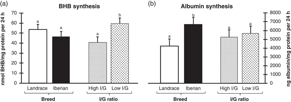
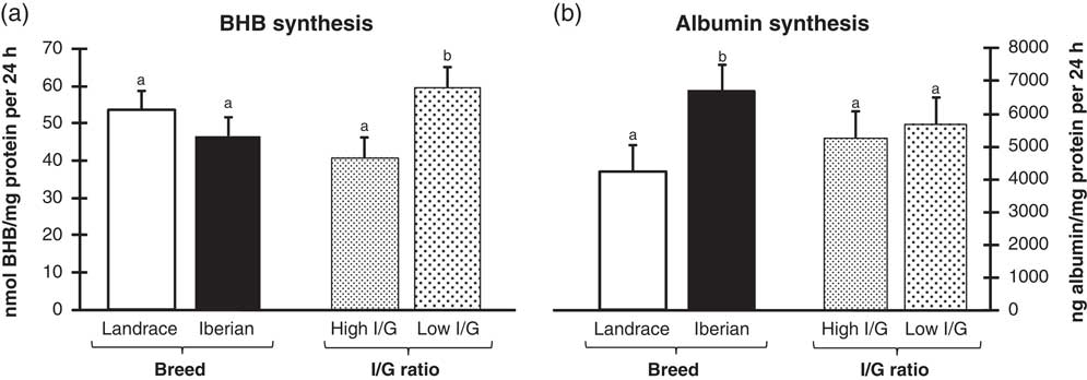
No difference in BHB synthesis was found when hepatocytes from Iberian and Landrace pigs were compared (P>0.10; Figure 3a). Nevertheless, glucagon stimulated BHB synthesis (47%, P<0.05; Figure 3a) relative to insulin. No significant interaction was found. As expected, BHB production was low as it is known that ketogenic capacity of liver from newborn pigs is much lower than in other mammalian species (Duée et al., Reference Duée, Pégorier, Quant, Herbin, Kohl and Girard1994) and low ketone production persists in the adult pig (Duée et al., Reference Duée, Pégorier, Quant, Herbin, Kohl and Girard1994). BHB levels in the present study were in the lower range to previously reported values for hepatocytes isolated from 60 kg pigs (Fernández-Fígares et al., Reference Fernández-Fígares, Shannon, Wray-Cahen and Caperna2004).
Influence of breed (Landrace or Iberian) and insulin/glucagon (I/G) ratio on β-hydroxybutyrate (BHB; nmol/mg protein per 24 h; a) and albumin (µg/100 µg protein per 24 h; b) synthesis. For BHB synthesis, cells were cultured for 48 h in a medium with high insulin (17.3 nM)/glucagon (0.287 nM) or low insulin (0.173 nM)/glucagon (28.7 nM) ratio and media collected at the end. For albumin synthesis, hepatocytes were cultured for 24 h in a medium with high I/G ratio, shifted to a bovine serum albumin- free medium with high or low I/G ratio and media collected. No significant interactions were found between factors. Data are least square means and s.e.m. for duplicate cultures from 8 (Landrace) or 10 (Iberian) pigs. Means with different letters differ (P<0.05).
The low rate of β-oxidation capacity has been ascribed to a propensity for fatty acid esterification v. oxidation in piglets (Pégorier et al., Reference Pégorier, Duée, Girard and Peret1983). As ketone bodies are the result of partial free fatty acid oxidation, no differences in fatty acid oxidation would be expected between pig breeds at the hepatic level. In vivo, early studies with starved miniature pigs (Müller et al., Reference Müller, Paschen and Seitz1982) suggested that mild ketosis encountered was modulated by insulin and glucagon. Indeed, similarly to the present experiment, where glucagon stimulated and insulin inhibited BHB production, it has been reported that ketone bodies synthesis in hepatocytes from 60 kg BW pigs are sensitive to long-term regulation by insulin, glucagon and dexamethasone (Fernández-Fígares et al., Reference Fernández-Fígares, Shannon, Wray-Cahen and Caperna2004), while short-term glucagon addition has been shown to have no effect on ketogenesis in pig hepatocytes (Pégorier et al., Reference Pégorier, Duée, Girard and Peret1982).
IGF-1 synthesis
Nutritional and hormonal factors determine animal growth and growth hormone (GH), by its stimulatory effects on IGF-1 production, is one of the major regulators of growth (Brameld, Reference Brameld1997). As IGF-1 increases protein synthesis (Tomas et al., Reference Tomas, Knowles, Owens, Chandler, Francis, Read and Ballard1992), part of the reason for the increased growth rate and efficiency in pigs may be because of differences in circulating levels of IGF-1. Serum concentrations of IGF-1 are positively correlated with growth rate in pigs (Buonomo et al., Reference Buonomo, Lauterio, Baile and Campion1987), although some workers found no difference when comparing pig breeds with different growth and body composition characteristics (Louveau et al., Reference Louveau, Bonneau and Salter1991). In the present study, synthesis of IGF-1 was decreased (16%) in hepatocytes of Iberian compared with Landrace (647 and 771 pg/mg protein as average, respectively; P<0.05). Previous studies with growing Iberian pigs demonstrated their low genetic potential for growth and lean tissue deposition (Nieto et al., Reference Nieto, Lara, Barea, García-Valverde, Aguinaga, Conde-Aguilera and Aguilera2012) compared with conventional breeds, which is in line with the lower hepatic IGF-1 synthesis in the present study. Although higher fasting serum level of IGF-1 with no difference in serum GH were encountered in Iberian compared with Landrace gilts, a single blood sampling was made (Fernández-Fígares et al., Reference Fernández-Fígares, Lachica, Nieto, Rivera-Ferre and Aguilera2007).
When Meishan and Large White intact boars were compared, blood IGF-1 concentrations were lower in the former (Weiler et al., Reference Weiler, Claus, Schnoebelen-Combes and Louveau1998). Nevertheless, others found no differences in plasma IGF-1 level (Louveau et al., Reference Louveau, Bonneau and Salter1991) when pigs with different growth potential were compared. Although the liver is a primary source of circulating IGF-1, extrapolation of in vitro to in vivo conditions should be made with great care, as elements such as other hormones, receptors or binding proteins (IGFBP) regulate its activity. Furthermore, while IGF-1 has a central role mediating GH biological actions, there exist GH-independent actions for IGF-1 and IGF-1-independent actions for GH (Butler and Le Roith, Reference Butler and Le Roith2001). GH and amino acid supply are synergistic stimuli of IGF-1 secretion (Wheelhouse et al., Reference Wheelhouse, Stubbs, Lomax, MacRae and Hazlerigg1999). Compared with Landrace, Iberian pigs have lower protein requirements (Nieto et al., Reference Nieto, Lara, Barea, García-Valverde, Aguinaga, Conde-Aguilera and Aguilera2012) and capacity of GH release (Rodríguez-López et al., Reference Rodríguez-López, González-Valero, Lachica and Fernández-Fígares2013) implying less IGF-1 release stimuli in vivo.
It has been reported that leptin can inhibit IGF-1 expression and secretion (Ajuwon et al., Reference Ajuwon, Kuske, Anderson, Hancock, Houseknecht, Adeola and Spurlock2003) while IGF-1 inhibits leptin receptor expression (Ramsay et al., Reference Ramsay, Richards, Li and Caperna2010). This would agree with the higher serum leptin levels (Fernández-Fígares et al., Reference Fernández-Fígares, Lachica, Nieto, Rivera-Ferre and Aguilera2007) and lower IGF-1 synthesis in the present study in Iberian compared with Landrace pigs.
IGF-1 decreased expression and activity of gluconeogenic enzymes pyruvate carboxylase and phosphoenolpyruvate carboxykinase (Wang et al., Reference Wang, Zhu, Chen, Li, Gao, Li, Zhang, Long, Wang and Liu2012) in calf hepatocytes, which would agree with the increased GNG in hepatocytes of Iberian (with lower IGF-1 synthesis) compared with Landrace pigs in our experiments.
Albumin synthesis
Albumin is synthesized in liver and represents from 12% to 18% of the total protein synthesized by the liver in rat hepatocytes (Peavy et al., Reference Peavy, Taylor and Jefferson1978). Increased albumin synthesis was found in Iberian compared with Landrace hepatocytes (58%, P<0.05; Figure 3b). Furthermore, no effect of insulin/glucagon ratio (P>0.10; Figure 3b) was found on albumin synthesis, independent of breed. In vivo liver fractional synthesis rate was similar in Iberian and Landrace pigs fed diets with different protein content (Rivera-Ferre et al., Reference Rivera-Ferre, Aguilera and Nieto2005). As synthesis of proteins for export and constitutive proteins constitute liver protein synthesis, it is possible that the former was affected by genetic background (present study) while total liver protein synthesis was not. Furthermore, there seems to be a compartmentalization where proteins for export are synthesized on the rough endoplasmic reticulum while constitutive proteins are synthesized on polysomes within the cell cytosol (Connell et al., Reference Connell, Calder, Anderson and Lobley1997). However, insulin was not involved in the stimulation of protein synthesis in the liver of neonatal pigs (Davis et al., Reference Davis, Fiorotto, Beckett, Burrin, Reeds, Wray-Cahen and Nguyen2001). Furthermore, no difference in albumin synthesis was found in humans with increased plasma insulin (Ballmer et al., Reference Ballmer, McNurlan, Essen, Anderson and Garlick1995) compared with controls.
Urea synthesis
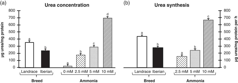
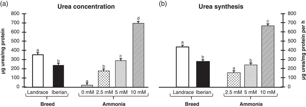
Blood ammonia derives from tissue catabolism and intestine lumen (Huizenga et al., Reference Huizenga, Glips and Tangerman1996). Most portal ammonia is removed from the blood by the liver through urea synthesis preventing hyperammonemia, which would disrupt the central nervous system. Urea concentration in the media (µg/mg protein) and urea synthetic capacity (µg/mg protein per hour) was decreased in hepatocytes from Iberian compared with Landrace pigs (32% and 37%, respectively, P<0.05; Figure 4a and b). Also, hepatocytes were sensitive to increasing amount of ammonia (0 to 10 mM) in the media, which induced augmented urea concentration (6.5, 11 and 28× relative to 0 for 2.5, 5 and 10 mM ammonia, respectively, P<0.0001; Figure 4a) and synthesis (55% and 325% for 5 and 10 mM compared with 2.5 mM ammonia, respectively, P<0.0001; Figure 4b). In the absence of added ammonia, urea synthesis is due to ammonia production from amino acid catabolism. In the present experiment, the basal media contained a total of 8 mM free amino acids and hepatocytes synthesized on average 1.3 µmol urea/mg protein per hour. When ammonia addition was regressed v. urea concentration in the media of Iberian hepatocytes, the following linear model was established: urea concentration (µg/mg protein)=28.08+37.87×ammonia concentration (mM) models coefficient of correlation was significant (r=0.702). A better alternative was the exponential model (urea concentration (µg/mg protein)=e(3.251+0.328×ammonia concentration (mM))), where r=0.826. Similarly, for Landrace both the linear (urea concentration (µg/mg protein)=54.59+50.64×ammonia concentration (mM)) and exponential (urea concentration (µg/mg protein)=e(3.728+0.316×ammonia concentration (mM))) models coefficient of correlation were significant (r=0.777 and 0.814, respectively).
Influence of breed (Landrace or Iberian) and media ammonia concentration (0, 2.5, 5 and 10 mM) on media urea concentration (a) and estimated urea synthesis (b) after 3 h of cultivation. Hepatocytes were cultured for 24 h in medium with high insulin (17.3 nM)/glucagon (0.287 nM) ratio and shifted the same medium with different concentrations of ammonia for 3 h. No significant interactions were found between factors. Data are least square means and s.e.m. for duplicate cultures from 8 (Landrace) or 10 (Iberian) pigs. Means with different letters differ (P<0.05).
GNG from amino acids results in ammonia production, which adds extra ammonia load for the liver to detoxify. In that sense, a greater gluconeogenic capacity of hepatocytes in Iberian (present work) compared with Landrace pigs would imply a greater ammonia load for the liver to detoxify in conditions that favor GNG (e.g. fasting).
In vivo, blood urea N concentration can be inversely related to the efficiency of N utilization (Coma et al., Reference Coma, Zimmerman and Carrion1995) and is associated with dietary protein intake (Yang et al., Reference Yang, Pettigrew, Johnston, Shurson, Wheaton, White, Koketsu, Sower and Rathmacher2000). No difference in serum urea was found when Iberian and Landrace gilts fed isonitrogenous diets were compared, although serum urea was diminished when the animals were fed with lower CP diets (Fernández-Fígares et al., Reference Fernández-Fígares, Lachica, Nieto, Rivera-Ferre and Aguilera2007). In contrast, pigs selected for lean growth efficiency (Fabian et al., Reference Fabian, Chiba, Kuhlers, Frobish, Nadarajah and McElhenney2003) had lower serum urea N concentration compared with control pigs as a consequence of decreased use of amino acids for energy, owing to their greater protein requirement. However, arterial urea concentrations are influenced by several factors such as renal function, fluctuations in the amount of total body water and hydration state. Although it is considered that the liver capacity for urea synthesis exceeds necessities of ammonia detoxification in most physiological situations, it is conceivable that for a very short time early during a meal, pigs may synthesize urea near the potential maximum velocity (Carey et al., Reference Carey, Cheung, Cohen, Brusilow and Raijman1993), especially in Iberian pigs with lower capacity of urea production.
The present study provides differences in hepatic metabolism between pig genotypes that highlight the importance of considering genetic background when in vitro studies are performed.
Acknowledgments
The authors wish to thank Dr L. Lara for statistical advice and Dr T. J. Caperna for critically reading the manuscript. This work was supported by grants AGL2006-5937 from the Ministry of Science and Innovation and AGL2009-08916 from the Ministry of Economy and Competitiveness of Spain. The assays described are part of the PhD thesis of Lucrecia González-Valero, Official Doctorate Program of Nutrition and Food Sciences of the University of Granada.