So-called “absence” of the leaflets of the arterial valves, usually the pulmonary valve, is a rare congenital malformation, reported in 0.2–0.4% of live-born infants.Reference Ferencz 1 When involving the pulmonary valve, it is found most frequently in the setting of patients diagnosed with tetralogy of Fallot.Reference Lev and Eckner 2 , Reference Nagao, Daoud, McAdams, Schwartz and Kaplan 3 Typically associated with microdeletion of 22q11.2 and agenesis of the arterial duct, the lesion can also present as an isolated abnormality. In addition to its association with tetralogy of Fallot, when the arterial duct is typically absent, so-called absence of the pulmonary valve can also be found when the ventricular septum is intact and the arterial duct is persistently patent. Although all leaflets of the valve can be absent, remnants of leaflet tissue are often observed as rudimentary and dysplastic structures, then being disposed in annular fashion at the ventriculo-pulmonary junction.Reference Szwast, Tian and McCann 4 The median gestational age at previous diagnosis of the lesion using intrauterine ultrasound is ~24 weeks, with a low rate of detection within or before the mid second trimester.Reference Wertaschnigg, Jaeggi and Chitayat 5 In contrast to the postnatal course, which is often dictated by the pulmonary complications of the lesion, the prenatal course is dictated by the impact of severe pulmonary insufficiency on right ventricular performance. This can manifest as fetal heart failure due to volume overload, with fetal hydrops and resulting fetal death occurring in up to one-sixth of recognised cases.Reference Szwast, Tian and McCann 4 , Reference Volpe, Paladini and Marasini 6 – Reference Razavi, Sharland and Simpson 8 We have now encountered four fetal cases in which we found so-called “absence” of the leaflets of not only the pulmonary valve but also the aortic valve and, in one case, the common truncal valve. Moreover, one of us has recently had the opportunity to collaborate in the analysis of the processes involved in development of the valvar leaflets in mouse embryos,Reference Anderson, Chaudhry and Mohun 9 – Reference Spicer, Bridgeman, Brown, Mohun and Anderson 11 using high-resolution episcopic microscopyReference Mohun and Weninger 12 and immunohistochemistry.Reference Phillips, Mahendran, Singh, Anderson, Chaudhry and Henderson 13 It is axiomatic that disruption of the normal processes of development must underscore the various pathologies involving the arterial roots, including the occurrence of so-called absence of the valvar leaflets. With this likelihood in mind, we have sought to correlate our insights obtained from normal valvar development with the findings from the autopsies performed in the four human fetuses.
Materials and methods
Comprehensive autopsies were performed in each of the four cases, documenting the cardiac malformations and all other associated malformations. These findings were compared with our current understanding of the development of the arterial valves, which is now based on the examination of over 300 episcopic data sets prepared from developing mouse embryos,Reference Anderson, Mohun and Brown 14 along with other studies using immunohistochemical techniques.Reference Phillips, Mahendran, Singh, Anderson, Chaudhry and Henderson 13 The opportunity to section the data sets in any desirable plane using the episcopic material has greatly facilitated the capacity to understand the origin and gradual appearance of the arterial valvar leaflets and their supporting valvar sinuses, as well as providing new insights regarding the process of separation of the arterial roots.Reference Anderson, Chaudhry and Mohun 9 – Reference Spicer, Bridgeman, Brown, Mohun and Anderson 11 We have prepared new sequences of images to show these changes, permitting us to draw inferences relative to the findings from the fetal autopsies.
Results
Case 1 (Fig 1)
The fetus was at 14–15 weeks of gestation, with the karyotype being positive for trisomy 13. At autopsy, the heart was enlarged, and was positioned within the mediastinum with the apex pointing to the midline. The right atrium, right ventricular outflow tract, and pulmonary trunk were dilated. The leaflets of the pulmonary valve were absent, without any recognisable valvar remnants at the ventriculo-pulmonary junction. The right and left pulmonary arteries were not dilated, and the arterial duct was widely patent. The ventriculo-pulmonary junction, marking the boundary between the right ventricular muscular infundibulum and the arterial walls of the pulmonary trunk, nonetheless, was clearly visible as an annular entity. The heart was otherwise normal, with a probe patent oval foramen and intact interventricular septum. There was arrhinencephaly, but all other organs were normally formed, although the liver was large and the fetus was hydropic.
Case 1. ( a ) The heart is enlarged and the apex is to the midline. The right ventricle, subpulmonary infundibulum, and the pulmonary trunk are dilated. ( b ) The right ventricular outflow tract and the pulmonary trunk are opened, demonstrating complete absence of the pulmonary valve. The red arrows mark the anatomic ventriculo-arterial junction.

Case 2 (Fig 2)
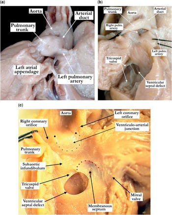
The gestational age of this severely macerated fetus was determined by foot length, and was consistent with 12–13 weeks. The tissue submitted for karyotyping failed to grow, but there was a clinical history of a DNA screening test positive for Trisomy 18. There was a disrupted omphalocele, along with several abnormal phenotypic features. These included a broad nasal bridge with midline depression of the nose, short philtrum, low-set ears, clinodactyly of the right fifth finger, and the second finger of the left hand overlapping the third finger. The intestine was poorly fixed and rotated, with the appendix at the mid-abdomen, a bicornuate uterus, and suggestion of ascites with rounded margins of the liver. The leaflets of both arterial valves were absent, with a slightly prominent ridge marking the superior border of one of the aortic valvar sinuses. The right ventricular outflow tract and pulmonary trunk were dilated, but the right and left pulmonary arteries were narrowed. There was a window-type patent arterial duct, with mild dilation of the ascending aorta. In addition, there was a perimembranous ventricular septal defect, a completely muscular subaortic infundibulum, dysplasia of the leaflets of the tricuspid valve, a defect within the oval fossa, and distal origin of the right subclavian artery. The presence of moderate deposition of haemosiderin in the liver, revealed by microscopic investigation, was suggestive of chronic right heart failure. Thymic involution was also present, suggesting chronic fetal stress.
Case 2. ( a ) The apex of the heart has been directed rightward to show the left aspect of the pulmonary trunk, arterial duct, and the left pulmonary artery. Note the dilated pulmonary trunk compared with the non-dilated left pulmonary artery. The arterial duct is short. ( b ) The right ventricular outflow tract has been opened, demonstrating complete absence of the pulmonary valve. The right and left pulmonary arteries are not dilated and the arterial duct is short. ( c ) The left ventricular outflow tract is opened showing the perimembranous ventricular septal defect with the red dots marking the posterior and inferior border of the membranous septum. The large black dots mark the level of the sinutubular junction and the small black dots outline a slightly prominent ridge marking the borders of a valvar sinus. The true ventriculo-arterial junction is easily appreciated in this area, with a crescent of muscle incorporated into the base of the sinus. Note the completely muscular subaortic infundibulum.

Case 3 (Fig 3)
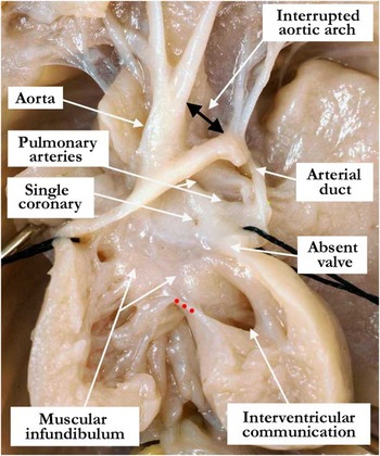
This markedly macerated fetus, based on the external measurements, was of 12–13 weeks of gestation. There was an omphalocele, with malrotation of the bowel. Tissue submitted for karyotyping failed to grow. There was a pulmonary dominant common arterial trunk, with interruption of the aortic arch between the left common carotid and the left subclavian arteries. The leaflets of the truncal valve were absent, without any identifiable remnants of valvar tissue at the ventriculo-truncal junction. The trunk was supported by a completely muscular infundibulum, but the interventricular communication was bordered postero-inferiorly by fibrous continuity between the leaflets of the tricuspid and mitral valves. The left ventricle was hypoplastic, with a parachute mitral valve. There was a single coronary artery and deficiency of the floor of the oval fossa. Microscopic examination of the liver revealed focal hepatic calcifications, along with marked deposits of haemosiderin, predominantly in hepatocytes.
Case 3. The right ventricular outflow tract is opened revealing the pulmonary dominant common arterial trunk. The truncal valve is absent and the anatomic ventriculo-arterial junction is appreciated where the fibrocollagenous wall of the common arterial trunk is supported by the musculature of the completely muscular infundibulum. The double-headed black arrow marks the interrupted aortic arch. The tricuspid and mitral valves are in fibrous continuity along the posterior–inferior border (red dots) of the interventricular communication rendering it a perimembranous defect.

Case 4 (Fig 4)
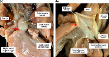
This case sample was received in multiple fragments as a result of a dilation and evacuation procedure secondary to an intrauterine fetal demise. Multiple fragments of the placenta, body parts, and organs were identified, along with the heart and lungs. Gestational age of 13–14 weeks was estimated by foot length. The pericardial sac was intact, with an attached thymus, and the lungs were normally lobed. The aorta and the pulmonary trunk were normally related as they exited the ventricular mass, with evident pulmonary stenosis at the ventriculo-arterial junction. The pulmonary trunk was dilated distal to the stenotic area. There was absence of the anticipated leaflets of the pulmonary valve adjacent to the aortic root, but a thickened and dysplastic leaflet was noted in the non-adjacent position. The right and left pulmonary arteries branched normally from the pulmonary trunk, and were minimally dilated. The brachiocephalic arteries branched normally from the aortic arch, and a patent arterial duct was partially torn from the under surface of the arch. The ventricular septum was intact. Chromosomal analysis revealed a normal male karyotype. Fluorescent in situ hybridisation for deletion of 18qter and 22q11.2 was also performed, but was negative.
Case 4. ( a ) This anterior and leftward view shows the dilated pulmonary trunk with stenosis (red arrow) at the ventriculo-arterial junction. The left pulmonary artery is not dilated. ( b ) The majority of the pulmonary valve is absent with one, thickened, dysplastic leaflet (red arrow) in the anterior position. There is stenosis at the ventriculo-arterial junction with post-stenotic dilation of the pulmonary trunk. The right and left pulmonary arteries are not dilated.

Summary of results
The four described fetal cases presented as early intrauterine losses, with one evacuated from the uterus by dilation and evacuation, all cases being between 12 and 15 weeks of gestation. In the first case, there was complete absence of all the leaflets of the pulmonary valve. In the fourth case, there was absence only of the leaflets anticipated to guard the valvar sinuses adjacent to the aortic root, with a dysplastic non-adjacent pulmonary valvar leaflet being present in this heart. The second case demonstrated absence of the leaflets of both arterial valves in the setting of a perimembranous ventricular septal defect and a window-type arterial duct. This fetus also exhibited a completely muscular subaortic infundibulum. The third fetus had a pulmonary-dominant type of common arterial trunk, with absence of all the truncal valvar leaflets. This fetus had a completely muscular subtruncal infundibulum and a single coronary artery.
Developmental considerations
Most of those who continue to investigate the development of the outflow tract of the heart, when describing its components, continue to follow the suggestion of Kramer,Reference Kramer 15 and recognise the “truncus” and “conus”. To the best of our knowledge, there is no consensus as to the boundaries of these components or whether the arterial roots should be considered as belonging to the “truncus” or the “conus”. As KramerReference Kramer 15 had shown in his stellar investigation, however, subsequent to the appearance of the intercalated cushions in the developing outflow tract, it is possible to describe discrete distal, intermediate, and proximal components of the developing outflow tract. This means that the developing outflow tract can be analysed in tripartite, as opposed to bipartite, fashion. The data now available from episcopic microscopyReference Mohun and Weninger 12 show the value of adopting the tripartite approach, as the arterial roots along with the valvar leaflets develop within the intermediate part of the outflow tract.Reference Anderson, Chaudhry and Mohun 9 – Reference Spicer, Bridgeman, Brown, Mohun and Anderson 11 The distal part of the tract separates into the intrapericardial components of the aorta and pulmonary trunk, with the branches of the aortic sac forming the extrapericardial parts of the arterial channels. The proximal part is transformed into the ventricular outflow tracts.Reference Anderson, Chaudhry and Mohun 9 – Reference Spicer, Bridgeman, Brown, Mohun and Anderson 11
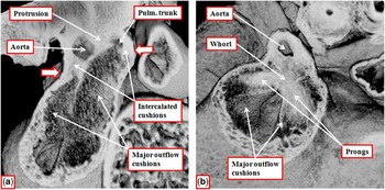
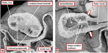
The intrapericardial components of the arterial trunks are initially separated from each other in the mouse during the eleventh embryonic day (E11.5). This is achieved by growth into the distal cavity of the outflow tract of a protrusion from the dorsal wall of the aortic sac (Fig 5a). The protrusion then fuses with the distal ends of the major outflow cushions. At the time of this fusion, the cushions occupy only the intermediate and proximal parts of the outflow tract. It has been suggested that it is an “aortopulmonary septal complex” that divides the intrapericardial arterial channels.Reference Bartelings and Gittenberger-de Groot 16 The episcopic sections, however, show that this complex, made up of the so-called “whorl” and its “prongs”, is not seen until E12.5, when the columns of condensed mesenchyme extend into the unfused proximal components of the major outflow cushions (Fig 5b). The so-called “septal complex”, therefore, is responsible for separating the developing arterial roots and the proximal ventricular outflow tracts, rather than the intrapericardial components of the aorta and the pulmonary trunk.
The images prepared from episcopic data sets from developing mice sacrificed at embryonic day (E) 11.5 ( a ) and 12.5 ( b ) show how the distal part of the outflow tract is separated by growth of a protrusion from the dorsal wall of the aortic sac into the intrapericardial aortic and pulmonary (pulm.) channels. The protrusion fuses with the distal ends of the major outflow cushions, which remained encased within a sleeve of outflow tract myocardium (white arrows with red borders). By E12.5 ( b ), the aortic root has separated from the pulmonary root. The columns of condensed mesenchyme that form the so-called “aortopulmonary septal complex”, made up of a whorl and two prongs, can now be seen to occupy the major outflow cushions. They divide the intermediate and proximal parts of the outflow tract, although the proximal parts of the cushions remain unfused at this stage. They do not separate the intrapericardial components of the aorta and the pulmonary trunk.

Fusion of the central components of the distal parts of the major outflow cushions during E12.5, therefore, serves to separate the aortic root from the pulmonary root. After the central fusion, however, the parietal margins of the cushions remain unfused. The interposition of the aortic and pulmonary intercalated cushions, first observed within the intermediate part of the outflow tract during E11.5 (Fig 5a), between the parietal unfused margins of the central cushions then produces the primordiums for subsequent development of the arterial valves (Fig 6a).
The images are from the same episcopic data set prepared from a developing mouse embryo sacrificed at embryonic day 12.5. ( a ) The short-axis section of the intermediate component of the outflow tract as viewed from its cranial surface. The interposition of the intercalated cushions with the unfused peripheral margins of the major outflow cushions has produced the primordiums of the developing arterial roots. As seen in ( b ), however, which is a longitudinal cut across the developing pulmonary root, the cushions themselves extend to the distal margin of the myocardial sleeve that surround the developing roots, with this level marking the effective ventriculo-arterial junction (white arrows with red borders).

At the stage of fusion of the central parts of the major cushions, the proximal and intermediate parts of the outflow tract retain their myocardial walls. The boundary between the myocardial and arterial components of the outflow tract at this stage, therefore, is at the distal border of the intermediate component of the outflow tract. At this stage, furthermore, although the obvious template for formation of the valves is evident when images are viewed in short axis, there is no discrete formation of either valvar leaflet. As the distal extent of the cushions, including the intercalated cushions, remains level with the distal margin of the myocardial walls surrounding the intermediate part of the outflow tract, it also follows that, as yet, there has been no formation of the arterial valvar sinuses (Fig 6b). At this stage, therefore, it is the bulk of the cushions themselves that serves as a valvar mechanism.
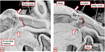
It is during the next day of embryonic development (E13.5) that we begin to observe the formation of discrete valvar leaflets. This occurs by a process of excavation of the distal surfaces of the unfused parietal margins of the major cushions, with comparable excavation involving the distal surfaces of the intercalated cushions. By E14.5, the excavation has extended towards the ventricular cavities in both the developing aortic root and the pulmonary root (Fig 7a). As the distal margins of the cushions undergo this excavation, there is ongoing proximal growth of the arterial walls of the outflow tract. The newly formed arterial tissues fill the semilunar spaces that are produced in the distal edges of the cushions by the process of excavation. Subsequent to the excavation, the distal extents of the cushions at their zones of apposition remain at the levels of the initial boundary between the intermediate component of the outflow tract and the arterial walls of the distal outflow tract. As the arterial walls have now extended proximally between these sites of apposition, which will form the eventual valvar commissures, the level of the myocardial wall of the intermediate component is effectively translocated in the proximal direction. By E16.5, the continuing process of excavation, with ongoing filling of the spaces within the excavating cushions by the ingrowth of arterial tissues, means that the junction between the walls of the newly formed arterial valvar sinuses and the persisting myocardial boundary is now closer to the basal hinges of the newly forming valvar leaflets rather than the level of the commissures. The newly -formed leaflets, however, remain bulky at this stage of development. It is barely possible to recognise the spaces that eventually, on the ventricular aspect, become the interleaflet triangles (Fig 7).
The images are from episcopic data sets prepared from developing mice sacrificed at embryonic day (E) 14.5 ( a ) and 17.5 ( b ). They show the gradual excavation of the peripheral components of the major outflow cushions in the pulmonary root (compare with Fig 6b). The distal margins of the cushions (red dotted line) now form the developing sinutubular junction, with effective regression of the ventriculo-arterial junction (white arrows with red borders) proximally towards the ventricular cavity. By E17.5 ( b ) it is possible to recognise the developing arterial valvar sinuses, with the peripheral margins of the excavated cushions now forming the semilunar hinges of the valvar leaflets. As shown in ( a ), the distal attachments of the semilunar leaflets continue to mark the sinutubular junction. The inference can be made that, in absence of formation of the leaflets, it will not be possible to recognise a discrete sinutubular junction, as was the case in our fetal specimens.

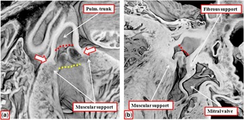
By term in the mouse heart, which takes place at embryonic day 18.5, there has been further thinning of the leaflets such that, particularly in the aortic root, it is now possible to recognise the well-formed sinutubular junction at the level of the valvar commissures. This level had initially coincided with the distal extent of the myocardium at the stage of appearance of the intercalated cushions (Fig 6b). By E18.5, there has also been attenuation of the muscular inner heart curvature. At earlier stages, this muscular fold interposes between the leaflets of the developing aortic and mitral valves in the roof of the left ventricle, providing the developing aortic valve with a muscular infundibulum. It is only shortly before term that it becomes possible to recognise the aortic-to-mitral valvar continuity that is the feature of the postnatal heart. The pulmonary valvar leaflets, in contrast, continue to be supported exclusively by the subpulmonary muscular infundibulum. In this regard, the muscular support of the leaflets of the pulmonary valve adjacent to the aortic root is provided by muscularisation of the surface of the proximal fused major outflow cushions.Reference Ya, Van Den Hoff and De Boer 17 The basal extents of the semilunar valvar leaflets, formed by excavation of the cushions, remain proximal to the anatomic junction between the infundibular musculature and the arterial walls in all three sinuses of the pulmonary valve. This means that the base of each sinus is myocardial. Subsequent to the development of aortic-to-mitral valvar continuity, it is only the two adjacent sinuses of the aortic root that have such myocardial support at their bases (Fig 8).
The images show the pulmonary ( a ) and aortic ( b ) arterial roots at embryonic day 18.5. They are from the same episcopic data set. It is now possible to recognise the full length of both roots, with the so-called echocardiographic annulus, formed by joining together the basal attachments of the arterial valvar leaflets, best seen in the pulmonary root (yellow dotted line in ( a )). Note that this is a virtual plane, and does not correspond with any anatomic entity. The sinutubular junctions (red dotted lines) are now well-formed in both arterial roots. The ventriculo-arterial junction is now much closer to the plane of the echocardiographic annulus than to the sinutubular junction (white arrows with red borders in ( a )).

Discussion
It is axiomatic that recognition of parallels between the development of the human and mouse heart may be relevant and even inspiring. We recognise, nonetheless, that in the setting of our present study, our examination of the normal development of the valvar leaflets in the mouse heart cannot directly inform the mechanisms of altered valvogenesis in humans. At best, we can make only inferences, rather than providing information that would more directly reflect cause and effect. We are also aware that, so as to achieve these latter goals concerning morpho-functional correlations, it is generally recommended that investigators should use a mouse model of the human disease. To the best of our knowledge, no such model currently exists for absence of the leaflets of the arterial valves. Should a model eventually emerge, it is our hope that our current inferences will provide the necessary yardstick for comparisons with normal development.
The normally developed arterial roots are made up of the valvar leaflets, supported by the valvar sinuses. Within each root, one pulmonary and the other aortic, the semilunar hinges of the leaflets extend from the proximal bases of the supporting ventricular outflow tracts to the distal level of the sinutubular junctions. On the ventricular aspects of the sinuses, the spaces between the hinges are filled by interleaflet fibrous triangles, with their apices also extending to the sinutubular junctions.Reference Sutton, Ho and Anderson 18 As the valvar hinges are semilunar, it follows that there is no true anatomic ring-like structure that can justifiably be defined as an “annulus”.Reference Anderson, Mohun and Spicer 10 It is perhaps paradoxical that a relatively annular attachment of the valvar leaflets is found only in the setting of a critically stenotic valvar orifice. In many cases of so-called “absence” of the arterial valvar leaflets, furthermore, particularly those involving the pulmonary valve, it is well recognised that rudimentary valvar remnants can be found attached in truly annular manner.Reference Lincoln and Garg 19 Taken together, these features emphasise the significance of the semilunar nature of the hinges of the leaflets in allowing the normal arterial valves to open and close in competent fashion.
The images prepared for this study using episcopic microscopy serve to endorse the previous analyses of the developing arterial roots.Reference Anderson, Chaudhry and Mohun 9 – Reference Spicer, Bridgeman, Brown, Mohun and Anderson 11 They show that the leaflets of the arterial valves are formed, in the mouse, from embryonic day 12.5 to term. The process involves gradual excavation of the distal surfaces of the major outflow cushions, along with excavation of the intercalated cushions formed within the intermediate component of the outflow tract. It is the process of excavation that eventually produces the semilunar configuration of the valvar leaflets. The distal sites of apposition of the valvar hinges at the commissures are then formed at the initial boundary between the intermediate and distal components of the developing outflow tract – this in the postnatal heart being the levels of the sinutubular junctions. The spaces produced proximal to these commissural attachments at the sinutubular junctions are filled during development by the continuing growth of arterial tissues, thus forming the arterial valvar sinuses. During development, therefore, the junction between the ventricular and arterial tissues in both arterial roots is effectively transferred from the levels of the sinutubular junctions to the levels much closer to the so-called echocardiographic “annulus”. This latter entity is a virtual structure. It is created by joining together the proximal margins of the excavated cushions, which at term represent the nadirs of attachment of the valvar leaflets.
Despite the fact that there is no anatomic entity that corresponds with the “annulus” forming the proximal boundary of both arterial roots, the term is uniformly used by echocardiographers. A recent questionnaire answered by cardiac surgeonsReference Sievers, Hemmer and Beyersdorf 20 revealed that some define the “annulus” as the overall semilunar configuration of the hinges of the arterial valves. Others such as echocardiographers considered the annulus to be the entrance to the arterial roots. The only true circular anatomical structure found within the roots – namely, the junction between the muscular ventricular outlet and the arterial walls of the valvar sinuses, best seen in the pulmonary root – was not nominated as an “annulus”. This ring is best demonstrated when the valvar leaflets are removed. It is not surprising, therefore, that the ring, which represents the true ventriculo-arterial junction, was obvious in all our fetal autopsy cases. What was surprising is that in only one of our four cases was there any formation of a valvar leaflet, this being a remnant of the non-adjacent pulmonary valvar leaflet. It could be pertinent that this leaflet is exactly the one that receives only a minor contribution of cells derived from the neural crest.Reference Phillips, Mahendran, Singh, Anderson, Chaudhry and Henderson 13 When seeking inferences from our knowledge of development, therefore, it is tempting to argue that, at least in this particular case, the problem is related to the contributions made by tissues derived from the neural crest. This is unlikely, as it is now accepted that the neural crest cells are required for appropriate septation of the outflow tract. This had occurred in normal fashion in the fetus with the dysplastic non-adjacent pulmonary valvar leaflet. Septation had occurred normally, however, in only three of our four cases, as the fourth case had absence of the leaflets of a common truncal valve. Common arterial trunk is known to be the consequence of failure of fusion of the major outflow cushions.Reference Van Mierop, Patterson and Schnarr 21 We can infer therefore that formation of the valvar leaflets from the outflow cushions is independent of the role of fusion of the major cushions in dividing the initially common outflow tract into its aortic and pulmonary components. We can also infer that formation of the leaflets is selective for the aortic and pulmonary valves, as the pulmonary valve was exclusively involved in two of our cases, with both valves involved in a third case. All leaflets of the common truncal valve, furthermore, were absent. The inference can also be made that presence of a completely muscular infundibulum is significant. It is most usually the pulmonary valve that shows so-called “absence” of the leaflets, and the normal pulmonary valve is always supported by an infundibulum. In our cases with absence of the leaflets of the aortic and common truncal valves, both valves were unusually supported by completely muscular infundibulums.
It may also be pertinent that, although usually described as “absent valve syndrome”, the pulmonary variant of the lesion is typically found with verrucous remnants of the leaflets formed in annular fashion at the level of the ventriculo-pulmonary junction. No such remnants were found in any of our fetal cases, apart from the dysplastic non-adjacent leaflet, nor were valvar remnants found in aortic or common truncal positions. We must presume that the remnants seen most times in postnatal cases would have appeared with ongoing gestation. This inference points to the need for additional studies to identify the molecular cues that underscore the ability of the outflow cushions not only normally to septate the developing outflow tract but also to form the leaflets of the arterial valves.
Conclusions
Our developmental and pathological findings lend support to the contention of the surgical team responsible for the recent questionnaire,Reference Sievers, Hemmer and Beyersdorf 20 namely, that there is need for consensus on the terms used to describe the arterial roots, including what is to be defined as the annulus. As was suggested by Kramer long ago,Reference Kramer 15 description of the components of the developing heart and of their malformations is preferred to using letters or numbers for classification. Kramer acknowledged that, on occasion, the descriptive terms are long, but this in turn simplifies understanding.Reference Kramer 15 If surgeons, cardiologists, echocardiographers, pathologists, and molecular biologists can reach consensus in naming the components of the arterial roots and recognising the part of the outflow tract from which they develop, then recognising the normal arrangement and distinguishing it from the abnormal will no longer be problematic. The developmental mechanisms, in turn, will also be easier to understand.
Acknowledgements
The authors are indebted to Dr Timothy J. Mohun, from the Crick Institute, London, who prepared all the episcopic data sets used to establish the mode of formation of the arterial roots. They also express their thanks to Professor Nigel Brown, St George’s Medical University, London, who made it possible for them to examine and prepare the data sets for publication.