Case report
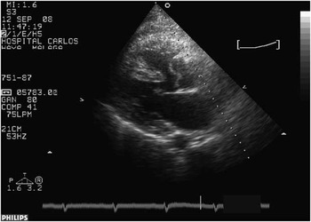
First case: A 50-year-old man, weighing 90 kilograms with a body surface area 2.07 metre square, was scheduled for elective repair of tetralogy of Fallot. He had previously refused the procedure twice, and now decided to undergo surgery because of increasing shortness of breath (New York Heart Association functional class II–III). On echocardiography, the aorta was overriding the ventricular septal defect (Fig. 1), the aortic annulus was 40 millimetres in diameter, the right ventricular outflow tract gradient was 110 millimetres of mercury, and the pulmonary annulus diameter was 23 millimetres. Pre-operative angiography revealed an anomalous origin of the left coronary artery from the right one, crossing the right ventricular outflow tract.
Aorta overriding the ventricular septal defect.

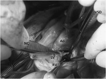
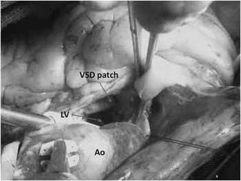
The pulmonary artery was opened transversely. The pulmonary valve was displastic, with fused commissures. Triple commissurotomy was carried out, and the fibrotic tissue under the valve was removed and parietal bands were incised. With a Hegar dilator no. 22, the right ventricular outflow tract was probed. Attention was then turned to the aorta, which was opened in an oblique manner towards the non-coronary sinus. The ascending aorta was not dilated, the valve was trileaflet, and the muscular crest of the ventricular septal defect (Fig. 2) was just underneath the valve, as the aortic valve itself was overriding the ventricular septal defect. The right ventricular outflow tract was again assessed through the ventricular septal defect. A Dacron patch, with the same size of the aortic annulus, was trimmed and sutured in a running fashion to the ventricular septal defect (Fig. 3). The caudal stitches were anchored slightly to the right aspect of the muscular crest of the ventricular septal defect – so as not to impinge the conduction tissue – and cephalad to the annulus of the right coronary sinus – between both commissures. At the end of the procedure, an echocardiography showed a pinpoint residual ventricular septal defect with no aortic regurgitation and a right ventricular outflow tract gradient of 30 millimetres of mercury, in sinus rhythm and absence on right bundle branch block.
Aortotomy (Ao) showing the aortic valve overriding both ventricles: right ventricle (RV) at the front and left ventricle (LV) at the back. Forceps grasping the muscular crescent of the ventricular septal defect (VSD) and the non-coronary aortic leaflet (NC).

After correction as seen from the aortotomy (Ao): the left ventricle outflow tract (LV) and the patch (VSD) closing the ventricular septal defect.

After 2 years, the patient is in New York Heart Association class I, with no residual ventricular septal defect, no aortic regurgitation, and complete relief of right ventricular outflow tract obstruction.
Second case: A 29-year-old woman from overseas, weighing 58 kilograms, presented with a tetralogy of Fallot. On examination, situs inversus with dextrocardia and bilateral superior caval veins were found. The aortic annulus was 26 millimetres in diameter, the pulmonary annulus was 23 millimetres, and the right ventricular outflow tract gradient was 80 millimetres of mercury. Surgery was conducted in a similar manner, cannulating the three caval veins. The pulmonary valve happened to be bicuspid and stenotic, and double comissurotomy was performed. Subsequently, parietal bands and fibrotic tissue under the valve were incised. On opening the aorta, the ventricular septal defect underneath was assessed, the right ventricular outflow tract revised, and a patch secured to close the defect. Echocardiography showed no residual ventricular septal defect, no aortic regurgitation, and 35 millimetres of mercury right ventricular outflow tract gradient.
After 6 months, the patient remains in New York Heart Association class I, with residual 20 millimetres of mercury gradient in the right ventricular outflow tract.
Discussion
Fallot repair in adult patients is very rare. On the other hand, aortic valve is seldom the route of choice for ventricular septal defect closure, either because of the small size or for the fear of distorting the aortic annulus. In the first case, the patient was diagnosed earlier in life but refused treatment twice, which eventually resulted in elective surgery at 50 years. The combination of dilated aortic root (40 millimetres) plus overriding aorta offered us a unique opportunity of approaching the ventricular septal defect through the aorta, easing the exposure and avoiding more usual ways such as atriotomy or ventriculotomy, as well as its side effects from scarring. Moreover, the enlarged size of the annulus and the location of the ventricular septal defect – just in front of the aortic valve – made the procedure a straightforward one. Even the issue of annular distortion was neutralised, as the patient's aortic root was not supposed to grow any longer. Owing to the fact that the pulmonary annulus diameter was adequate and patch augmentation of the right ventricular outflow tract was unnecessary, the anomalous coronary artery crossing the right ventricular outflow tract became irrelevant.
Situs inversus, with mirror image pattern of mediastinal structures, could have been cumbersome had the ventricular septal defect been approached through the left-sided right atrium in our second patient – hazard of residual ventricular septal defect and rhythm disturbances. Fortunately, the proper diameter of the pulmonary annulus and a short infundibulum – as in the first case – enabled us to carry out a fairly straightforward procedure through the aorta.
Ventricular septal defect closure through the aortic valve has seldom been reported. Yacoub et alReference Yacoub, Khan, Stavri, Shinebourne and Riddley-Smith1 published a series of ventricular septal defect closure and aortic valve resuspension by this approach in conal ventricular septal defect plus aortic regurgitation. In his report, a direct ventricular septal defect closure – without patch – was enough to deal with the ventricular septal defect and aortic insufficiency. Watanabe et alReference Watanabe, Sakai, Song, Shimomura, Tajima and Yasuura2 and Belli et alReference Belli, Houyel, Serraf, Lacour-Gayet, Petit and Planché3 reported the closure of residual ventricular septal defect in corrected Fallot patients through the aortic approach. The paper from Belli et alReference Belli, Houyel, Serraf, Lacour-Gayet, Petit and Planché3 was based on the lessons learnt from the natural history of patients previously operated for double outlet right ventricle who eventually developed subaortic stenosis or residual intramural ventricular septal defect. Literature about primary closure of ventricular septal defect through the aortic valve in Fallot patients is scarce. Leão et alReference Leão, Buffolo, Coto, Maluf and Andrade4 reported 124 patients with ventricular septal defect closure through the aortic valve (including six primary Fallot and five residual ventricular septal defect in Fallot patients. Watanabe et alReference Watanabe, Sakai, Song, Shimomura, Tajima and Yasuura2 mentioned this approach in the discussion of his paper.
The chances of reproducing this procedure in typical cases of tetralogy of Fallot are negligible, as the attachment of the ventricular septal defect patch to the aortic annulus will undoubtedly distort the aotic root in the future. The same statement rules for the double outlet right ventricle with subpulmonary ventricular septal defect – Taussig–Bing – in which the closure of the ventricular septal defect through the neo-aortic valve endangers its function on predisposing it to regurgitation. On the other hand, as previously stated, these grown-up patients presented us with the real chance of a straightforward procedure through a well-developed aortic root, avoiding “spatial” problems in our situs inversus case.